MRI is a powerful diagnostic imaging tool, with more than 100 million scans performed worldwide each year. While MR signals contain rich information from multiple molecules and numerous physical and biological processes, current clinical MRI exams rely solely on signals from water molecules in tissues and generally only obtain one tissue biomarker at a time. But MRI could do so much more.
A research team headed up at the University of Illinois Urbana-Champaign has done just that, devising a new MRI technique – multiplexed MRI (MRx) – that enables simultaneous mapping of multiple molecular signals using a standard clinical 3 T MRI scanner.
The barrier to performing multiparametric imaging with conventional MRI lies in the “curse of dimensionality”, in which high-dimensional imaging requires prohibitively long scan times. Multimolecular MRI, meanwhile, is limited by weak signals from brain metabolites and neurotransmitters (typically 1000–10,000 times weaker than proton-based signals from water molecules), which often overlap, making them difficult to detect and separate.
“MRx overcomes these challenges through specialized data acquisition and processing strategies,” explains study leader Zhi-Pei Liang. “During data acquisition, MRx simultaneously excites and encodes all detectable molecular signals with sparse sampling to achieve high imaging speed. During data processing, MRx employs physics-driven machine learning methods to separate and quantify the different signal components.”
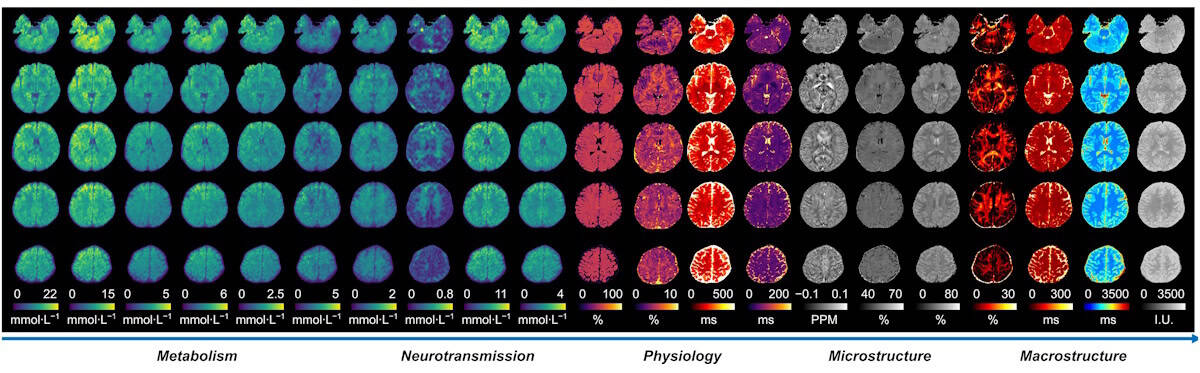
Reporting their findings in Nature, the researchers demonstrate high-resolution mapping of 22 quantitative biomarkers of the whole brain in a single scan. They also show how a new sparse sampling scheme enables acquisition of these biomarkers in just 14 min – significantly shorter than clinical multi-contrast MRI protocols that can take up to an hour.
“Our main motivation was to develop an ‘omni’ imaging technology that fully harnesses the rich biological information embedded in magnetic resonance signals, enabling us to unravel the structural, physiological and molecular fingerprints of brain function and diseases,” says Liang.
In vivo studies
MRI is widely used within brain tumour diagnosis to evaluate tumour location, size and extent, and blood–brain barrier disruption. However, standard MRI scans do not directly reveal the underlying pathophysiological changes and tumour heterogeneity. MRx, on the other hand, can acquire a wide range of biomarkers that provide valuable information on processes such as neuronal loss, energy metabolism, axonal damage, hypoxia, demyelination and many more.
To test the technique, Liang and colleagues performed MRx imaging on patients with clinically diagnosed brain tumours, using machine learning to combine the measured biomarkers into a single variable defining the tissue state at each pixel. This MRx “tissue state index” could differentiate eight distinct tissue states: grey matter; white matter; cerebrospinal fluid; oedema (fluid build-up); meningioma; low and high-grade oligodendroglioma; and glioblastoma. Standard multiparametric MRI failed to separate these states.
This ability to accurately characterize tissue states could enable a range of essential clinical tasks, such as grading low- versus high-grade brain tumours, for example, or separating glioblastoma from oedema during radiation therapy planning.
MRx could also prove invaluable for lesion characterization in multiple sclerosis (MS), a critical process for stratifying patients, planning treatment and predicting disease progression. The researchers demonstrated that MRx of patients with MS could differentiate active and chronic MS lesions without requiring contrast agents (as in current practice), attributed to the technique’s ability to visualize biomarkers specific to individual pathophysiological processes.
Such MRx biomarkers also helped to predict lesion progression, by capturing key pathophysiological features that cannot be revealed by conventional MRI, a feature that could enable early interventions and improve patient outcome.
Beyond cancer and MS, many other brain diseases could also benefit from MRx, including stroke, epilepsy and Alzheimer’s disease, for example. “MRx is expected to open up new opportunities for brain mapping and for precision healthcare of brain diseases, including neurological and neurodegenerative disorders,” says Liang.
Metamaterial antennas enhance MR images of the eye and brain
For the proton-based studies reported in this latest study, MRx was performed without needing any modifications to the MRI scanner hardware. Instead, the method is implemented using a new pulse sequence for data acquisition plus custom software for data processing. Liang notes that extending MRx to include multiple nuclei – such as sodium, phosphorus, and deuterium – will require specialized multinuclear RF coil hardware.
“Our current efforts are focused on further improving the robustness and reliability of MRx under practical clinical imaging conditions, to facilitate both scientific studies and clinical translation,” he tells Physics World, noting that MRx has already been licensed (through Siemens) to imaging centres worldwide for evaluation of its clinical potential. “We are also expanding the technology to map additional molecular species and, ultimately, to enable multinuclear multiplexed imaging beyond protons.”


