Applying physics to the properties and underlying structures of the molecules of life offers an insight into the mechanisms that make living beings tick. But even seemingly simple actions like muscle contraction involve a wide array of biological interactions, which has shrouded the dynamics and function of individual molecules behind a curtain of complexity.
The scientific community has developed a powerful toolbox of methods to probe these interactions over the past two decades, uncovering previously hidden information about the structure, dynamics and function of individual biomolecules. And Miklós Kellermayer, who heads up the Department of Biophysics and Radiation Biology at Semmelweis University, Hungary, has been at the forefront of this rapidly growing field, called single-molecule biophysics, since the very beginning.
“In 1995/6, I was a postdoc at Washington State University, where we collaborated with single-molecule visualization pioneer Carlos Bustamante,” Kellermayer recalls. “We built an optical trap and succeeded in pulling the muscle protein titin – the first protein molecule to be mechanically manipulated in the history of biophysics.”
Following this success, Kellermayer’s career can be thought of as a microcosm for how the wider single-molecule biophysics landscape has evolved. He returned to his country of birth in 1997, and since then has both invented novel techniques and branched out to explore and expose the inner workings of a wide range of proteins.
Precision positioning
One of these techniques represented a step change in capabilities for the community. In 2006, Kellermayer and collaborators used a Mad City Labs objective piezo translator – a device that allows ultra-precise positioning – to focus a high magnification objective within a total internal reflection fluorescence (TIRF) microscope; a powerful method whereby molecules are usually made to fluoresce via chemical or genetic means so that they can be selectively imaged.
The piezo-electric stage allows us to obtain much more precise measurements, and manipulate structures like titin, DNA and longer proteins
Professor Miklós Kellermayer, Semmelweis University, Hungary
They then combined the enhanced TIRF microscope with an atomic force microscope (AFM) – a high-resolution technique that ‘feels’ a surface with a mechanical probe – for the first time. This unique spatially and temporally synchronized set-up allowed the researchers to track the molecular topography and manipulate individual biomolecules, while at same time monitoring changes in fluorescence – even in living cells.
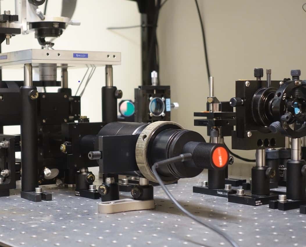
Kellermayer and his team continue to explore the bleeding edge of what is possible with today’s technology. For instance, they have built unique optical tweezers that allow them not only to trap different molecules using radiation pressure from a focused laser beam, but also to manipulate and measure how much movement is necessary to pull a molecule with a certain force. “The central piece of this instrument is a hyper-strength Mad City Labs xyz piezo-electric stage,” explains Kellermayer. “It allows us to obtain much more precise measurements, and manipulate structures like titin, DNA and longer proteins.”
Flexing muscles
Kellermayer’s group has applied these and other techniques to a host of previously intractable problems in recent years, including further important contributions to understanding the role of the titin molecule inside muscle. Titin is the largest protein ever discovered, measuring more than 1 µm in length, and it is known to act as a molecular spring that imbues muscle with its elasticity. But whether titin unfolds during muscle extension, and how this unfolding might contribute to muscle contraction, are questions that have been hotly debated among the biophysics community.
To answer these questions and illuminate the molecule’s folding mechanisms, Kellermayer and colleagues have used high-resolution optical tweezers to manipulate individual titin molecules that were purified from back muscle taken from rabbits. They discovered that some protein domains in titin can unfold under very low physiologically relevant forces, and in later work they were able to partially unfold the molecule and then allow it to refold. This was one of the first experiments to reveal that titin refolding assists muscle contraction by generating mechanical force.
Tapping at viruses
Beyond titin, the group is also highly active in elucidating how DNA is ejected by a virus when it infects a host cell. A virus consists of a nanoscale shell called a capsid that encapsulates the hostile genomic material. After binding to surface receptor sites, most viruses transfer their genome inside the host cell while leaving the capsid outside. This material fools the host organism into manufacturing the viral structural elements, which spontaneously reproduce the virus particle by self-assembly.
Given viruses are already being used as a blueprint for targeted drug delivery, surprisingly little is known about the dynamics of capsid behaviour, let alone how a virus releases its genetic material. “Using AFM, we studied the virus bacteriophage T7 – a virus that infects susceptible bacterial cells, such as E. coli – and found a very interesting structural change in the virus’s capsid wall when we pushed it with the AFM cantilever,” explains Kellermayer. More specifically, force from the cantilever prompted the capsid to buckle in discrete steps that were integer multiples of around 0.6 nm.
Then, when the researchers retracted the cantilever, the capsid recovered its structure in the same discrete steps. “It’s a reversible structural change,” Kellermayer says. “The bacteriophage can heal itself, can recover from mechanical distortion.” This discovery offers an insight into how capsids protect viruses from harsh environmental impacts.
Yet Kellermayer and his team did not stop there. In their most recent paper, the group again used AFM on T7, but instead of applying constant force on the capsid, they gently tapped it by oscillating the AFM cantilever. To their surprise, when they knocked on the virus’s door, it answered by opening up and releasing its DNA. “We still don’t understand the mechanism behind this, but it is certainly remarkable,” explains Kellermayer, who also found that increasing the cantilever force accelerated the DNA release process. “Hopefully, this will lead us to a better understanding of how viruses eject their DNA into the host cell.”
With further insights likely to come from current attempts to measure the force required to pull out the DNA from the viral capsid, as well as studies of how different ligands might influence the behaviour of titin and numerous other topics the team is investigating, Kellermayer’s research is testament to how single-molecule experiments can solve the ‘unsolvable’ and unlock an incredible wealth of hidden information about the molecules of life.