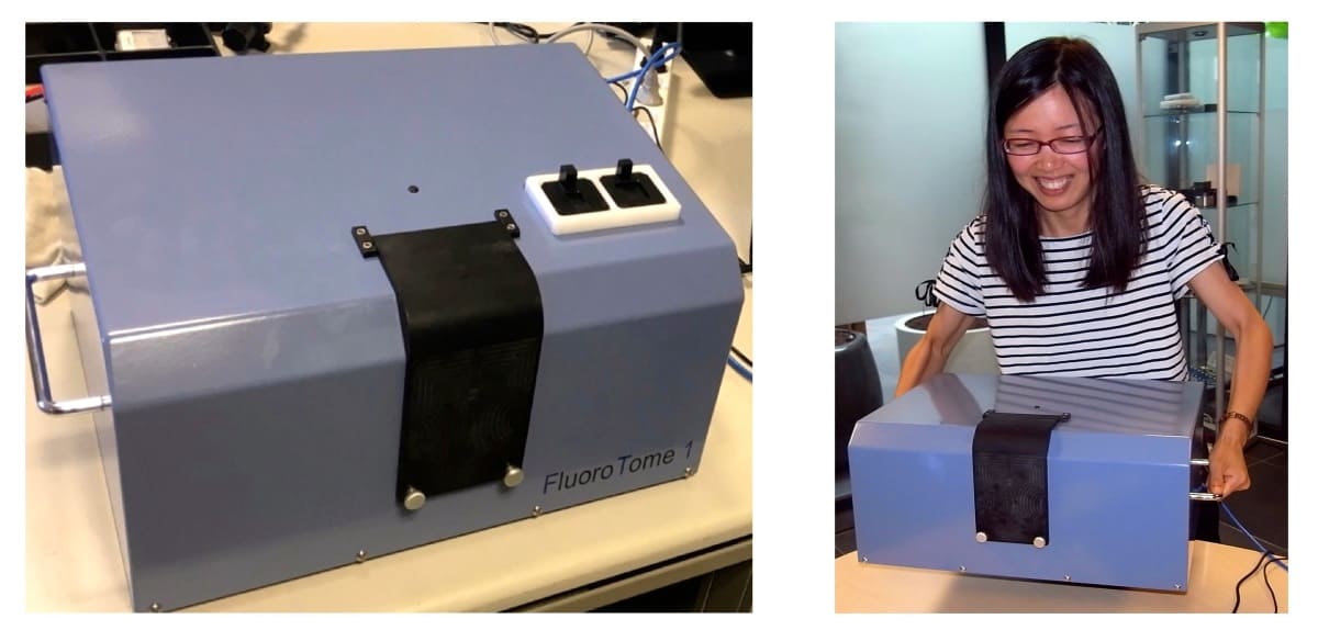
The ability to create 3D images of radio-fluorogenic (RFG) gels following radiation exposure could enable visualization of the dose deposited during a radiation treatment. To achieve this, researchers in the Netherlands have created a portable, user-friendly device – called FluoroTome 1 – that can rapidly read out an irradiated gel to produce a series of fluorescence images (Polymers 10.3390/polym11111729).
RFG gels become permanently fluorescent when exposed to high-energy photon or particle radiation, with the emission intensity proportional to the local absorbed dose. One important application of this property is use in high-resolution radiotherapy dosimetry. FluoroTome 1, developed by John Warman and researchers at TU Delft, enables on-site scanning of RFG gels, creating multiple tomographic slices of the fluorescence signal.
“Scanning of irradiated samples and data analysis can be carried out, using user-friendly software, within minutes of radiation exposure,” Warman explains. “This possibility of rapid, on-site feedback is particularly important for equipment testing, protocol control and educational applications, and is a feature that is not generally possible with other polymer-gel dosimetry equipment.”
Originally developed as a bench-top system, the new device is compact, portable and apart from two 220 V outlets, requires no special on-site facilities or a dark room. FluoroTome 1 was constructed in collaboration with Netherlands-based 4PICO BV, a firm with expertise in fabricating functional UV-optical devices.
FluoroTome 1 works by creating a thin sheet of uniform UV light, transporting the irradiated RFG gels through this sheet using a remotely controlled translation stage, and creating multiple digital images of the UV-excited fluorescence. The team successfully tested the FluoroTome 1 by using it to record images of a 20 x 20 mm square RFG gel irradiated with four 3 mm square 300 kVp X-ray beams.
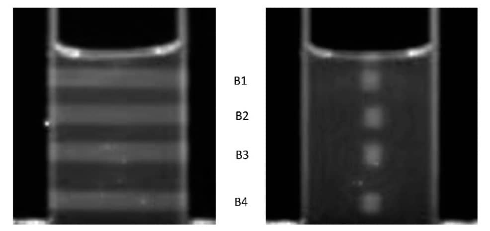
The system can also easily compile the recorded images into a 3D video of radiation dose deposition, enabling dosimetric verification of complex radiation fields.
Warman notes that FluoroTome 1 was specifically designed for application in collaboration with other radiotherapy or radiation physics groups. The portability of the apparatus, as well as its independence of special on-site facilities and user-friendly software, should enable it to be used in most high-energy radiation facilities. “The actual irradiation set-ups and specific gel containers used will of course need to be designed and constructed separately,” he notes.
The team’s immediate plans are to use FluoroTome 1 to monitor proton beams at the Holland Proton Therapy Centre, recently constructed on the grounds of TU Delft. “My own particular interest is in the fundamentals of radiation interactions with materials,” Warman tells Physics World. “These include problems such as the differences between chemical, physical and computational estimates of energy deposition in proton beams and the relative biological effectiveness at the Bragg peak. The application of proton pencil beams to ocular cancers is another interesting practical problem for which we recently performed preparatory measurements using a 3 mm X-ray beam and a gel phantom.”
More general applications of RBG gel studies include quality control testing of radiotherapy equipment and computer protocols. “The FluoroTome 1 could also become a fast-feedback teaching aid for students and clinical personnel. And, it could provide visual images for radiation oncologists to explain to patients the differences between different types of radiotherapy procedures.” Warman adds.


