
Here’s a sobering thought: children born to mothers who consume large amounts of alcohol during pregnancy have been found to have altered connectivity in their brain networks. Specifically, abnormal functional connections between sensorimotor areas and hubs of the “default mode” and “salience” networks (involved in motor function, cognitive control, emotions and consciousness) suggest that children with pre-natal alcohol exposure (PAE) recruit additional brain regions for sensorimotor tasks, which could imply poorer brain network efficiency. This work was performed by researchers at the Universities of Calgary and Alberta, and will help further investigations concerning sensorimotor alterations in youths with PAE (Human Brain Mapping doi: 10.1002/hbm.24004).
Foetal alcohol spectrum disorders (FASD) describe a range of effects and symptoms caused by PAE. Sufferers may have difficulties in cognition, such as learning or remembering, motor coordination problems, and sensory processing deficits. They are also at greater risk of developing mental health illnesses such as ADHD and depression. Previous research has shown abnormalities in brain function and structure, but there are only a few articles, which investigated resting-state (rs) networks in PAE, using functional MRI (fMRI).
Everything is connected
Typical fMRI studies involve the participant performing a task, or experiencing a stimulus passively. A blood-oxygenation level dependent (BOLD) signal is measured, which itself is an indirect measure of neural activity over time. In rs-fMRI, there is no task, but instead, researchers measure stimulus-free brain-states, and by using connectivity analyses, can observe background functional networks (spontaneous BOLD signal fluctuations between regions that are temporally correlated). In this study, the researchers performed rs-fMRI on 59 participants with PAE and 50 controls, with data acquired across multiple centres.
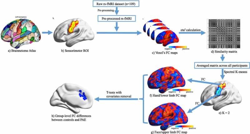
The researchers calculated functional connectivity (FC) maps for each participant, based on a seed region in sensorimotor cortex. They generated an average similarity matrix of FC between each voxel and every other voxel within the sensorimotor region of interest (ROI). They classified two cluster ROIs, corresponding to the hand/upper limb area and the face/lower limb area. Finally, they generated FC maps between these two regions and the rest of the grey matter of the brain.
Alterations in functional connectivity
The organization of somatosensory cortex in controls and PAE participants was very similar, suggesting that there is no large disruption to sensorimotor organization, regardless of motor skill deficits.
There were, however, significant group differences in functional connectivity. For PAE participants, the researchers observed higher FC from the seed regions in sensorimotor regions to the insula and anterior cingulate (the salience network – how we perceive certain stimuli relative to other background stimuli). They also saw a decrease in FC from the seed to the superior temporal gyrus and precuneus (hubs of the default mode network – a network that activates during wakeful resting).
These FC abnormalities could imply abnormal interactions between cognition and motor functions, and could affect areas associated with face processing, emotions and memory. The decreases in FC were observed in regions that are involved in manual coordination and sensorimotor processing.
The team performed a comparison of FC across age. While they observed an increase in FC in controls (between facial sensorimotor regions and the insula) across time, this was absent in PAE participants. The authors suggest that altered development could be related to altered cognitive-motor development, noting its potential use as a biomarker.
The findings of this study show that although the topographic organization of sensorimotor areas is seemingly unaffected in PAE, there is higher FC between sensorimotor seed regions and salience and executive networks. This could be explained by an overcompensation mechanism, whereby more regions in sensorimotor networks are recruited than usual, implying a less efficient network.


