A new quantum sensor developed by scientists at the University of Sussex in the UK could help clinicians identify diseases such as dementia, Alzheimer’s and Parkinson’s by tracking patients’ brain waves and monitoring how their speed changes over time. The sensor, which is based on a real-time, high-spatial-resolution neuroimaging technique known as magnetoencephalography (MEG), uses an array of quantum devices called optically-pumped magnetometers (OPMs) to map the tiny magnetic fields generated when neurons in the brain send out electrical signals. If used to monitor patients over a period of several months, the researchers say the new sensor could identify declines in brain signal transmission speed that may be associated with pathology.
Non-invasive neuroimaging techniques have improved significantly over the past few decades, greatly expanding our knowledge of the brain’s inner workings by providing information about neural responses and processes. Previous studies have shown that the ways these processes are distributed within the brain, and how they change over time, are reliable biomarkers for anomalous brain activity associated with neurodegenerative diseases. If clinicians could detect these biomarkers accurately, and with high enough precision, they might be able to predict how a disease will evolve or how a patient will respond to treatment.
The problem is that current clinical methods cannot simultaneously resolve brain signals in space and time. Functional Magnetic Resonance Imaging (fMRI), for example, can map brain regions with high spatial resolution, but its temporal resolution is low (around 1 s) because it measures changes in local blood flow, which lag substantially behind electrical brain activity. Electroencephalography (EEG), for its part, detects these electrical signals directly, and thus works in real time. However, its spatial resolution is low, especially for high-frequency brain waves.
Evaluating cortical signals
In principle, MEG offers the best of both worlds, making it possible to measure the postsynaptic potentials of brain cells located just below surface of the scalp non-invasively, in real time and with high spatial resolution. Indeed, recent research has used MEG to evaluate abnormal cortical signals in patients with Alzheimer’s, Parkinson’s, autism spectrum disorder and even severe cases of post-traumatic stress disorder.
The drawback is that MEG has to be performed in special magnetically-shielded rooms to reduce magnetic noise from the environment, which is often many orders of magnitude higher than neuromagnetic fields (which are in the femtotesla to picotesla range). In addition, most of today’s MEG systems detect these tiny fields using superconductive quantum interference devices (SQUIDs), which require bulky cryogenic refrigeration. This means they cannot be placed close to the skull, limiting the spatial and temporal resolution of SQUID-based MEG scanners.
Around the turn of the millennium, researchers developed an alternative known as a “spin-exchange relaxation-free” (SERF) OPM. The version used in the current study contains a gas of rubidium atoms, and Peter Kruger, who leads the Quantum Systems and Devices group at Sussex, explains that when these atoms experience changes in their local magnetic field, they emit light differently. Hence, when researchers shine beams of laser light at the atoms, fluctuations in the emitted light reveal changes in the magnetic activity in the brain.
Better at brain signal tracking
In the new work, Kruger, PhD student Aikaterini Gialopsou and other members of the team used their OPM-MEG to record the spatio-temporal patterns of neuronal signals in volunteers responding to visual stimuli. They then compared these patterns to those obtained by conventional SQUID-MEG, demonstrating that the new sensor is better at tracking brain signals in both space and time. “We discovered that this quantum sensing technique can combine high spatial and temporal resolution,” they explain. “While previous techniques were able to locate signals in the brain, this one is the first to record the precise timing of brain signals.”
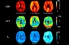
Neuroimaging identifies early-stage dysfunction of the blood–brain barrier
According to Kruger, the new quantum sensor is accurate to within milliseconds and has a spatial resolution of just millimetres. He and his colleagues now aim to improve the quality of their images still further by increasing the number of sensors in their OPM-MEG scanners. At the moment, this is done by squeezing individual sensors closer together. While this approach is straightforward, it is quickly reaching its limits because of cross-talk between sensors, overheating and other difficulties in scaling up individual sensors to whole imaging arrays, Gialopsou explains. “We are tackling this problem in a fundamentally different way by integrating high-density arrays of OPM sensors based on standard microfabrication techniques and shared resources,” she tells Physics World. “The first modular arrays developed in our group can be easily reconfigured, allowing for quick prototyping of novel sensing schemes and optimisation of sensor components and control systems.”
The new OPM-MEG is detailed in Scientific Reports.