
A computational technique developed to process seismic images of the Earth’s subsurface could allow for high-resolution human brain imaging, reports a new study by researchers from Imperial College London. Although presently in the simulated, proof-of-concept stage only, the development could pave the way towards a cheaper, portable and more universally applicable method for rapid diagnosis of stroke and head trauma, and continuous monitoring of a wide range of neurological conditions (npj Digit. Med. 10.1038/s41746-020-0240-8).
Both of the leading conventional techniques for performing imaging on the brain come with inherent limitations. MR imaging is unsuitable for use on patients who have – or are suspected could have – metallic implants or harbour foreign bodies. It is also impractical for use on severely obese, claustrophobic or uncooperative patients. X-ray CT, meanwhile, involves exposure to harmful ionizing radiation, ruling out its use with young patients or for continuous monitoring. Both modalities also require large, expensive, high-powered machines that cannot practically be set up outside of hospital or laboratory settings.
In contrast, ultrasound imaging is universally safe for use and can be made portable – but traditional applications have not been able to scan within the human skull. This is because the bone attenuates, scatters and reflects the waves in complex ways that cannot be undone by simple algorithms.
In a new study, physicist Lluiís Guasch and colleagues turned to a computational technique known as full-waveform inversion (FWI), which is used by geophysicists to extract three-dimensional images of the Earth’s subsurface from data collected by seismometers on the passage of waves underground. A nonlinear data-fitting procedure, FWI works by using real-world seismic data to create a rough model of subsurface conditions from which wave equations can be solved to produce mock data. The model is then iteratively improved until this output provides the best fit for the real-world data.
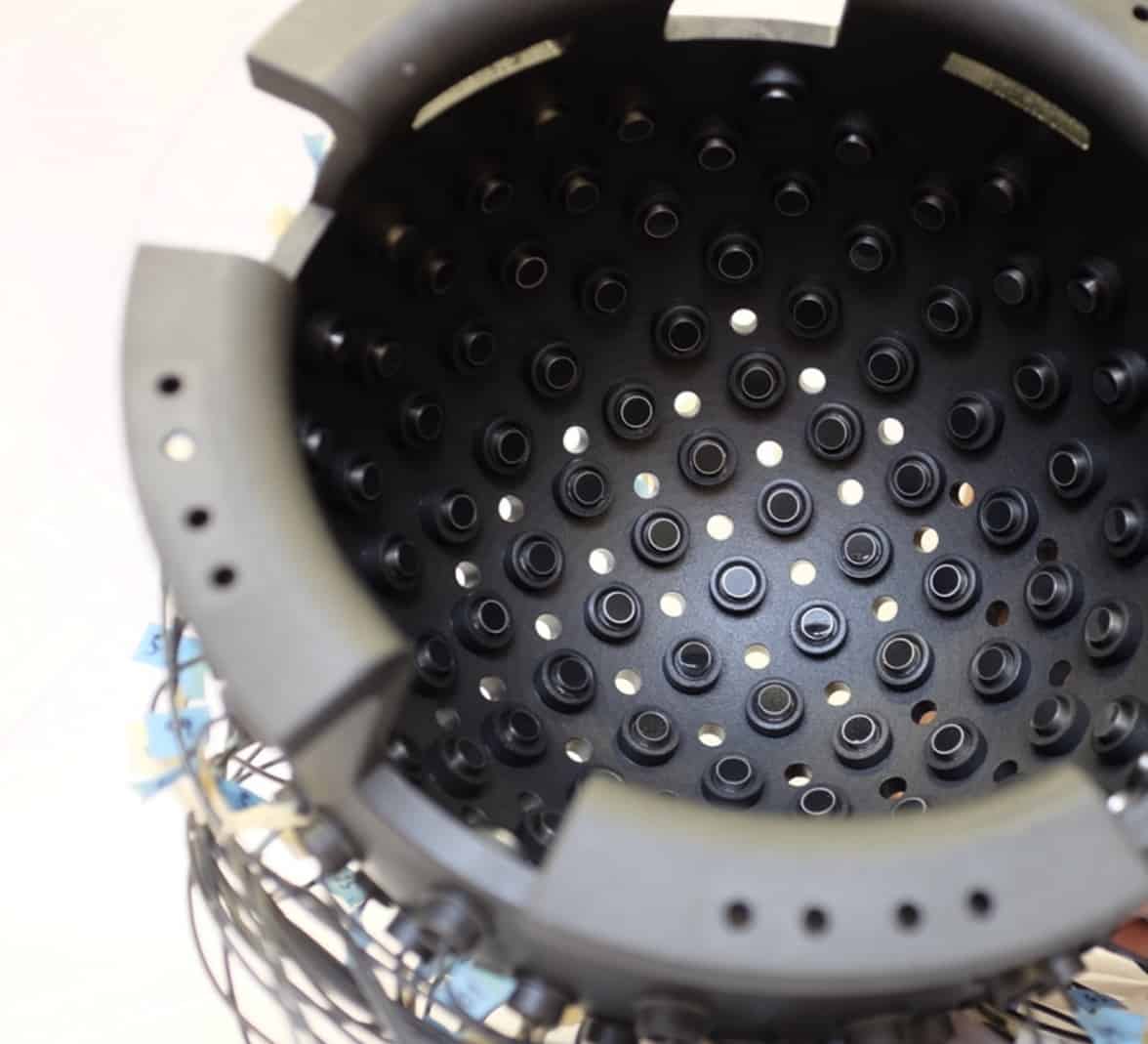
Instead of using seismometers across the Earth’s surface, the researchers instead envisage using a helmet-like mesh of 1024 ultrasound transceivers. Through simulation, they show that in such a set-up, FWI is indeed capable of reconstructing high-resolution images of the brain like an MRI scan – one in which grey matter, white matter, ventricles and structure can be clearly seen. They also demonstrate in the lab that ultrasound transceivers are able to record signals from within a human skull with the required signal-to-noise ratio for processing with their FWI algorithm.
“This is the first time FWI has been applied to the task of imaging inside a human skull,” says Guasch. “In many ways, it is easier to apply FWI in medical imaging than in geophysics.” This, he explains, is because – unlike when dealing with the unique nature of different subsurface images – individual skulls have commonalities that can help guide the image reconstruction process.
“Neurology has been waiting for a new, universally applicable imaging modality for decades; FWI could well be the answer,” adds co-author Parashkev Nachev.
Furthermore, the researchers say that it should be possible to eventually realize a clinical version of their scanner that is portable – sized to fit on a motorbike or within an ambulance – that could allow scans to be undertaken on patients in advance of reaching hospital. Similarly, the device could be mounted on a frame to perform bedside imaging. The one drawback of the approach, however, is that it presently takes considerable time – as the helmet produces 1024 × 1024 individual ultrasound signals, which take around 32 hours to process on a conventional server.
“This is an important piece of work, as most imaging physicists would have assumed that, quite apart from the large signal attenuation that the skull produces, the multiple internal reflections and scattering of the soft tissue signals as they hit the bone interface would render any hope of reconstruction impossible,” says Stephen Williams, an imaging scientist from the University of Manchester who was not involved in the present study. “The paper provides compelling evidence that a physical realization of the concept should be possible, provided that the computer processing time can be reduced by around 200 times compared to the simulations reported in the article.”
The researchers note that three-dimensional ultrasound tomography using FWI could find particular relevance for rapid diagnosis and treatment of stroke. With their initial study complete, they are moving to further develop their prototype system with the goal of producing the first brain image of a live human subject – alongside improving the robustness of their image generation algorithm and lowering computational costs.


