Using conventional semiconductor manufacturing techniques in a clean room at Ohio State University, scientists have devised a convenient method for inserting genetic material into a selected patch of tissue. The new technique – termed “tissue therapy” – uses strong local electric fields focused by an array of nanoscale channels on a silicon wafer. Pressed against the skin, the device rapidly transports reprogramming factors to where they are needed. The treatment takes less than a second, and was shown to induce the growth of new blood vessels and neurons in mice, saving badly damaged limbs and doubling the chances of stroke recovery.
Many of the hardest conditions to treat, such as heart disease and stroke, promise to be treatable by cell or tissue therapy. Beneficial cells can be injected into the patient or delivered directly to damaged tissue. Here they facilitate healing by secreting therapeutic factors or through integration into the dysfunctional tissue.
Despite a lot of investment, limited cell resources and the need for pre-processing has kept this treatment from widespread use in the clinic. These issues can be circumvented by inserting sections of genetic code into the patient’s own cells (a process called transfection), reprogramming the tissue to change function. Although cell reprogramming has been accomplished before in vitro, injecting cells into organisms can cause unwanted interactions, so the ability to generate them in vivomakes the process both more efficient and more effective.
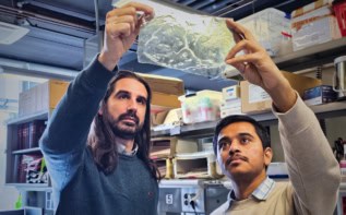
The transfection tools available to scientists range from viruses to chemicals, nanoparticles, and microscale needles, but they are either too untargeted, or they are too selective and time-consuming to be used practically in specific tissues in large organisms. Writing in Nature Nanotechnology, lead author Chandan Sen and colleagues at Ohio State University report a highly convenient method with which to topically transfect a region of tissue in an animal, affecting only a small area, and without the need for a viral vector.
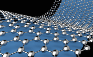
The team’s device consists of a 200 μm-thick silicon wafer with T-shaped arrays of nanochannels. These channels serve to focus the electric field, which causes pores to open in the target cells. The nanochannels also deliver the reprogramming agents, which are then electrophoretically transported through the transient pores.
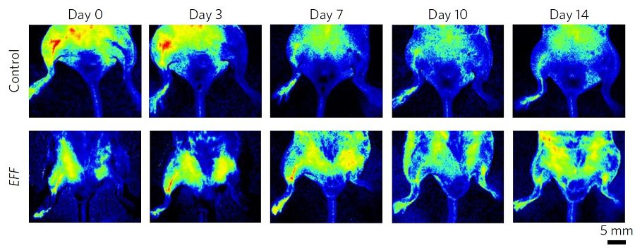
The researchers conducted experiments using mice to confirm that their approach had clinical potential. Mice with a transected femoral artery were administered factors that encourage blood vessel growth in the area of the cut. Within seven days of the treatment the limbs began to recover their blood flow, rather than withering away. Factors that induce neuron development were also applied to the skin of mice that had suffered a stroke. An extraction from the treated tissue was transferred to the brain, making the mice twice as likely to recover.
Although the field of cell therapy is fraught with complexity, the group’s thorough study presents a promising outlook for tissue therapy as it takes the next step on the path to clinical application.