I just came out of a medical physics press conference that presented three very different ways that physics can be put to use saving lives.
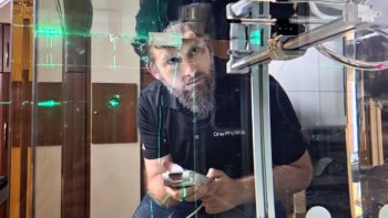
The first presentation was from David Nolte of Purdue University who has created a very simple but effective way of measuring motion inside cancer cells. The technique involves splitting a laser beam, reflecting one beam off a tumour and then recombining the two beams at a detector. The two beams interfere and motion within the cancer cells causes the interference to change.
The result is an image of the tumour covered in speckles that change rapidly as the organelles inside the cancer cells move. A moving cell is a healthy cell, so the technique can be used to study how some anti-cancer drugs slow down the movement within cells, ultimately killing them.
Andre Brown of the University of Pennsylvania described his work on fibrin, which are molecular chains that create web-like structures that aid in the clotting of blood. Blood clots cause heart attacks and strokes so it is very important to understand the mechanical properties of fibrin — particulalry how it stretches.
Brown used a technique developed a few years ago whereby the tip of an atomic force microscope (AFM) is used to pick up one end of a fibrin chain and tug on it. He discovered that fibrin was made of a chain of coiled proteins, with each coil unfolding 23nm when pulled hard enough by the AFM. The next step is to work out how this unfolding affects larger fibrin structures.
Finally, Michael Deem of Rice University explained how he has used statistical physics to develop strategies of multiple vaccination to keep the body’s immune system one step ahead of a rapidly mutating virus.
And a big thanks to the APS press office for a fantastic lunch today!