Physicists in Germany have created a practical source of femtosecond-duration electron pulses for electron microscopy. The pulses, each of which usually contains just one electron, emanate from the tip of an extremely sharp metal needle that is illuminated by femtosecond laser pulses. The source has already been used to image a nanometer-sized trench and could be used in time-resolved electron microscopes that follow the motion of individual atoms and molecules as they rearrange themselves during structural transitions or chemical reactions (Phys. Rev. Lett. 98 043907).
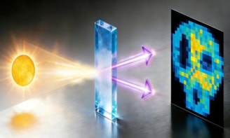
Electron beams are easily focused and have wavelengths much shorter than visible light – making them a powerful tool for studying matter down to atomic length scales. However, electrons are charged particles and their mutual repulsion means that they avoid bunching together in very short pulses.
In the past few years, physicists have tried to create very short electron pulses by irradiating metal surfaces with powerful femtosecond laser pulses. However, each laser pulse creates a pulse containing a large number of electrons, which broaden to a few hundred femtoseconds’ duration thanks to mutual repulsion. While physicists have tried to limit the number of electrons in a pulse by focusing the laser onto a very fine metal tip, they have so far struggled to understand and ultimately control the emission process.
Now, Christoph Lienau and colleagues Max Born Institute for Nonlinear Optics and Short Pulse Spectroscopy (MBI) in Berlin have found a way to avoid repulsive broadening by designing a tip that emits about one electron per pulse. In their source they use a 80 MHz Ti:sapphire laser to irradiate the tip of an extremely fine gold needle with light pulses that are 7 femtoseconds long, producing electron pulses less than 20 femtoseconds’ duration.
The tip is extremely sharp – it has a curvature radius of less than 20 nm – which means that the intensity of electromagnetic field associated with the laser pulse is enhanced by about a factor of ten. As a result, the electron pulses leave the tip without the need for large bias voltages. This is an important breakthrough because high bias voltages had previously prevented such tips from being used in imaging systems. Bias-free operation also makes the electrons a very sensitive probe of matter.
Although the needle emits about one electron per pulse, Lienau told Physics Web that the high pulse rate of the laser ensures that there are enough electrons to perform electron-diffraction experiments.
The researchers also demonstrated the practicality of their technique by creating a tip-enhanced electron emission microscope (TEEM) based on a tungsten (rather than gold) tip that is illuminated by the same laser pulses. The TEEM was able to image a 100 nm-wide groove in a metallic surface with a spatial resolution of tens of nanometres.