In less than 100 seconds, Andrea Morello introduces the concept of a quantum bit or “qubit”. He explains why the quantum properties of qubits can be used to create computational systems that are exponentially more capable than classical computers for performing certain tasks. Morello – who is based at the University of New South Wales in Australia – discusses some of the ways in which researchers are creating qubits, covering superconductors, spin systems and ion-trap methods. Morello is a member of the editorial board of Quantum Science and Technology, a new journal published by the Institute of Physics, which also publishes Physics World.
Music and physics might seem like polar opposites, one having great emotional potency and the other being a cerebral subject of equations, theories and deductions. Both, however, benefit from improvisers – people who stand on the shoulders of giants, taking earlier triumphs and building on them to create something new. For me, analogies like these, which draw parallels between physicists and jazz musicians, are the most fascinating revelations in Stephon Alexander’s book The Jazz of Physics – despite the book’s eye-catching promise to reveal “the secret link between music and the structure of the universe”.
The book begins autobiographically, with a description of Alexander’s early childhood in Trinidad and his formative years in New York’s Bronx neighbourhood. Writing in a fluent and easy-to-read style, Alexander describes how he turned down the opportunity to pursue fame and fortune through hip-hop and jazz in favour of his other passion: theoretical physics. Now a professor at Brown University in the US, he remains an amateur saxophonist, and has been known to take his latest theoretical musings to jazz gigs, to scribble down ideas between sets and so improvise new ideas in physics.
Similarly, my reading of The Jazz of Physics was accompanied by music mentioned in the book, such as My Favourite Things by John Coltrane and Ambient 1: Music for Airports by Brian Eno. For those unfamiliar with the latter’s work, suffice to say that Eno has been one of the world’s most influential electronic musicians and music producers since the 1970s; the most exciting message I received on Twitter all last year was a photo showing my popular-science book on a bookshelf in the toilet of Eno’s studio. Alexander, though, trumps my anecdote by actually taking us into that studio. He also discusses improvisation with great saxophonists, such as the controversial free jazz pioneer, Ornette Coleman.
A common misconception of scientific research is that it is all about people working alone to achieve dramatic “eureka!” moments. As the large cast of supporting characters in The Jazz of Physics shows, this is not how most of science progresses. To begin with, it seemed to me that Alexander was overly keen to namedrop well-known musicians and physicists, but then I realized that this is just a reflection of how he works. Whether Alexander is playing on his sax or doing physics, the role of the other musicians/scientists supporting his improvisation is vital.
Another misconception is that scientific research is a linear process with a simple story line. Throughout the book, Alexander demonstrates how important it is for physicists to play with ideas, to take things that might at first seem daft and see what they reveal. The book is very much a personal story, but it also illuminates some of the current arguments about the value of “blue skies” research versus work with more immediate impact. Jazz doesn’t move forward if every improvisation is just a pastiche of past maestros. It needs to build on the framework of past players to take new and unexpected avenues. The same is true of science.
There is plenty of science in this book as well, with Feynman diagrams, string theory and cosmology being just some of the areas covered. Sometimes, though, the autobiographical narrative means that topics are introduced and then not fully developed because Alexander’s career moved in a different direction. This is certainly a faithful portrayal of how most scientists work: just as a good jazz improviser draws on many different influences, Alexander’s science has drawn on a broad range of fields in theoretical physics. And it is fine for those who have some familiarity with the subjects, as I expect most readers of Physics World will. But at times, I found it a bit unsatisfactory, since it meant that some topics are covered quite briefly, and some important concepts aren’t fully explained. If a reader with little physics background were to approach this book, it would be like someone brought up on 1950s rock and roll going to hear an Ornette Coleman free jazz gig for the first time.
Putting this aside, though, the book’s attempt to bring together modern jazz and modern physics strikes me as admirable. The Jazz of Physics riffs on the idea of a rhythmic universe, where the cosmos is following a cyclical pattern of expansions and contractions interspersed with big bangs. After each big bang, a new universe is created that may or may not have the capability to develop life. Alexander’s musical analogy is the cycles of improvisation in Coltrane’s Giant Steps, where the start of each cycle gives Coltrane an opportunity to develop a new idea and form a new musical universe. It’s an intriguing comparison, and it certainly seems fresher than drawing analogies between classical music and classical physics, where there is a long history of scientists telling stories about Pythagoras and the “music of the spheres”. Time to put on some Coltrane and riff some new research ideas?
Professionally speaking, biologist Stuart Firestein is a fan of failure. Done right, he argues, failures in science are much more than mere mistakes, and better even than the “painful but character-building” sort of failure described in self-help and business books. In Failure: Why Science is So Successful, Firestein explores the complex role that failure plays (or should play) in how science is done, taught, perceived and funded, focusing on problems that transcend disciplinary boundaries. The best example of his cross-disciplinary reach is probably the section on education. Here, the author decries what he calls “the bulimic model of science education”, in which teachers cram “gobs of facts into [students’] heads so that they can puke them back up on an exam, then move on to the next unit with no measurable gain”. Few teachers (or students) will dispute that stomach-churning critique, but Firestein isn’t finished. In his view, the source of the problem isn’t just the current vogue for targets and tests: it’s the scientific method, at least as it is commonly understood. The idea that science proceeds by making observations, formulating hypotheses and testing them sounds great, he writes, “except that no scientist that I know of actually follows this prescription”. Telling a science student to form a hypothesis, he explains, is “like giving an art student a brush and the direction to ‘do painting’ ”. Much better, he argues, to teach that science begins with curiosity and proceeds through a series of failures. Firestein’s perspective on the so-called “replication crisis” in science (see feature “No result, no problem?”, May 2016 print and digital magazine only) is similarly unorthodox. While he acknowledges that many published papers contain irreproducible results, in his view this is only natural in a discipline where failure is both common and necessary. Moreover, some of the measures proposed to remedy the situation would, he argues, be counter-productive. Deeply thought-provoking and (as this review shows) frequently quotable, Firestein’s book is nevertheless better at diagnosing problems than at offering solutions. The author’s remedy for overly cautious grant-funding procedures, for example, amounts to skewing application criteria in favour of “the merit of their science and the creativity of their approach”. A good idea, of course, but one does not need to be a bureaucrat to see that “merit” and “creativity” are extremely hard to define. In this sense, one could call Failure a failure – but only in the best possible sense of the word.
2016 Oxford University Press £14.99/$21.95hb 304pp
Seismic shifts in history
In 132 AD the Chinese court mathematician and astronomer Zhang Heng ordered the construction of a curious instrument. The device (now sadly lost) incorporated eight dragon heads, eight squatting toads and a set of heavy brass balls. Its purpose was earthquake detection: distant tremors would cause one or more balls to fall from the dragons into the open mouths of the toads, creating a noise that would alert the authorities and (in theory) indicate the direction of the shaking. The story of Heng’s seismometer is one of many gems in Andrew Robinson’s book Earth-Shattering Events: Earthquakes, Nations and Civilization. The book is organized as a series of case studies on specific earthquakes, from the 1755 shock that devastated Lisbon, Portugal to the 2011 Tōhoku earthquake off Japan’s north-east coast. In each case, Robinson – a veteran author equally at home with history and science – draws on previously published accounts to summarize what happened, and to analyse the earthquake’s effect on the country that experienced it. Individually, these case studies make vivid reading, and collectively they tell a fascinating story about how seismology developed as a science. However, the author’s broader goal is to explain how earthquakes have influenced the course of human history, and occasionally, this ambitious effort leads him – if you’ll pardon the pun – onto shakier ground. One of Robinson’s arguments is that historians and archaeologists tend to ignore or downplay the role that earthquakes play in initiating political change. Yet as his own case studies show, even when major tremors do spark significant changes (and they often don’t), the nature of those changes is highly dependent on long-term sociopolitical trends. With such weak evidence of causality and few clear rules, it’s hardly surprising that historians prefer to focus on other things (including, of course, long-term sociopolitical trends). The bottom line is that if predicting earthquakes is hard, predicting their effects on the grand sweep of history is nigh-on impossible – although as this very entertaining book shows, it can still be interesting to try.
An artificial material with the eye-catching optical properties of the Callophrys rubi butterfly has been created by researchers in Australia. Using a special lithography technique, the team was able to make photonic “gyroid” nanostructures similar to those found in the butterfly’s wings. The artificial structures, which outperform their natural counterpart in many ways, could find use in a variety of photonics and optics technologies.
Gyroids are 3D periodic structures made up of intertwining, curved surfaces. They have lattice constants that are comparable to the wavelength of visible light, which means that they have a range of optical properties, such as structural colour. This is what gives the Callophrys rubi butterfly a beautiful blue-green sheen on its wings. Thanks to their cubic symmetry and the fact that they are mechanically strong, gyroids could also be ideal for making photonic crystals and other optical metamaterials. These are artificial structures that have a number of desirable optical properties that make them ideal for controlling light in technologies such as optical communications and displays.
Stronger and better defined
However, the techniques employed to make optical metamaterials have been unable to produce artificial gyroid structures with lattice constants comparable to those found in butterfly wings. Now, a team led by Min Gu of RMIT in Melbourne has used a technique called optical two-beam super-resolution lithography to create 3D gyroid structures with lattice constants of 360 nm. This gives the artificial gyroids a similar blue-green colour to the butterfly’s wing. However, the artificial structures are mechanically stronger than natural gyroids, and have long-range periodicities and well-defined crystalline boundaries. The latter two properties are lacking in natural materials, which suffer from uncontrolled structural disorders.
Another unique feature of the artificial gyroids is that they have chiral properties that are lacking in the imperfect natural structures. An artificial structure, for example, will contain only left- or right-handed single gyroid enantiomers, while the natural version contains a mixture of both. This means that the artificial gyroids are much more suitable for applications such as photonic crystals with optical band gaps and miniature chiral beam splitters. Metamaterials made from the artificial gyroids are expected to have tuneable nonlinear optical properties and respond to light at ultrafast speeds, so making them ideal for high-speed optical switches.
Apart from applications in photonics, the new gyroid structures could help make more compact optoelectronics because, thanks to their smaller size, larger numbers of devices can be integrated onto a single chip. According to Gu, the superior mechanical strength of the artificial material makes it well-suited for high levels of integration.
Despite the best intentions of conservators in preserving works of art, the treatments they apply could turn out to have damaging effects in the future. Back in the 17th and 18th centuries, for example, people used to rub Michelangelo’s famous frescoes in the Sistine Chapel with bread or linen dipped in wine. Thankfully, we now have more sophisticated tools such as laser cleaning or selective cleaning using hydrogels or micro-emulsions, which can be fine-tuned to remove unwanted deposits without affecting the underlying painted substrate.
In principle, any intervention must be reversible such that a work of art can be restored to its previous untreated condition. In practice, this is extremely difficult or even impossible to achieve, and it is often more realistic to adopt a principle called “re-treatability and compatibility”. Conservators therefore continuously have to balance the risks of doing nothing against doing something. Indeed, the fear of being responsible for devaluing a piece of cultural heritage has forced some conservators to lay down their swabs and spatulas, and to go in search of better tools.
This challenge is particularly relevant for the preservation of modern art, where few established conservation protocols exist. Traditional conservation ethics are difficult to apply when artists have used ephemeral materials in their work, such as plastics. Once degradation has been initiated in plastics via thermal, photo-oxidative or hydrolytic means, the process proceeds auto-catalytically and very little can be done to prevent the art work’s imminent demise. In some cases replication is the only alternative for preserving heritage, which is reminiscent of conservation efforts in the dark ages.
Collectors and auction houses are increasingly seeking advice from conservators on how to prevent their treasured pieces from decaying or on how to restore a modern artwork. Unfortunately, they are too often met with a bad prognosis as pieces lose their vibrancy, start cracking and yellowing or lose an inherent material property such as flexibility that could be intrinsic to the meaning of the work. The scale of the problem can perhaps be better appreciated in a financial context: in 2014 the global art market was worth a little over €50bn ($56bn), of which post-war and contemporary art accounted for 48% and modern-art sales 28% of all auction sales, respectively.
Nanotechnology to the rescue
Thanks to a multi-partner European Commission (EC) project called Nanorestart, which kicked off last year, nanotechnology could soon provide conservators with novel ways to future-proof modern works of art. Nanotechnology is certainly not new to the art world. Since the Bronze Age, artisans have used the “magical” properties of nanoparticles in their wares that seemingly defy the laws of physics. The dichroic properties of the fourth-century Roman Lycurgus Cup, which is on display in the British Museum in London, were a mystery until as recently as 1990, when scientists used transmission electron microscopy to discover that clusters of silver and gold nanoparticles measuring 50–100 nm across changed the optical appearance of the glass from a deep red to opaque green, depending on whether viewed in transmitted or reflected light.
Even throughout the Middle Ages and into the Renaissance, the optical and reflective properties of metal nanoclusters were exploited to create the beautiful iridescent quality of medieval and renaissance lusterware, whereby their colour changes depending on the viewing angle. Thankfully, many of these artefacts have already withstood the test of time. But many other cultural heritage objects, particularly those made from organic materials, risk being lost to future generations if not treated carefully.
Today, heritage science is a cross-disciplinary domain that aims to conserve and interpret cultural artefacts and improve access to them. While there has been a steady growth in our understanding of preservation techniques for classical artworks, preserving modern art is much more challenging. These works are often made from novel materials, some of which are intentionally transient. Indeed, artists may unintentionally use materials that have an inherent vice such as polyurethane foam, cellulose nitrate, cellulose acetate and polyvinyl chloride. These are known as “malignant” plastics, not just because they are intrinsically unstable, but also because the degradation mechanisms they suffer produce harmful products, such as corrosive volatile organic compounds (VOCs) that could damage other plastics or objects in their vicinity.
The idea of using nanotechnology to protect heritage artefacts was pioneered by Piero Baglioni and colleagues at the Centre for Colloid and Surface Science (CSGI) at the University of Florence, Italy. It was built on groundbreaking conservation work by Enzo Ferroni, who – following the Florence floods in 1966 – helped develop a method to remove salts threatening frescoes that had been immersed in flood water. The method is a two-step process, using ammonium-carbonate and barium-hydroxide solutions to remove sulphur from – and thus consolidate – the contaminated frescoes. Unfortunately, the penetration depth of the consolidant was limited by the particle size and, in some cases, risked the formation of a white bloom on the surface of the painted layer.
The team at the CSGI therefore developed more efficient methods based on inorganic particles with smaller sizes. “Nano-lime”, which is based on dispersions of calcium-hydroxide nanoparticles in non-aqueous solvents, has similar physical and chemical properties to the original lime plaster used in frescoes and so behaves and ages in a similar fashion, without introducing stresses. Through the EC’s FP7 “Nanoforart” project, which was completed in 2014, inorganic nanotechnology-based consolidation alternatives for frescoes were refined, as were systems that could remove past treatments, such as polymer-based consolidation treatments used in wall paintings. Nanosystems were also developed for the consolidation, de-acidification and controlled removal of unwanted deposits from organic materials such as paper, wood and canvas.
Plastic concerns
Today, one of conservation’s biggest challenges concerns contemporary artworks made from plastics. Plastic’s reputation as an everlasting material has disappointed, with devastating degradation processes now observed in plastic collections dating from the 1920s to the 1960s. Iconic pieces from the Bauhaus and Pop Art movements are ticking time bombs. Russian sculptor Naum Gabo’s early and now disintegrating pieces made from cellulose nitrate (another malignant plastic releasing acidic nitrogen-dioxide gasses) are another prominent example. The situation is complicated because plastics vary enormously in their make up. They span a range of synthetic/semi-synthetic polymers with many different chemical compositions, all of which follow different degradation pathways such as hydrolytic, thermal and photochemical processes, including thermal and photo-oxidation.
Fortunately for these materials, Nanorestart – launched under the EC’s Horizon 2020 programme – is by far the biggest international research effort to develop nanotechnology-based solutions to the conservation of modern and contemporary materials. The project pools the expertise of enterprise and academic leaders across 27 partner institutions in nanoscience and conservation, and is divided into four distinct themes: cleaning of contemporary painted and plastic surfaces; stabilization of modern canvases and painted layers in contemporary art; removal of unwanted modern materials; and advanced protection of modern artworks in museums and outdoors.
The Institute for Sustainable Heritage at University College London (UCL), where I am based, is investigating whether polyfunctional active and passive coatings developed by Nanorestart partners would extend the lifetime of polymer-based rapid prototype (RP) artworks, which are starting to enter museum collections. Since RP objects are not end products and so are not designed to last, they present a bigger conservation challenge than that of the early industrial plastics of the 1950s and 1960s. Indeed, RP artefacts are extremely complex materials with unpredictable chemical and physical properties caused by unknown formulations and non-standard processing parameters. Some RP materials, particularly photopolymers, have already been shown to be chemically unstable and could benefit the most from nanotechnology-based coatings, particularly if applied at the point of creation.
Once again, the “magical” properties of metal nanoparticles could hold the key to preserving RP artworks. Nanorestart partners are working on active coatings that act as radical scavengers to deactivate radicals formed during oxidation and thereby stop the autocatalytic process, for example. Passive coatings are another target, offering gas barrier and ultraviolet (UV) blocking properties to protect works from oxygen, humidity and ultraviolet light, which are the main agents of polymer degradation.
A crucial test of such treatments, and another focus of the UCL team’s work, is to compare the stability of artworks pre- and post-application to help conservators make decisions. Microfadeometry, which involves measuring the colour change in a tiny spot while a sample is continuously irradiated with high-intensity light, is one way to achieve this. The technique is fast, making it a promising tool with which to test the effectiveness of coatings on polymeric surfaces, and the spot is so small that it cannot be noticed after the test is complete.
Breathalyser test
Characterizing materials at the microscopic scale in a non-destructive manner is a mantra in heritage science. Ultimately, Nanorestart colleagues at UCL would hope to develop nanotechnology-based sensors that can detect VOCs emitted by polymers in situ during degradation. These “heritage breathalysers” would provide an alert when harmful VOCs such as acetic acid produced by cellulose acetate are emitted and action is needed, for example changing the display or storage conditions to slow down the process before it damages nearby objects.
These are just a few examples of the rapid technological progress occurring in modern heritage science, which often feels like a game of cat and mouse when trying to keep up with artists constantly at the forefront of experimentation. Some artists are even creating nano-art directly. Jonty Hurwitz, for example, uses two-photon lithography to print sculptures on the nanoscale. These sculptures perfectly illustrate what conservators might have to deal with in the future as art and science enjoy increasingly close connections.
A tiny magnetic needle just 10 μm long could be used to create a magnetic-field sensor that – if built – would be 1000 times more sensitive than the best available magnetometers today. That’s the claim of physicists in the US, who say that this performance could be achieved by having the needle wobble in the presence of very weak magnetic fields – in the same way that atoms wobble during nuclear magnetic resonance (NMR) measurements.
A compass needle is a bar magnet that will simply align itself along the Earth’s magnetic field. Particles such as electrons, muons and some atoms also behave like bar magnets, but when they are in a magnetic field their magnetic moments rotate around the direction of the field – much like how a wobbling top rotates around the direction of Earth’s gravitational field. This process is called Larmor precession and it plays an important role in nuclear magnetic resonance. By measuring this precession frequency, atom magnetometers are able to detect magnetic fields as tiny as one trillionth of the Earth’s magnetic field.
Falling out of phase
While atom magnetometers are extremely sensitive, quantum mechanics imposes a “standard quantum limit” – or SQL – on their performance. In an atom magnetometer, measurements are made on many atoms in a gaseous sample to boost the signal and reduce the noise. However, because each atom is independent, the atoms will fall out of phase with each other and this puts an SQL on the precision of the measurement. One way around this is to ensure that the individual magnetic moments are strongly coupled to each other and are therefore not able to fall out of phase. This strong coupling is exactly what exists in a compass needle, in which all of the magnetic moments point in the same direction.
While conventional compass needles don’t wobble, Derek Jackson Kimball of California State University, East Bay, Alexander Sushkov of Boston University and Dmitry Budker of the University of California, Berkeley, have done calculations that show that a tiny needle will undergo Larmor precession as long as the spin angular momentum associated with its magnetic moment is greater than the orbital angular moment associated with the precession. The trio reckon that a needle of cobalt that is about 10 μm long with a radius of about 1 μm would do the trick. Such a needle would comprise a single magnetic domain in which all atomic spins add together to give a very large spin angular momentum. The orbital angular moment of the needle would, however, be relatively low because of its small size.
The researchers have calculated that the precession frequency of the needle could be measured using a superconducting quantum interference device (SQUID), which itself is a very sensitive detector of magnetic fields. According to the team, the sensitivity of their needle magnetometer would be limited by the performance of the SQUID, rather than that of the needle. But even so, it would be about 1000 times more sensitive than the best existing atom magnetometers and capable of detecting fields smaller than about 1 fG, which is less than one hundred trillionth of the Earth’s magnetic field.
Daunting technical challenge
However, Jackson Kimball and colleagues admit that building a practical magnetometer would be a significant challenge. The tiny ferromagnetic needle would have to be cooled to near absolute zero (0.1 K) while being suspended in a vacuum chamber. Writing in Physical Review Letters, they say: “Perhaps the most daunting technical challenge…is the problem of suspension.” Their calculations suggest that hanging the needle from a very fine wire or levitating it using light would both result in the magnetometer being overwhelmed by external noise. One possibility, they say, would be to levitate the needle above a superconductor using the Meissner effect. Another solution would be to have the needle free-fall in a drop tower or in microgravity on a satellite – although they admit that a laboratory-based set-up would be more convenient.
One potential application for the magnetometer could be high-precision testing of some aspects of the Standard Model of particle physics. The device could, in principle, look for exotic spin-dependent interactions between electrons at much lower energy scales than possible today. The researchers also point out that their calculations may be of interest to astrophysicists. This is because micron-sized ferromagnetic needles could be present in the interstellar medium, where it may be possible to observe them wobbling around interstellar or intergalactic magnetic fields.
Photons can have half-integer values of angular momentum when they are confined to fewer than three dimensions. That is the conclusion of physicists in Ireland, who have revived an experiment first done in the 1830s to show that photons are not limited to having just integer values of angular momentum. The discovery could have applications in quantum computing and could also boost the capacity of optical-fibre data transmission.
The angular momentum of light comes in two varieties: spin and orbital. Spin is associated with optical polarization, which is the orientation of light’s electric-field oscillations. Orbital angular momentum rotates a light beam’s wavefront around its propagation axis, giving it a corkscrew shape.
Individually, the two types of angular momentum come in multiples of the reduced Planck’s constant, ħ. For spin, those multiples are either +1 or –1, while the orbital variety can take any integer value. To date, physicists have assumed that a photon’s total angular momentum is simply the sum of these two parts and that it therefore comes in integer multiples of ħ. But in the latest research, Paul Eastham of Trinity College Dublin and colleagues have shown that the total angular momentum can in fact take on half-integer values.
Hollow cylinders
Inspiration for the work, says Eastham, came from celebrations of the 200th anniversary of the birth of Irish mathematician William Hamilton in 2005. Hamilton and physicist Humphrey Lloyd showed, in the 1830s, that a beam of light passing through a “biaxial” crystal takes on the shape of a hollow cylinder. The void at its centre is now known to be caused by the light acquiring orbital angular momentum. The bicentennial prompted renewed interest in this effect among physicists in Ireland, says Eastham, who joined Trinity College in 2009 and then started to think about exactly how such beams behave quantum-mechanically.
Eastham drew on work from the early 1980s regarding matter particles confined to two dimensions, in particular Frank Wilczek’s prediction that electrons travelling on a plane around a magnetic flux could have non-integer angular momentum. Eastham and colleagues Kyle Ballantine and John Donegan realized that a similar effect could occur within a beam of light having spin and orbital momentum. Given that Maxwell’s equations require rotational symmetry in three dimensions for the normal summing of a photon’s angular momentum, and noting that the symmetry of a beam in a biaxial crystal is limited to rotation about its axis of propagation, they worked out that the beam’s photons should have half-integer angular momentum.
Topological defects
“The vortex of a beam with orbital angular momentum is a topological defect; it is a knot that you can’t untie,” he says. “We realized it is possible to make beams with a more complicated topological defect, where both phase and polarization vary across the beam.”
To demonstrate light’s fractional angular momentum experimentally, the team shone a laser beam through a biaxial crystal preceded by a polarizer and then split the beam inside an interferometer. Employing a technique devised by Miles Padgett at the University of Glasgow in the UK, they rotated the beam in one arm of the interferometer before recombining it with the (un-rotated) beam travelling through the other arm, and then measured the output.
To analyse the beam’s total angular momentum, the researchers rotated the orbital and spin components by different amounts: 180° and 90°, respectively. This enabled them to sort photons into two groups with half-integer values: those having +ħ/2 and others having –ħ/2. To make sure individual photons had angular momentum of ħ/2 – rather than half of them carrying ħ and the other half zero – they measured the beam’s “shot noise”. This noise will be lower if the quantum of angular momentum flow is smaller, which is what they observed.
Quantum computing
“In my undergraduate physics lectures I learnt that light has integer angular momentum, but we have now shown that it doesn’t have to,” says Eastham, who adds that he hopes the research will encourage others to “look more at the implications of low dimensions in optics”. He also points, somewhat tentatively, to possible applications of the work, including an optical analogue of “topological” quantum computing and a new way of exploiting angular momentum to increase bandwidth in optical-fibre communications.
Michael Berry of the University of Bristol describes the demonstration as “a new wrinkle on the old subject of the angular momentum of light, supported by a clever experiment”. Padgett says that the Trinity group has provided a “lovely treatment of light transmission through biaxial crystals, particularly as regards the angular momentum content of the light”. However, he adds that it is not clear whether the new findings could be applied to fibre-based communications.
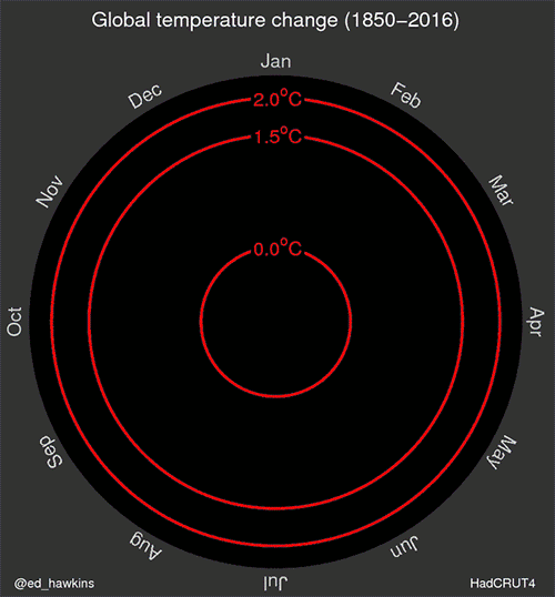
Ring of fire: spiralling global temperatures. Created by climate scientist Ed Hawkins of the University of Reading.
By Tushna Commissariat
As we face up to the realities of global warming and see the effects of climate change become apparent, it’s more important than ever that people the world over truly grasp its impact. With this in mind, University of Reading climate scientist Ed Hawkins has created the above animated spiral, which shows how the global temperature has changed over the past 166 years. Using data from the Met Office’s Hadley Centre observations datasets, Hawkins’ animation presents data in a a clear and artistic way. “The pace of change is immediately obvious, especially over the past few decades. The relationship between current global temperatures and the internationally discussed target limits are also clear without much complex interpretation needed,” says Hawkins, who is based at the university’s National Centre for Atmospheric Science. Take a look at his webpage to learn more about the project and for a list of specific weather events that are noticeable in the data.
Researchers have found no evidence of sterile neutrinos in two years’ worth of data from the IceCube Neutrino Observatory at the South Pole. The international scientific collaboration that runs the detector says that the results cast serious doubt on the existence of these hypothetical particles.
Buried beneath the ice at the Amundsen–Scott South Pole Station, IceCube is designed to search for high-energy particles from space, including cosmic rays and neutrinos. It consists of 5160 light sensors suspended on 86 strings in 1 km3 of ice. When a particle interacts with the ultra-clear ice, it can create flashes of light that are then detected. In 2013 the IceCube collaboration announced the first ever detection of cosmic neutrinos.
Neutrinos are particles with no electrical charge and are known to come in three “flavours”: electron, muon and tau. They were originally thought to be massless, but the discovery that they can change, or “oscillate”, between different flavours suggests that they do have mass. There is much that physicists do not understand about neutrinos, and some experimental results are difficult to reconcile with the three-neutrino model. But the existence of a fourth type of neutrino, a sterile neutrino, could provide an explanation.
Harder to detect
Sterile neutrinos would only interact with other matter via gravity – making them even harder to detect than other neutrinos. If they do exist, they would be able to help answer important questions about neutrinos, such as why they have mass and whether they are dark-matter particles. Several experiments are probing the existence of sterile neutrinos, but so far none of the particles have been detected.
In the latest research, the IceCube collaboration performed independent analysis on two sets of data from the observatory, looking for sterile neutrinos in the energy range between approximately 320 GeV and 20 TeV. If present, light sterile neutrinos with a mass of around 1 eV/C2 would cause a significant disappearance in the total number of muon neutrinos that are produced by cosmic-ray showers in the atmosphere above the northern hemisphere and then travel through the Earth to reach IceCube. The first set of data included more than 20,000 muon-neutrino events detected between 2011 and 2012, while the second covered almost 22,000 events observed between 2009 and 2010.
As neutrinos travel through space, they oscillate from one flavour to another. Physicists know that this oscillation is modified when neutrinos travel through dense matter because the neutrinos interact slightly with surrounding electrons and nucleons. Sterile neutrinos, however, would not interact with matter, and this would result in a resonance effect on the oscillations of neutrinos at energies around a few TeV. Francis Halzen, IceCube’s principal investigator and a particle physicist at the University of Wisconsin–Madison in the US, says that this “should be clearly seen as a sharp depletion in our measured muon-neutrino spectrum”, and should also produce “a characteristic structure in the zenith-angle distribution of these neutrinos”. “Neither one is seen,” he adds. “The absence of this resonance is pretty striking and cannot be missed.”
Previous sightings ruled out
According to the IceCube collaboration, these results do not rule out sterile neutrinos completely, but they do exclude much of the parameter space in which they could exist. In particular, the results exclude, with a confidence level of approximately 99%, the allowed parameter space for several experiments that had observed anomalies in neutrino oscillations, which had been interpreted as possible signs of sterile neutrinos.
Halzen says that not finding the characteristic sterile-neutrino signal “really calls into question their existence, or their role in explaining any anomalies in the neutrino data that may exist”. Discussing the anomalies seen in neutrino oscillations, he says that “We know that neutrino oscillations call for physics beyond the Standard Model. It looks now less likely that sterile neutrinos are part of it.”
However, Patrick Huber, of Virginia Tech in the US, says that although “it is a beautiful result”, it provides “little we didn’t know” and “qualitatively does not change much”. “To address the sterile-neutrino question, a decisive experiment is needed. One possibility would have been to use stored muon beams, but this has been ruled as too expensive. I am, however, doubtful that this question can be settled without such an experiment, unless we are very lucky and for instance the next generation of short-baseline reactor experiments finds a signal for sterile neutrinos,” he told physicsworld.com.
The measurements and analysis are described on the arXiv preprint server.
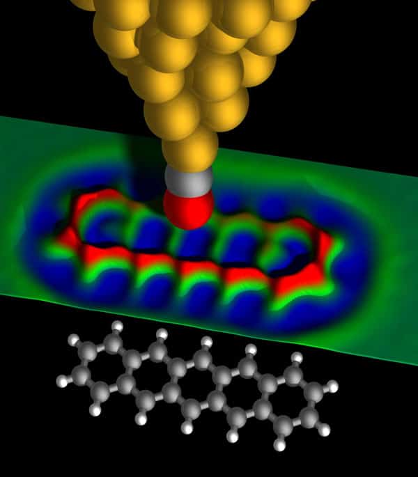
Feeling the force Schematic representation of an AFM tip terminated by a CO molecule, with the carbon atom (grey) attached to the last copper atom of the metallic tip (gold) and the oxygen atom (red) pointing towards the sample. The map in the central part of the image shows colour-coded data recorded above a pentacene molecule (C22H14), which is shown as a ball-and-stick model below. (Courtesy: Nat. Chem.3 273)
The past three decades has seen a revolution in our understanding of nature at the nanoscale. This immense progress owes much to the invention of advanced scanning probe microscopes that let us visualize and manipulate nanoscale objects. The scanning tunneling microscope (STM), which was developed in the early 1980s, achieved an unprecedented spatial resolution comparable with the size of an individual atom. Invented by Gerd Binnig and the late Heinrich Rohrer of IBM Research Rüschlikon in Switzerland, the pair received the 1986 Nobel Prize for Physics for the groundbreaking work. Then, in that same year, Binnig – together with IBM’s Christoph Gerber and Calvin Quate at Stanford University in the US – implemented the first atomic force microscope (AFM). Together, these tools have shaped the trajectory of nanotechnology ever since.
At the heart of all scanning probe microscopes is the probe tip, which scans the surface of a sample using piezoelectric translators. High-resolution maps of the topology and other surface properties can be achieved if the tip is sharp enough and held near to the sample. In contrast to scanning and transmission electron microscopes, which were invented many years earlier, scanning probe microscopes do not require sophisticated electron-focusing optics. The diffraction limits and optical aberrations that plague these and visible microscopes therefore do not apply, meaning that the resolution of scanning probe microscopes is mostly limited by the sharpness of the probe tip.
Importantly, scanning probe microscopes can both image the surface of a sample and carry out various types of spectroscopy; they can even modify surfaces directly. Atoms can be pushed around, for instance, as shown for the first time by Don Eigler and co-workers at IBM Research Almaden in the US in 1990 (Nature344 524). A decade after this breakthrough, a team from the Free University of Berlin used an STM to dissociate and fuse molecules and thus trigger chemical reactions on individual molecules in a controlled manner, marking an important step towards the synthesis of molecules from atomic building blocks (Phys. Rev. Lett. 85 2777).
The STM and AFM differ in how they measure the interaction between the sample and the tip. The STM is based on quantum-mechanical tunnelling, whereby an electron has a finite probability to pass through the potential barrier between the tip and the sample, which is classically forbidden. The tunnelling current decreases exponentially with the tip–sample distance, resulting in high spatial resolution, but the STM is restricted to small tip heights and also to samples that are electrically conducting.
The AFM, in contrast, relies on measuring the force between tip and sample. Consequently, the AFM is not limited to studying conducting samples and also works extremely well outside vacuum conditions and even in liquids. Moreover, the various interaction forces between the sample and the AFM tip have very different origins and decay lengths. For these reasons, the AFM has become a more widely used tool than its STM ancestor.
The AFM has undergone a number of different physical implementations over the years. Common to each is a cantilever that holds the tip, which is deflected because of the interaction between the tip and the sample. In the first implementation of an AFM 30 years ago, an STM was used to measure the deflection of a metallized cantilever. Later, this approach was superseded by optical-beam deflection, fibre interferometry and piezoelectric detection. Indeed, the particular operational mode of an AFM is chosen depending on the conditions under which the measurement is conducted, the materials being studied, and the properties being investigated. Since the interaction forces between the tip and the sample include components from electrostatic, magnetic, capillary, Van der Waals or chemical forces, in addition to effects caused by friction, mechanical properties and heat conduction, the AFM can provide an enormous range of microscopes and spectroscopes with a plethora of different applications.
Atomic resolution
Some of the most exciting applications of the AFM today demand instruments with the highest possible lateral resolution. For this purpose, an AFM tends to be operated in “non-contact” mode, as introduced in 1991 by Dan Rugar and co-workers at IBM Research Almaden (J. Appl. Phys.69 668). Unlike the more conventional contact mode, where the tip physically touches the surface and the force on the tip leads to a static deflection of the cantilever, when operating in non-contact mode, the tip and sample never touch.
The interaction between them can still be measured, however, by making the cantilever oscillate and detecting how much its resonance frequency becomes detuned as it moves across a surface. In order to obtain the highest possible resolution, the AFM will also typically operate under extreme conditions: an ultrahigh-vacuum environment leads to atomically defined surfaces with as little contamination as possible, while cryogenic temperatures immobilize molecules on the surface, thus decreasing thermal drift and increasing the stability of the system.
In 1995 Franz Giessibl, now at the University of Regensburg in Germany, used an AFM to obtain atomic-resolution images of the silicon “7 × 7” surface (Science267 68). Imaging this particular surface at the atomic scale had been the defining breakthrough for the STM 10 years earlier, and achieving it with an AFM proved that the technique had finally caught up in spatial resolution. About a decade after this milestone, however, another great leap forward for the AFM was achieved by Yoshiaki Sugimoto and colleagues at Osaka University in Japan based on studies of the surfaces of semiconductor alloys, demonstrating both elemental sensitivity and atomic manipulation with an AFM even at room temperature (Nature446 64; Science322 413). Then, in 2007, Alexander Schwarz and co-workers at the University of Hamburg in Germany demonstrated atomic contrast for the first time by using an AFM to directly detect the magnetic exchange force (Nature446 522).
A great advance in high-resolution AFM came even earlier, with the introduction of the “qPlus” sensor in 1998 (Appl. Phys. Lett.73 3956). This piezoelectric cantilever – based on a quartz-crystal tuning fork – allows oscillation amplitudes smaller than an atomic diameter, which is crucial for measuring short-range forces at the atomic scale. In 2008 Markus Ternes and co-workers at IBM Research Almaden used this detection scheme to slide single atoms across a surface using an AFM and also to directly measure the forces involved (Science319 1066). The following year, at IBM Research Zurich, the research group of the present authors used a similar set-up to resolve the atomic structure of individual molecules for the first time using an AFM (Science325 1110). The trick, which we discovered by chance, was to modify the tip itself using atomic manipulation. It turned out that after the pick-up of a single carbon monoxide (CO) molecule by the AFM tip, the resolution on molecules was dramatically increased.
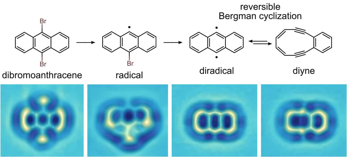
Chemical control A chemical reaction scheme (top) observed for an individual molecule using an AFM with CO tip functionalization, with each reaction step induced by bias voltage pulses across the AFM tip. (Courtesy: Nat. Chem.8 220)
Such tip functionalization is important for several reasons. First, it makes the tip passive and thus stops the molecule under study from being picked up. Second, the CO molecule effectively produces a very sharp tip because of the small atomic radius of the attached oxygen atom. Finally, since the CO molecule is flexible, it may tilt and cause an apparent increase in resolution because chemical bonds appear sharpened and the relative variations caused by bond-order differences are exaggerated (Science337 1326). The measured force variations corresponding to the interaction between the CO molecule at the tip and the molecule being imaged are on the order of piconewtons (10–12 N). Several other tip functionalizations, for instance using a single xenon, krypton or chlorine atom, have also been shown to achieve atomic resolution. However, because of the stability of CO tips and the apparent sharpening thanks to the CO tilting, CO tips are the most popular to date.
Addressing single molecules
This trick for AFM imaging paved the way for studying the structure and chemistry of individual molecules. It meant that the AFM could be used to identify unknown compounds (Nature Chem.2 821) and to investigate products formed by on-surface synthesis (Science 340 1434), for instance. Another unique advantage of an AFM for this purpose is that it offers the possibility of studying complex molecular mixtures molecule by molecule. Alternative protocols also allow the charge distribution within a molecule to be mapped, as well as the adsorption geometry to be measured with unprecedented resolution and tiny differences in the bond order to be detected within individual molecules.
Such detailed knowledge about complementary molecular properties is essential for understanding and predicting the reactivity, stability and interaction of molecules with respect to their environment. After all, single molecules and their functionalities are promising for applications in organic photovoltaic devices, molecular electronics and the synthesis of novel compounds.
CO tip functionalization is now widely used in the AFM community to achieve atomic-level resolution where it was not possible before. Recently, for example, an AFM was used to visualize molecular networks based on hydrogen (Science342 611) and halogen bonding (ACS Nano9 2574) with atomic resolution, and last year a team used an AFM to resolve the atomic arrangement of metal clusters relevant for fundamental studies of catalysts (Science 348 308). AFMs fitted with CO tips have also turned out to be highly suited for the investigation of graphene sheets and nanoribbons, with recent studies determining the exact atomic termination (Nature Commun.4 2023), precise position of dopant atoms (Nature Commun. 6 8098) and atomic resolution of graphene edges and junctions (Nano letters15 5185). The performance of eventual devices based on such 2D materials depends crucially on their exact atomic structure, which an AFM can determine better than any other tool. Moreover, the AFM allows such systems to be atomically modified.
Indeed, the combination of high-resolution AFM with atomic manipulation now offers the unprecedented possibility of custom-designing individual molecules by making and breaking bonds with the tip of the microscope and directly characterizing their products on the atomic scale. Very recently, our group was able to form diradicals by dissociating halogen atoms and then reversibly trigger a ring-opening and ring-closing “Bergman” reaction via atomic manipulation, allowing us to switch and control the molecule’s reactivity, magnetic and optical properties (Nature Chem.8 220). This offers novel functionalities for molecular-based logic and opens new routes for the creation of designer molecules by single-molecule radical chemistry.
Importantly, a high-resolution AFM offers opportunities to understand and control physical, chemical and biological processes at the level of individual molecules. Ongoing improvements in force sensitivity plus temporal and spatial resolution will push the frontiers in nanoscience further. Perhaps in another 30 years the AFM might be further improved towards an atomic assembler as addressed by Richard Feynman in his famous 1959 talk “There’s plenty of room at the bottom”: a tool that might build arbitrary, 3D atomically precise devices, metamaterials and molecules. Either way, there is no doubt that the AFM will continue to promote discoveries from fundamental physics all the way to chemistry and the life sciences, unravelling nature’s most enigmatic mechanisms at the nanometre scale and beyond.