Two years ago, a six-volume paean to molecular gastronomy made a surprise appearance in Physics World‘s annual list of the year’s best physics books. Written by Nathan Myrhvold – a PhD-level physicist and a former Microsoft executive whose CV also includes a one-year stint as Stephen Hawking’s postdoc – Modernist Cuisine made our list thanks to its descriptions of scientific cookery, including explanations of how to use standard laboratory equipment such as water bottles and centrifuges in food preparation (see “Cooking up a storm“).
Myrhvold’s follow-up effort, The Photography of Modernist Cuisine, lacks the scientific heft of its predecessor, but as these photos illustrate, it is breathtakingly beautiful and full of surprises.
Intricate patterns in a romanesco cauliflower. (Courtesy: Nathan Myhrvold/Modernist Cuisine LLC)
The image at the top, for example, may look like a distant planet viewed through the porthole of a spacecraft, but it is actually the bottom end of a blueberry: the “planet” is part of the berry’s ovary, while the rough-edged lobes of the “porthole” are the remains of its blossom, or calyx. The next image shows the fractal-like patterns characteristic of a romanesco cauliflower, while the delicate folds of bright-pink material are found on the surface of a cabbage.
Bright-pink folds on the surface of a cabbage. (Courtesy: Nathan Myhrvold/Modernist Cuisine LLC)
In the final photo, below, oil is ignited as it bursts from an orange as it is peeled.
Setting the oil in orange peel on fire. (Courtesy: Ryan Matthew Smith/Modernist Cuisine LLC)
With a “wingspan” of more than a third of a metre, a mass of nearly 6 kg and a hefty price tag, The Photography of Modernist Cuisine is not the sort of book that can be tucked discreetly into a Christmas stocking. Still, the book – which includes a “how we did it” chapter that delves into the photographic techniques and the back stories of some of the book’s 405 photos – is a visual feast, and we think it will appear under a few trees this season.
Quantum entanglement between two distant parties could be achieved using a non-entangled or classical information carrier. That is the conclusion of three independent teams of physicists that have done quantum-optical experiments backing up their claims.
Entanglement is a purely quantum-mechanical phenomenon that allows two particles, such as photons or electrons, to have a much closer relationship than is predicted by classical physics. Over the years, it has played a central role in creating in quantum-information systems such as quantum cryptography.
In most quantum-cryptography protocols two parties – normally referred to as Alice and Bob – wish to exchange information securely using a source that creates pairs of photons that are entangled in terms of their polarization. While any measurements Alice makes on the polarization of a succession of photons will be random, Bob’s results will be correlated to hers. This means that when compared on a photon-by-photon basis, both Alice and Bob’s photons are likely to have the same polarization.
An important challenge facing physicists creating such systems is “decoherence” – the destruction of entanglement that occurs when a quantum system interacts with its surroundings. To avoid this, researchers will normally try to keep an entangled system completely isolated from its environment, which can be quite a difficult task.
Separable states
Now, research published in three separate papers in Physical Review Letters suggests another surprising scheme for preserving entanglement in quantum cryptography – the use of an information carrier that is not itself entangled.
The experiments make use of separable states, which are quantum states that show certain correlations but are not entangled. These states were first defined in 1989 by physicists who were trying to understand the precise nature of entangled states. In this case, Alice polarizes a photon in a particular direction, say vertically, and then uses a classical communication method (such as a telephone) to tell Bob to polarize his photon in another direction, say horizontally. Both Alice and Bob’s photons are separable states and it was concluded that they are not entangled. However, in 2003 Toby Cubitt, then at the Max Planck Institute of Quantum Optics in Germany, and colleagues, calculated that, in some cases, separable states can be used to distribute entanglement.
Classically carried
Now, Alessandro Fedrizzi of the University of Queensland in Australia and colleagues have confirmed this in the lab. Their experiment begins with Alice and Bob sharing an entangled pair of photons (A and B). Then, this entanglement is destroyed by the introduction of other random states. This effectively makes A and B separable states because they still carry distinct correlations but are no longer entangled. A third photon, C, that is also a separable state serves as the carrier. Alice then sends A and C through a quantum gate. This causes A and B to interfere, thus creating another correlation (but not entanglement) between them. Then, C is sent along to Bob, who combines it with his B.
The surprising result is that A and B are entangled, even though C was never entangled with either of them. Rather, C is simply communicating the entanglement. The researchers also say that “distributing entanglement with separable carriers is resilient to noise and in some cases becomes the only way of distributing entanglement through noisy environments”.
Uncertain states
The second group to have done work along similar lines includes Christian Peuntinger of the Max Planck Institute for the Science of Light in Germany and colleagues. Peuntinger’s team applies the Heisenberg uncertainty relation between the amplitude and phase of the photons in two of its beams (A and C). Once again, C then carries the information of the entanglement to B but is itself never entangled with either A or B. The researchers say that their result “highlights the utility of quantum correlations beyond entanglement for the establishment of a fundamental quantum-information resource and verifies that its distribution by a dual classical and separable quantum communication is possible”.
Hidden states
Christina Vollmer, Roman Schnabel of the Albert Einstein Institute in Germany and colleagues are the third group to follow a very similar protocol. The only difference being that they begin with an entangled state that is hidden by mixing one part of the system with the external environment.
All three papers show that separable states that are not entangled can be used to carry entangled states. The technique could be particularly useful in quantum-communication systems involving more than two parties, while also serving as a tool to better understand entanglement and its responses to noise and classical communication.
All of three papers are published in Physical Review Letters.
Tiny “tags” made of dye molecules stuffed into carbon nanotubes have been used to develop a high-resolution imaging technique based on Raman scattering. Created by researchers in Canada, the tags boost the weak Raman signal of molecules about one million times. The new approach could lead to improved medical diagnostics and treatments, and could even be used to fight counterfeiting.
Raman spectroscopy involves shining a beam of light onto a solid or a liquid to identify its molecular composition. While most of the photons will scatter from the sample with no change of energy, a small number of photons will exchange a tiny amount of energy by causing molecules in the sample to vibrate. This is called Raman scattering, after the Indian physicist Chandrasekhara Venkata Raman who first observed it in liquids in 1928 and won the 1930 Nobel Prize for Physics for his discovery.
“One can imagine Raman scattering as the process by which photons shake a molecule,” explains Thomas Szkopek of McGill University, who is part of the research team. The vibration of each type of molecule is unique and so is the amount of energy exchanged with photons. By measuring the energy spectrum of the scattered photons, it is possible to determine what molecules are present in the sample.
Major limitation
Raman spectroscopy is widely used in medicine, chemistry and drug development. A major limitation, however, is that the scattered light is very weak. This makes the technique impractical for many applications, especially those involving high-resolution imaging of biological samples, because it takes so much time to collect the weak signal from each pixel.
Increasing the strength of the interaction between the light and the molecules does not help much, as it leads to the Raman light being hidden by much brighter fluorescence. “To be practical in high-resolution imaging, there is a need to boost the Raman signal by about a million times or more,” says Richard Martel of the University of Montreal, who led the research team.
There have been previous attempts at amplifying the Raman effect, but most of these involved manipulating the optical field with metal nanostructures. This requires putting the molecules of interest in direct contact with metallic particles, but this has proved difficult because the particles are difficult to stabilize and control.
We can turn up the brightness of the Raman light while leaving the fluorescence light turned off
Thomas Szkopek McGill University
Martel’s team, which includes postdocs Étienne Gaufrès and Nathalie Tang of the University of Montreal, took a different approach and turned to dyes and carbon nanotubes, which are hollow tubes of carbon with walls as thin as just one atom. “The carbon nanotube is very effective at turning off the undesired dye-molecule fluorescence while allowing the desired Raman scattering process to continue,” explains Szkopek. “We can turn up the brightness of the Raman light while leaving the fluorescence light turned off.”
Tubes filled with dye
The researchers begin by using nitric acid to purify the nanotubes, cleaning them and opening their ends. They then dissolve dye molecules in a solvent, mix in the nanotubes and heat the solution for a few hours. The dye molecules fill the tiny nanotubes and align along the tube axis. The resulting “nanoprobes” are about 1 nm in diameter, 500 nm long and contain about 500 dye molecules.
The next step involves adding a nanoprobe to the sample to be examined. The nanoprobes can be chemically grafted onto any object, even bacteria or proteins, therefore becoming a sort of “Raman tag”, says Martel. “Attaching the nanoprobes to a target is like printing a barcode into the object, allowing it to be identified even if it’s not Raman active or visible,” he says.
A measurement then proceeds like a normal Raman spectroscopy procedure. A laser beam inserted in the microscope objective shines onto the sample and its scattering is probed with a spectrometer. The only difference is a huge increase in the intensity of the Raman signal.
Protected from photobleaching
“Once the dye molecule absorbs a photon, the energy is passed on to the nanotube before the molecule has the opportunity to change the energy back into a photon that would contribute to fluorescence,” says Szkopek. Carbon nanotubes therefore suppress the fluorescence that would otherwise overwhelm and hide the Raman signal, making the signal from the nanoprobes so bright that it completely dominates the spectrum – one million times stronger than the Raman signal of other molecules, once the fluorescence is supressed. The nanotubes also protect the dye from the environment, especially photobleaching by the laser beam. This tagging approach is already used in fluorescence, Szkopek adds, but the fluorescence signal is not a great barcode compared with the Raman signal – many more complex signals can be generated with the dyes in a Raman nanoprobe.
There are other advantages to using the technique, says Mark Hersam of Northwestern University, who was not involved in the study. For instance, the ability of the nanotubes to supress fluorescence and at the same time to protect the dyes from the environment allows for broad use of such Raman tags – in particular, “for multispectral analysis in applications ranging from protein detection to biomedical imaging”, Hersam says. He also thinks that nanoprobes could be added to banknote ink could help eliminate the problem of counterfeiting.
Nobel laureates Steven Chu (left) and David Gross talk energy in Gothenburg. (Courtesy: A Mahmoud)
By Michael Banks
Yesterday I joined more than 1000 people attending the day-long Nobel Week Dialogue event in Gothenburg, Sweden. The delegates battled the cold winter weather to make it to the Swedish Exhibition and Congress Centre, just south-east of the city centre (and next to a theme park, of all things).
This is the second such Nobel Week Dialogue and the first time it has been held in Gothenburg. Last year the theme for the event in Stockholm was the “genetic revolution” and this year it was on “exploring the future of energy”.
A new type of thermionic generator that turns heat or light into electrical energy has been developed by researchers in Germany and the US. The new design overcomes the “space-charge problem” that has plagued previous attempts at developing practical devices. The device is about four times more efficient than previous generators and the new technology could find use in a range of applications including solar power and the harvesting of waste heat.
Thermionic generators convert heat or light into an electric current by using the temperature difference between two metallic plates that are separated by a vacuum. The “hot” plate is heated either by incident light or thermal conduction and this causes electrons to evaporate from its surface. These electrons then condense on the surface of the cold plate. This creates a charge difference between the two plates, which can drive a usable electric current.
Because they convert heat or light directly into electrical energy, thermionic generators have considerable potential for practical applications. If used in coal-fired power stations, for example, thermionic converters would, in principle, be more efficient than steam turbines. Thermionic generators could also be applied to a variety of lower-temperature applications, such as the collection of solar energy or the recycling of waste heat in car engines.
The space-charge problem
While this potential for highly efficient energy conversion has been known since the late 1950s, the practical applications have been severely limited by what is called the space-charge problem. For plates separated by more than about 3–5 μm, the negative charge of the “cloud” of electrons that forms in the gap inhibits subsequent electrons from being emitted from the hot plate. This effectively puts a halt to the flow of electrons between the plates. While reducing the gap between plates would help, making a generator with plates that maintain a separation of less than 3 μm at high temperatures is extremely challenging.
Hot and cold: the thermionic generator at a high temperature. (Courtesy: J Mannhart/MPG)
Previous efforts to solve the space-charge problem have mainly involved inserting caesium ions into the space between the two plates. The positive ions act to neutralize some of the offending charge, thus allowing more electrons to be released from the hot plate. While this approach has been used in the lightweight TOPAZ nuclear reactors that powered some Soviet satellites, it results in a drop in power output of about 50%.
Now, Jochen Mannhart of the Max Planck Institute for Solid State Research in Stuttgart – along with colleagues at the University of Augsburg and Stanford University – has come up with a new way of solving the space-charge problem by creating an electric field in the space between the plates. This field first accelerates the electrons leaving the hot plate and then slows them down as they approach the cold plate. The charge cloud is therefore moved along and does not repel subsequent electrons – allowing for a continuous current. The field itself is created by a honeycomb-patterned gate with hexagonal holes in it. The electrons are guided through the holes by applying a magnetic field between the plates.
A 40% efficiency could be possible
“Practical thermionic generators have reached efficiencies of about 10%. The theoretical predictions for our thermionic generators reach about 40%,” says Mannhart. This figure also incorporates the energy needed to create the electric field.
Mannhart and colleagues believe that commercialization of their design could take a further 5–20 years, depending on whether the application is high temperature or low temperature. For the latter, Mannhart says that further optimization of the converter would be needed.
Nicholas Melosh of Stanford University, who was not involved in this study, describes the work as a “clever new technique” to address the space-charge problem, adding that “while more development still needs to be done, this principle could create new devices to convert waste or solar-generated heat into useful electricity”.
Planar triode revival
Neil Fox of the University of Bristol in the UK points out that the new generator has similarities to a planar triode design tested at the Massachusetts Institute of Technology (MIT) in the late 1950s. This previous design had suffered from energy loses caused by electron–electron collisions and scattering. “[Mannhart and colleagues] have come up with a rather neat vertical triode structure that seeks to improve on the MIT device, by incorporating beam collimating concepts similar to those used in particle accelerators,” explains Fox. “The data presented…show that this magnetic triode is a significant improvement over a closed-spaced diode, but suggests that electron–electron collisions and scattering losses to the gate are still present.”
The team is now working to increase the efficiency of its generator design in two ways. First, it is building high-performance converters from existing semiconductor technologies. Second, it is optimizing its electrodes through the use of new materials, especially oxides, and nanotechnology.
A team of researchers in the US has achieved spin coherence times of more than 200 µs in nitrogen vacancy (NV) centres in tiny “nanodiamonds”, shattering the previous record for coherence times in this material. The nanodiamonds were made using a new mask and ion etching process that the team believes could be used to develop real-world applications of NV centres including magnetic-resonance probes and quantum computers.
The NV centre in nanodiamond could be ideal for use in a host of future quantum technologies, including quantum computing and nanoscale sensing. However, most nanodiamonds available today contain a high density of paramagnetic impurities that make the electron spins in NV centres extremely fragile – they cannot hold their spin direction for very long (microseconds at most), which means that they are unable to store quantum information for any practical length of time.
Atomic impurities, or defects, in natural diamond lead to the colour seen in pink, blue and yellow diamonds. One such defect, the NV, occurs when two neighbouring carbon atoms in diamond are replaced by a nitrogen atom and an empty lattice site. NVs in nanodiamonds could be ideal as biological probes because they are non-toxic, photostable and can easily be inserted into living cells. They are also capable of detecting the very weak magnetic fields that come from electronic or nuclear spins, and so can be used as highly sensitive magnetic resonance probes capable of monitoring local spin changes in a material over distances of a few tens of nanometres. And, in contrast to conventional magnetic resonance imaging techniques in biology in which millions of spins are required to produce a measurable signal, the NV defects can detect individual target spins with spatial precision measured in nanometres.
Annoyingly short coherence time
The main problem until now, however, was that the spin coherence time in these NVs was annoyingly short because of the high concentration of paramagnetic impurities (namely nitrogen) in diamond nanocrystals grown by conventional high-pressure high-temperature (HPHT) processes. A team led by Dirk Englund of the Massachusetts Institute of Technology (MIT) has now developed a top-down fabrication technique using a self-assembling porous metal mask and reactive ion etching process that produces extremely pure nanocrystals devoid of paramagnetic impurities. These nanocrystals contain NVs that are able to preserve their spin states for as long as 210 µs.
The defects also have record magnetic field sensitivities of 290 nT Hz–1/2, which means that they can be used as magnetic field probes that have a diameter of just 50 nm. “And that is not all: the NVs can be produced in their billions or more per shot, without much effort on our part, thanks to the simple ‘self-guiding’ or porous metal mask we employed,” MIT team member Matthew Trusheim said.
Single photon sources and solid-state qubits
As well as being ideal as magnetic field sensors, the NVs might be placed in photonic structures and used as single photon sources or as solid-state quantum bits (qubits) entangled with photons, he adds. Quantum computers exploit the counterintuitive idea that tiny objects can exist in more than one state at the same time. Rather than processing classic bits – which are either 0 or 1 – such devices instead manipulate qubits that can be 0 and 1 at the same time. Vast numbers of logic operations could then be possible in parallel, making these computers theoretically far faster than ordinary machines.
Until now, qubits made from nanodiamond NVs were incredibly fragile and the quantum information they held was rapidly destroyed by interactions with noise in the surrounding environment. The new result, published in Nano Lett. DOI: 10.1021/nl402799u, could go a long way in changing all this.
Gold and palladium mask
The researchers made their highly pure nanocrystals of diamond by first depositing a metal mask (made of gold and palladium) onto a high-purity bulk diamond substrate. The mask self-assembles into droplets that are tens of nanometres in size. “Next, we used an oxygen plasma etch, where reactive ions are accelerated onto the substrate surface to remove the diamond,” explaind Trusheim. “The metal mask blocks the incoming ions, forming small regions where the diamond is preserved. These regions are then removed mechanically and become our nanodiamond.”
The team, which includes scientists from Columbia University, the City College of New York and the University at Albany-State of New York, says that it would now like to use these NV-fluorescent nanodiamonds in real sensing applications. “For example, they could be used to detect the electric fields coming from neural action potentials in the body, or to detect magnetic proteins in living cells,” says Trusheim. “We are also integrating these diamond structures into photonic networks to efficiently interface photonic qubits with spin qubits,” he reveals.
An entangled state of two quantum bits (qubits) can be created and stabilized using interactions that are normally thought to be detrimental, according to two international groups of researchers. While the groups studied two different physical systems, they have both shown that an entangled quantum system can be produced and maintained by coupling it to an environment that dissipates energy.
External noise
Entanglement is a purely quantum-mechanical phenomenon and that plays a crucial role in quantum-information systems. It allows two particles, such as photons or electrons, to have a much closer relationship than predicted by classical physics, and it is this relationship that could be exploited in quantum computers and cryptography. Physicists are keen to create and maintain entangled states in a variety of quantum systems, but a common problem that arises while creating these states is decoherence – the destruction of entanglement that occurs when a quantum system interacts with its surroundings. To avoid this, researchers try to keep an entangled system completely isolated from its environment – a difficult task.
But in recent years, new theoretical work has shown that the dissipative interactions with the environment could instead be used to preserve coherence, and the method has already been used to stabilize the state of a single qubit. Now though, the two research groups have gone one step further by actually using dissipation to create and sustain an entangled state in two different systems – one team uses ions, while the other uses superconducting qubits.
Anders Sørensen and Florentin Reiter at the University of Copenhagen, along with Nobel laureate David Wineland, John Gaebler, and colleagues at the National Institute of Standards and Technology (NIST) in the US produced a stable entangled state between two trapped beryllium ions. They used a set of tailored interactions including “sympathetic cooling” by two magnesium ions in the same trap.
Excited states
The ions’ spins were entangled using two ultraviolet laser beams and induced to “leak” any “unwanted” quantum states to the environment through continuous application of microwaves and one laser beam. “The simple explanation is that it is similar to so-called optical pumping. The two qubits can be in four different states and one of them is the entangled state that we want,” explains Sørensen. “If they are in any of the three undesired states, a laser puts them to an excited state from which they decay down again.” This excitation is done until all the qubits end up in the desired state, where carefully chosen interactions and resonance conditions ensure that the excitation out of the state is suppressed.
Trap and hold: NIST’s ion trap. Electrical potential is applied through thin gold wires on a chip and used to trap ions in a narrow slot. (Courtesy: NIST)
The researchers found that after 12 ms, the spins were still highly entangled. “In principle, you can prepare a dissipative state where you are truly in a steady state so that the lifetime is as long as you wish,” says Sørensen. This was almost the case in the team’s experiment, but there is a small leak that the researchers do not correct. The team estimates that the lifetime of the entanglement is closer to 84 ms, which is very long compared with any other timescale in the experiment. The qubits approached the target state within a few milliseconds and were found to be in the correct entangled state 75% of the time. By applying about 30 repetitions of their excitation technique, the scientists boosted the success rate to 89% in a separate experiment.
According to Sørensen, the team purposefully designed its system so that it could be scaled up to include more atoms – something that could allow quantum computation. The researchers current experiment also let them investigate whether the method of allowing the system to talk to the environment actually represents an advantage for such real experiments.
Sørensen admits that, for now, the dissipative processes tend to be a bit slower than the more commonly used quantum gate – the basic building block of a quantum circuit that operates on a small number of qubits – but not necessarily much slower. He told physicsworld.com that one benefit of the new dissipative-state preparation is that the researchers can “now really begin to investigate whether there is an advantage of using dissipation as compared to gates. For the concrete system that we used, you probably could have done better with gates, but we can see that there are other scenarios such as optical cavities or if you have fluctuating lasers where using dissipation really helps.”
Feedback loops
The other team to achieve entanglement using dissipation included Shyam Shankar and Michel Devoret at Yale University, along with other colleagues. Their qubits are superconducting circuits, used along with a microwave cavity that serves as the dissipative reservoir. Shankar explains that, normally, a measurement-based feedback system is used to create entanglement. That involves first measuring the state of the qubit system with high fidelity, then using real-time electronics to determine whether the system is in the desired state or not. If it is not, a control drive would be applied to the system to bring it back to the desired state. Instead, the team uses an “autonomous” feedback scheme that does not require a measurement process to maintain coherence. The researchers apply six continuous drives (tones at carefully chosen microwave frequencies) combined with a specifically engineered coupling between the qubits and the dissipative reservoir. The researchers have engineered their system such that, in the presence of these drives, the ground state of the effective driven system is an entangled state.
Quantum building blocks
The fidelity of the entanglement is determined by the ratio of the feedback rate to the error rate. Currently, the researchers’ entangled state was maintained with an average fidelity of 67%, for a maximum of 500 μs. “This time is essentially indefinite…we could have taken the data for a longer time if we wanted to. The entangled state that we stabilize is, of course, a building block to make more complicated multi-qubit entangled states that are required for quantum-information processing,” says Shankar, further explaining that this method could allow for a “fully error-corrected logical qubits to be developed in the future”.
“Our autonomous method using dissipation eliminates this external feedback loop and is thus simpler to implement,” says Shankar. “We also do not have to worry about the latency of an external loop; in fact, the intrinsic dissipation of the system can correct the errors faster than any external loop could.” But he also points out that such an autonomous-feedback strategy has to be tailored to the particular system used and the particular state one is trying to stabilize, making it difficult to create any kind of general protocol.
In the coming months, Shankar’s team plans to combine the autonomous approach along with an external measurement-based feedback loop to get the best of both worlds. “The simplicity and quick response of autonomous feedback with the flexibility and reliability of measurement-based feedback,” says Shankar. On the other hand, Sørensen and colleagues are looking at extending their method to multi-particle entanglement so that they can see just how well and how many qubits they can entangle.
The Crucifixion by Puccio Capanna. (Courtesy: North Carolina Museum of Art/Gift of the Samuel H Kress Foundation)
Museum guards can get very nervous when inquisitive visitors step towards a work of art and lean in for a closer look. After all, someone crazy might want to attack the masterpiece with a knife or spray a load of paint over it. However, for true art lovers, getting right next to a work of art can offer a deeper appreciation of what the artist had in mind. Brushstrokes, colour composition and other detailed features can – once you step back and regard the artwork from its intended viewing distance – almost magically create a wealth of different impressions and emotions.
Quite simply, close-up views can reveal information about the artwork and its artist. Old Master paintings, for example, are very delicately layered objects with many features that are not immediately obvious. Our eyes mostly perceive the paint layers, which are a complex arrangement of coloured pigments, but these are embedded in “binders” that can alter the appearance of the pigments despite themselves being transparent. In fact, some Old Master painters used dozens of glaze layers to achieve the desired colour, gradient and even perceived depth. Leonardo da Vinci’s Mona Lisa is a prime example of such a layering technique, although very few visitors to the Louvre are ever allowed to get close enough to fully appreciate it.
Careful examinations of paintings can also reveal preparatory layers, initial sketches or under-drawings, with the latter being particularly useful to establish the true identity of an unknown artist. Sometimes even entire new art works can be discovered underneath the surface. However, such close-up analyses are best left to professional art conservators and conservation scientists, both equipped with microscopes and other imaging techniques. These people are experts in obtaining highly detailed structural and chemical information, which is crucial in identifying, authenticating, preserving and conserving works of art. Their aim is to identify individual pigments and pigment mixtures, work out how they are layered in 3D and establish how the artist applied them.
Valuable clues about an artwork’s authenticity can also come from checking if the pigments are really those that were used at the time the painting was supposed to have been created and/or seeing if the work has degraded in the manner expected for a painting of that age. Paints, glazes and varnishes all deteriorate naturally when exposed to light and humidity – in fact some early cadmium-yellow and lead-white paints react with each other and gradually turn black with age. Such degradation phenomena can be confined to a few microns of the surface but sometimes they can even encompass entire layers of paint. The challenge for conservators is to identify how much the painting has degraded, what products have been created and what can be done to stop things getting any worse. The specialists have several tools at their disposal, but our group at Duke University, led by Warren Warren, has been working on a new laser-based technique – pump–probe microscopy – that we think could help in that quest.
Tools of the trade
For art conservators, the easiest way to get detailed microscopic information about the composition and layering of pigments is to simply remove a small cross-sectional area of the art work with a scalpel. By laying this piece on its side, the layers can be seen and analysed, just like a sample extracted from an ice sheet or a tissue biopsy in medicine. However, sampling creates spots of damage and, although these marks are almost invisible to the naked eye, this technique should only be used sparingly, particularly on noticeable areas such as someone’s face or with paintings of great value. And even when samples can be taken, they provide information on only a few selected areas of the entire artwork. For some types of art, such as decorated or illustrated manuscripts, sampling is impossible because the pages are much too thin and delicate.
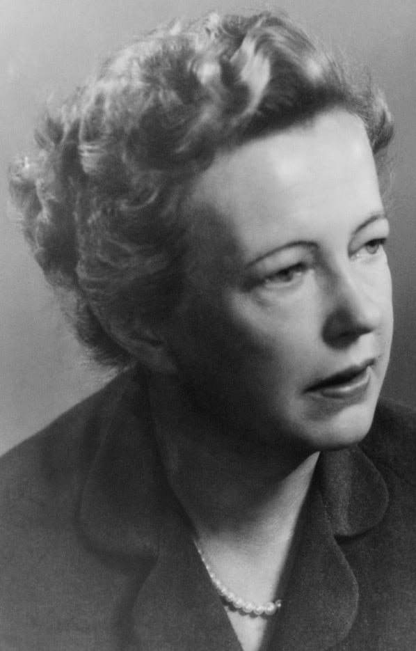
Nonlinear thinking Nobel laureate Maria Göppert-Mayer predicted an effect that lies at the heart of two-photon fluorescence. (Courtesy: Louise Barker/American Institute of Physics/Science Photo Library)
There are, however, some imaging methods that do not leave visible marks on the painting, each having its own strengths and weaknesses. X-rays are a particularly powerful tool, but they can only identify atomic elements and they cannot determine the individual pigments in a mixture. In addition, most X-ray imagers shine radiation through a sample and do not provide information in 3D, although spatially resolved (confocal) versions are being developed that can sample selected points. As for studying larger areas with this technique, that requires access to synchrotron sources, which is costly and not easily obtained.
Cheaper and easier to use than X-rays is light. Infrared reflectography, for example, can reveal under-drawings beneath a whole painting, while ultraviolet fluorescence photography can reveal areas covered with certain varnishes. Selected spots can also be probed with Raman, Fourier-transform-infrared or reflectance spectroscopy, all of which are good at identifying specific pigments, even if they are generally restricted to imaging surfaces, rather than providing 3D information.
In fact, for all mapping tools there is a trade-off between having a high spatial resolution, a large field of view, a good chemical specificity, and the ease (and cost) of use. We think that our new kid on the block – pump–probe microscopy – could find its place in conservation science by circumventing some of these limitations.
The art of being square
To see why pump– probe microscopy can be used in art imaging, it is worth first describing its precursor – a technique developed by physicists that was initially used in the seemingly unrelated field of biomedical imaging. Known as “two-photon fluorescence” (TPF), the technique involves shining infrared light onto a sample containing a fluorescent compound, which emits a photon only if it simultaneously absorbs two separate photons each of roughly half the energy needed to excite the molecule. As not one but two photons need to be absorbed for the molecule to fluoresce, the light emitted is not proportional to the intensity of the incoming light, but instead varies with the square of it. (This nonlinear effect was first predicted by the Nobel-prize-winning physicist Maria Göppert-Mayer back in the 1930s.)
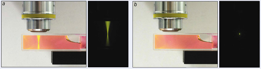
For a physicist, a linear response is generally a good thing, as it often leads to simple, more easily predictable systems. But in optical microscopy, linearity comes with one big drawback. As shown in figure 1a, although the fluorescence is strongest at the focus, it is also generated by out-of-focus light. Even though the fluorescence in these other regions is much dimmer, much of it comes from layers above and below the point to which the light has been focused, making the signal hard to decipher.
1 Hip to be square (a) In conventional fluorescence, one photon excites a fluorescent molecule, so that the amount of fluorescence is simply proportional to the intensity of incoming light. Here, a cuvette of dye is excited with continuous blue light and “linear” fluorescence can be seen throughout the entire cuvette, shown also in close-up. (b) But a fluorescent compound can also emit light if it absorbs two photons, each of roughly half the energy needed to excite the molecule. In this ‘two-photon fluorescence’, the light emitted is proportional to the square of the laser intensity, which is why the same dye exposed to pulsed infrared light only fluoresces at the focus because only here is the light intense enough. This localized excitation can be used to image the 3D distribution of fluorophores by scanning the beam across the sample. (Courtesy: Duke University)
But because the fluorescence scales more strongly with intensity in TPF microscopy than it does in linear techniques, the signal is mostly generated where the light is focused on a sample because here the intensity is at its highest (figure 1b). In fact, for good objective lenses, this region can be as small as 1 μm3.
This localization sounds great, but it comes at a price, which is that nonlinear processes are generally harder to induce than linear ones. In fact, all things being equal, TPF would need a light source about a million times more powerful than those used with ordinary microscopy. But we can make TPF work by turning to ultrafast lasers, which essentially lump a beam’s photons into very short but intense pulses, thereby creating large peak intensities without increasing the average power. Using this approach, in 1990 Watt Webb’s group at Cornell University was the first to implement TPF in a microscope and observe biological samples without damaging them.
Making an object fluoresce while matter surrounding it stays dark is, however, just one way to create the contrast needed to form an image and other groups swiftly developed similar nonlinear mechanisms that can also produce image contrast in biological media. These include second- (or third-) harmonic generation, which involves adding the energy of two (or three) photons to create a single photon of twice (or three times) the energy. Another example is coherent anti-Stokes Raman scattering, in which a photon is inelastically scattered, increasing its energy an amount equal to one of the molecule’s vibrational modes.
Pumping for information
By demonstrating these nonlinear contrast mechanisms, researchers opened the door for nonlinear microscopy to be used to study biological tissue. However, its potential remained largely untapped as it is easier to use techniques that generate light of a different colour to the incoming beam because the emitted light can be detected fairly simply using colour filters. Unfortunately, most nonlinear interactions do not generate such distinct colours and so are harder to detect. While physicists and spectroscopists are familiar with these interactions, current measurement strategies use far too much power to be applicable to tissue (or art).
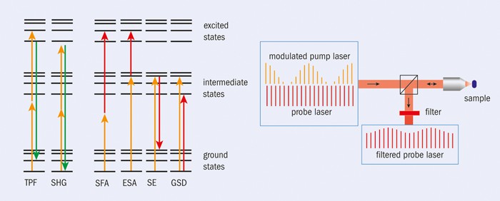
Nevertheless, some of these other nonlinear techniques can provide the contrast needed to create an image by measuring the intensity of light from different parts of a sample. These include sum frequency absorption (SFA) and excited-state absorption (ESA), both of which involve a sample absorbing two photons of different wavelengths – one from a “pump” laser and the other from a “probe” laser (figure 2). In SFA both photons need to arrive at exactly the same time, while in ESA the molecule first rests in an excited state after absorbing a photon from the pump laser, before only later absorbing a photon from the probe. Another nonlinear process is ground-state depletion (GSD), in which a pump photon excites molecules, with those remaining in the ground state then absorbing a photon from the probe. In stimulated emission (SE), meanwhile, excited molecules provide extra photons that add to the probe beam.
2 Two are better than one Nonlinear interactions between light and matter can be generated in many different ways, including two-photon fluorescence (TPF) and second-harmonic generation (SHG), which both involve a molecule absorbing two incident photons (orange) to create new colours of light (green) that are easy to detect. Also shown are four other powerful nonlinear two-photon interactions – sum frequency absorption (SFA), excited-state absorption (ESA), stimulated emission (SE), and ground-state depletion (GSD). Each interaction involves one photon from a pump laser (orange) and one photon from a probe laser (red). These interactions can yield more molecular information than TPF or SHG but because they do not produce a distinct colour they are harder to detect. We detect them by modulating the intensity of the pump laser and detecting synchronous variations in the reflected probe laser.
In all of these cases, the nonlinear interaction couples the pump and the probe beam – in other words, turning the pump laser on or off alters the intensity of the probe. So, for example, when the pump beam in an ESA experiment is switched on, the intensity of the probe drops after passing through a sample because the molecule absorbs one photon from both beams. In an SE experiment, in contrast, the intensity of the probe beam rises as an extra probe photon gets generated. And if the incident pump beam is turned on and off periodically, we can use a lock-in amplifier to measure the resulting variation in the probe beam (either from transmitted or back-scattered radiation), with its phase giving information about the type of nonlinear interaction (loss or gain in the probe) and the amplitude relating to the strength of the interaction (or the concentration of a given molecule).
In all cases, this localized excitation can then be used to image the 3D distribution of molecules by scanning the beam across the sample. However, additional details about the molecules can be obtained by introducing a time delay between pump and probe pulse because when the pump pulse excites a molecule, the population in a particular state can relax back to the ground state(s) with a certain time constant that depends on the nature of the molecule. So by switching on the probe pulse a certain time after the pump pulse, we can map out how fast ground-state or excited molecules decay, thereby providing clues as to the composition and structure of the molecule.
Into the art world
Laser spectroscopists have used such modulated pump–probe techniques for decades, but it is only recently, thanks to the development of highly stable, ultrafast dual-colour sources, that these methods have been used to image biological samples. In 2007 our group at Duke was the first to apply this technique to image melanin pigments in skin. Melanin is meant to protect the skin from Sun damage, but in melanoma – an extremely aggressive form of skin cancer – this pigment is involved in uncontrolled cell proliferation. The Duke team has since shown that pump–probe microscopy can provide valuable microscopic information on how this disease develops and spreads.
3 Dynamic probe These graphs show pump–probe dynamics in test samples with the pigments (a) lapis lazuli, (b) vermillion, (c) caput mortuum, (d) quinacridone crimson, (e) phthaloblue and (f) indigo. Each trace is the signal from the amplifier measuring changes in the probe beam as a function of the time delay (in picoseconds) between the pump and probe pulses – positive signals are from interactions that reduce the probe intensity, negative from effects that increase it. These samples, which include various colours and pigment types (mineral and organic, synthetic and natural), reveal a wide range of dynamics. Some responses are almost instantaneous, but most involve one or more molecular states that decay over times from picoseconds to fractions of nanoseconds. The insets show false-colour images of the amplifier signal (positive signals in yellow, negative in blue). For these images we chose a fixed pump–probe time delay (by an amount differing for each pigment) and scanned the beam over the paint sample. The images highlight structural features in the paint samples.
Although conventional, nonlinear contrast imaging is now widespread in biomedicine, it has been used much less in the art world because the inorganic pigments used in paintings rarely fluoresce. However, our group, which originally developed absorption-based contrasts for biomedical imaging of skin, later realized that these contrasts could also be applied to pigments in artwork. After all, pigments are rather good at absorbing light, which is why they are on a painting in the first place.
Biological tissue and paintings might seem very different objects, but they are in fact rather similar. Both are complex, delicate and microscopically heterogeneous arrangements that contain highly absorbing pigments that need to be identified and localized. In fact, the most obvious way to make a realistic-looking face is to make the paint structure match the absorption and scattering properties of skin – a point that Leonardo da Vinci understood empirically, even if he had no tools to measure those properties.
Armed with a biomedical microscope and tissue-imaging experience, our team therefore set out to test the pump–probe microscope’s capability to image paint pigments in 3D. Collections of paper strips with small paint samples from Kremer Pigments – a company specializing in the manufacture of historical pigments – provided a convenient testing ground for the new microscope application. To our surprise, about half of the tested pigments provided a signal strong enough for imaging, even with a pump and probe wavelength combination that was simply set to image melanin, rather than the pigments.
In fact, we showed that 3D images can be acquired, letting us extract “virtual optical cross-sections” – maps of the distribution of pigments – without optically or mechanically damaging the sample in any way (2012 Optics Letters37 1310). Figure 3 shows the pump–probe dynamics with a single-depth optical cross-section image for a representative selection of pigments, ranging from organic to mineral, and from synthetic to natural.
A leap of faith
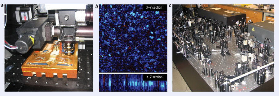
After obtaining contrast in art-pigment samples and demonstrating non-invasive 3D cross-sectioning capabilities in lab-made test samples, the time was ripe for an actual demonstration with real artwork. But where does one get access to relevant historic works of art? Fortunately for our group at Duke, we are only a short drive away from the North Carolina Museum of Art (NCMA), which not only houses a world-class art collection but also has enthusiastic and exceptionally skilled conservators and curators. In what proved to be a truly interdisciplinary collaboration – and with a certain leap of faith! – the conservators agreed to bring Puccio Capanna’s 1330 painting The Crucifixion to our lab and let us test it with our pump–probe microscope. Figure 4 shows this Renaissance master painting undergoing the procedure, from which the painting emerged visibly unscathed.
4 Under the microscope (a) Puccio Capanna’s historic 1330 painting The Crucifixion under Duke University’s pump–probe microscope, with acquisition parameters tuned for lapis lazuli. (b) The distribution of this precious pigment shown (top) as a ‘virtual slice’ – acquired parallel to the surface but at a depth of about 30 µm within the paint layer on the Virgin Mary’s robe – and (bottom) as a ‘virtual cross-section’ in the plane perpendicular to the surface. Image dimensions are 365 × 365 μm and 365 × 60 μm, respectively. (c) The lasers and associated optics of this research-grade microscope are bulky, but much more compact set-ups are under development. (Courtesy: Duke University)
In this demonstration, the lasers were tuned to map lapis lazuli, a precious blue mineral pigment. In the Renaissance age, lapis lazuli – being so rare – was actually more expensive than gold and was therefore used sparingly, primarily for iconic religious figures. Pump–probe microscopy was able to map the distribution of the pigment through the entire paint layer, which in this case was a surprising 60 μm thick, with micron-scale 3D resolution.
Being able to show that our microscope can non-invasively image a painting was naturally exciting. However, the current microscope system is bulky – requiring optics benches and other laser-lab paraphernalia – and was optimized for tissue imaging, rather than for works of art. Much more work remains to be done before pump–probe microscopy can be more widely used in the art world. In particular, the equipment would need to be portable so that it can be taken to conservation labs and easily adjusted so that it can be used to investigate many different parts of an artwork.
Fortunately, the Duke and NCMA collaboration has recently been able to secure funding from the National Science Foundation to bridge the gap between imaging skin and art. Among the aims of our research grant is to develop a much more compact microscopy system, which could provide conservation scientists and conservators with the opportunity to evaluate artworks, including not just painted or coloured works but also pottery and statues. One interesting possibility is to study iron oxide, the pump–probe signature of which seems to change permanently when heated – suggesting that firing history in terracotta could be measured. Nonlinear microscopy could also be used to detect paint fragments buried under the surface (for example, on Greek statues), or even read a delicate scroll without unrolling it.
Much work remains to be done, of course, yet the promise of pump–probe microscopy is genuine. What’s more, it reveals an unusual but fabulous spin-off from society’s investment in basic biomedical research.
It was fairly straightforward. I did maths, physics and chemistry at A-level partly because they were my strongest subjects, and partly thanks to the influence of my father, who said that science would always get you a good job. Then, when it came time to move on to university, I was just better at physics – I can’t say I had a huge desire to study the subject! But there were parts of physics that I really enjoyed, and I think I’d enjoy studying it much more now than I did when I was very young and it was all about passing exams.
How did you get into the energy industry?
I had done a “science and society” project on future energy issues as an undergraduate at the University of Bristol, so when I saw that Cambridge’s Cavendish Laboratory had a research group devoted to energy studies, it really attracted me. As part of my PhD in the group, I studied the residential energy market, so it seemed logical for me to move into the industry after I finished. Initially, I didn’t get the right job – I misread a job advert and thought it was more senior than it was – but I tried some different ones and eventually I moved into commercial negotiation for British Gas. My career really took off from there.
You became the first managing director at Amerada Hess Gas. What was that like?
I’d been doing commercial negotiation around gas purchasing and transportation, so when I was headhunted to start Amerada’s retail gas business from scratch, it was a big jump. Initially I was going to turn it down; I spoke to my father about it and he said he wasn’t sure I could really do it. But then, just as I was about to say “no”, my father died, and it was one of those moments where you think, “What am I afraid of, here?” If it’s not a great success, I’ll learn from that, but if I say no I’ll never know what I would have been capable of. So, oddly enough, his death sparked a flame of ambition in me, and I said yes. I guess it taught me to go out and pursue opportunities as they arise, and I’ve done that ever since.
How did you get to be chief executive at Sightsavers?
After we sold Amerada Hess Gas, I did some travelling and I decided that I would like to get into the not-for-profit sector. I thought it would be difficult to do that with my private-sector CV, so I applied to be a non-executive director of my local housing association in Notting Hill, London, because I thought it would give me useful experience (which it did). Then a few years later, Sightsavers advertised for a chief executive role and it was just one of those things where, very occasionally, you see something and you think, “That’s what I want to do.” I had blindness in my family (my father and uncle were both blind before they died), so it had some personal resonance for me. I didn’t actually think I’d get the job because I had so little experience either in international development or in the charity world, so I was hugely pleased when I got it.
That’s the second time you’ve mentioned feeling unqualified to do something. Are you prone to impostor syndrome?
I think so, occasionally. You sort of think, “My goodness, am I really doing this?” But since my father died, one of the things I have consciously done is that even if I think, “Oh, they won’t want me, I couldn’t possibly,” I make myself do it and that’s stood me in good stead. And I think women are particularly prone to it – there’s that classic story of how women will look at a job advert where there are six requirements and think, “Oh, I can’t do that because I only meet five of them”, whereas a bloke will say, “I meet three out of six, I’ll give that a go”. I’ve seen that in some of the women I’ve worked with and I’ve encouraged them not to be held back by it.
What’s been the biggest challenge for you in changing sectors?
In my first year, there was some resistance from some members of staff along the lines of “What could a woman coming from the oil and gas industry possibly know about development work?” I had to prove myself, and it took a while to do that, since you can’t do it like they tell you to do it in the private sector. There, the assumption is that as a chief executive, you have 90 days to get in there and stamp your authority on the organization. In this sector, unless you’re dealing with a financial crisis, that kind of behaviour would be regarded as much too corporate. So in the beginning, you have to make changes incrementally. Then as you establish yourself, listen to people and experience things, you can make more and more change, such that now I’m eight years in I’m making more radical change in our organization than ever. I’ve tried to speed up decision-making, for example, and to bring in new staff so that we’ve got a balance between people who are experts in the development field and people who bring more corporate skills such as project management. It’s important to maintain the balance between passion and professionalism in an organization like this; people primarily come to work for you because they want to make change in the world, but it’s also important to recognize that you need discipline to make sure that when you’ve committed to a donor to deliver a project, you deliver it when you said you would.
What are you working on at the moment?
We’ve got a very big push on tropical diseases, particularly trachoma, which is actually chlamydia in the eye. What happens is that people get infected as young children, over and over again, and their eyelids start turning inside out. Then the eyelashes scrape the cornea, which is absolutely agonizing, and they go blind. We are aiming to eliminate blinding trachoma from all the countries where we work by 2020. We’re in discussions with the Queen’s Diamond Jubilee Trust and also the UK Department for International Development (DFID) about a major grant that would be a huge scale-up for us, enabling us to work in collaboration with a range of other agencies across the world as well as the health ministries of various countries. That’s probably our most exciting project. Certainly for me, personally, the idea of helping to eliminate a disease from the world – and it’s not just trachoma, there’s several that we work on, including one called river blindness – I can’t really think of anything more exciting in terms of a legacy.
There’s been some controversy about the amount the UK spends on foreign aid recently. What’s your view on that?
There’s a classic saying that “charity begins at home”, but the fact is that 99.3% of the money the UK government spends is not related to foreign aid or development. Also, the cost-effectiveness of some of the things we do – whether it’s distributing drugs for trachoma and river blindness, or doing cataract operations, which cost something like £25 or £30 depending on what country you’re in – is incredible. I’ve been to hospitals in places like Mozambique and seen the impact on an old person who can see their kids again, and perhaps the girl who was forced to stay home from school can now get back to school because granny can see. You look at that and you think, actually, that’s not a lot to spend. And although you see a lot of negative coverage in the papers about foreign aid, if you look at something like Comic Relief, you’ll see that it produces an outpouring of money, mainly for Africa, in record amounts every year. That, and the fact that our own donations are increasing, suggests to me that this rejection of supporting people in poorer countries is not as widespread as the media says.
Any advice for today’s students?
Keep your options open and don’t assume that you have to follow an obvious treadmill, or that the first job you get is the one you have to do for life. And there’s one other thing, particularly for women. When I was studying physics, we were very much the minority, and I remember being really quite intimidated when some of my male classmates would go around saying, “Oh, these exams, they’re so easy it’s an insult to my intelligence.” I found them quite difficult, and I remember being really worried – only to discover that a lot of the guys who had been going around boasting actually didn’t do very well at all, whereas I did fine. So don’t be intimidated if you’re in the minority. You’re just as good as they are.