
Nature supports science bloggers

By Hamish Johnston

More embargo-related cheek from a prestigious journal…

There is an editorial in today’s Nature about the effect that science bloggers and digital camera owners are having on the discussion of preliminary results at conferences.

You may recall the physics paparazzi incident in which conference attendees photographed slides of preliminary data from an experiment and then posted their analyses on the arXiv.

The editorial asks if it is desirable — or even practical — to stop conference goers from publicizing someone else’s preliminary results?

Nature has made the case that blogging by researchers is good. Critical discussion of worthy results should not in principle be restricted to walls of a conference hall or even the pages of a journal”, says the editor.

But wait a minute, isn’t this the same journal that places embargoes on its papers, forbidding scientists from speaking to the public — and presumably blogging — about their work until the paper is published?

Indeed it is, and the editorial concludes that in some situations it is appropriate to ban blogging from conferences. The argument being that such closed shops would encourage the free exchange of controversial scientific ideas.

The same can’t be said about the journal’s embargo policy.

DNA sorts carbon nanotubes

Carbon nanotubes are relatively easy to grow, but sorting these tiny tubes according to their electronic properties is a time consuming and costly task. Now, however, researchers in the US have invented a way of isolating different types of nanotube by mixing them with DNA. Although the technique is currently too expensive to be commercially viable, the scientists believe that it could someday be used to create high-quality carbon nanotubes for electronics and other applications.

A single-walled carbon nanotube (SWNT) can be thought of as a sheet of carbon just one atom thick that has been rolled up into a tube with a diameter of about 1 nm. The atoms in the sheet are arranged in a hexagonal lattice and the relative orientation of the lattice to the axis of the tube — what is known as its “chirality” — dictates whether the tube is a metal or a semiconductor. Tubes that are about 1 nm in diameter have about 25 different possible chiralities.

The diverse properties of SWNTs have allowed researchers to build simple electronic devices such as transistors from these tiny tubes. Indeed, some scientists even believe that SWNTs could replace silicon in future electronic devices because they are tiny, but can still carry large amounts of current. An important challenge however, is that it has not yet proved possible to make just one type of SWNT at a time. Instead, mixtures of tubes with different chiralities have to be painstakingly sorted using expensive processes that are not commercially viable.

DNA to the rescue

Now, though, Ming Zheng and colleagues at the chemical firm Dupont have separated SWNTs by chirality by mixing them with a solution of DNA and then using a standard chemical separation process known as liquid chromatography. This technique involves sending molecules through a tube that is packed with beads. As every type of molecule reacts differently with the surface of the beads, the system can be adjusted so that one particular type of molecule travels rapidly through the tube, while the others lag behind.

The new technique involves mixing the SWNTs with a particular DNA sequence, which coats the surface of the nanotubes to form large, hybrid molecules. Zheng and colleagues found that the chromatography system could be adjusted so that hybrids containing SWNTs of a specific chirality are first through the tube — and could therefore be isolated. The DNA is then removed from the SWNTs, leaving a relatively pure sample.

SWNTs of a different chirality can be selected by using a different DNA sequence — with the team identifying a total of 12 DNA–SWNT combinations. This was not an easy task because they began with 1060 possible DNA sequences, which were winnowed down to 350 candidates using a series of chemical and structural criteria.

Chiral purity

Zheng’s team found, using an optical-absorption technique, that the chiral purity of the sorted nanotubes was between 70–90% (with an uncertainty of ±10%) depending on which chirality was targeted. When the electronic properties of one group of tubes with 90% ±10% purity were tested by using them to make field-effect transistors, 99% of them were semiconductors.

While this success rate suggests that the technique could be used commercially, Zheng told physicsworld.com that the high cost of making the DNA sequences means that it is not commercially viable. However, he believes that low-cost DNA sequences could become commercially available in the future if there is a demand from the nanoelectronics industry.

Another shortcoming of Zheng’s technique, according to Mark Hersham at Northwestern University, is that it is better at isolating semiconductor SWNTs than it is at finding metal nanotubes — something that the Dupont team says it doesn’t fully understand. “Further work is required to isolate both types equally using DNA”, says Hersham.

The research is published in the journal Nature .

Blacklisted?

vixra.jpg
The new arXiv?

By Hamish Johnston

Is the love affair with the arXiv preprint server on the wane?

In case you have never heard of it, the arXiv is a vast online repository of physics papers, most of which are uploaded before they have passed muster by peer review. It is seen by many as a shining example of how the Internet can be used to accelerate the dissemination of knowlege — and some see it as a key step in the evolution away from the traditional journal publishing model.

But not everyone is a fan of the Cornell University-based service.

Maverick blogger Tommaso Dorigo is leading a campaign to expose the alleged “blacklisting” of certain researchers, who claim that their papers are not being uploaded to the server — or being relegated to “graveyard” categories.

Oxford University’s Marni Sheppeard — who believes she may be on such a blacklist — has posted a link to an alternative to arXiv called viXra.org.

viXra has just been launched by Phil Gibbs (who I think is an independent physicist) and contains one paper — an essay entitled This Time – What a Strange Turn of Events! by Phil Gibbs.

Elsewhere in the blogosphere, David Bacon calls for more transparency in the arXiv’s editorial policies.

So, are Tommaso, Marni, Phil and David just a few dissenting voices in an otherwise happy physics community, or is the love affair with arXiv waning?

I’m sure this is a completely unrelated incident — but Bacon also asks has the arXiv been hacked?.

Thunderclouds accelerate cosmic electrons

As well as producing spectacular lightning shows, thunderclouds can also sometimes emit strange long-lasting bursts of gamma rays. Now, a group of researchers in Japan claims to have the best evidence yet that these gamma rays are produced by the clouds acting as enormous particle accelerators. The scientists reached this conclusion in the spirit of Benjamin Franklin by studying an electric storm from a mountaintop in Japan.

It has long been known that lightning strikes produce short bursts of gamma rays, but in the past few years scientists have also observed longer gamma-ray events, of up to 90 seconds, that cannot be explained by the same mechanism. Several research groups have linked these longer events with high energy electrons accelerating in the electric field of a thundercloud but there is little physical data to corroborate.

In 2007 Harafumi Tsuchiya of the Cosmic Ray Laboratory of Japan’s RIKEN research institute and colleagues set out to investigate the longer bursts experimentally. The scientists travelled to a nuclear power plant on the Japanese coast to take measurements of electromagnetic activity during a winter thunderstorm. They found — by analysing the energy distribution of the pulse — that the gamma rays do indeed originate from just a kilometre or so above the Earth’s surface.

Lightning strikes twice

With this latest research, Tsuchiya and his colleagues relocated to a Japanese mountaintop 2770 m above sea level in order to study this phenomenon in more detail. At this altitude the scientists were able to capture recordings of both photons and electrons and they say they have now confirmed the accepted theory of how gamma rays are generated.

The researchers observed cosmic electrons, which — after arriving at the Earth’s atmosphere — were accelerated to relativistic speeds in a thundercloud’s electric field. Occasionally these electrons collide with air molecules knocking off electrons which are also accelerated in the field. This can lead to a runaway or “avalanche” of electrons that can emit bremsstrahlung gamma rays if they reach relativistic speeds. “We made a simultaneous detection of gamma rays and energetic electrons arriving from thunderclouds” — the first such observation, Tsuchiya told physicsworld.com.

Researchers in the field are already excited by these new results. “This type of observation is very rare, thus any new observations further our understanding of the phenomena significantly,” said Jeremy Thomas a geophysical field researcher of the Bard High School Early College II, New York and the University of Washington.

Predicting the storms

David Smith at the University of California recognizes the historical significance of this study. “This work is extremely important to the field in that they have demonstrated that these “surges” of gamma rays have an energy spectrum that indicates they are caused by the acceleration of very energetic electrons — a phenomenon first predicted by [Nobel laureate] CTR Wilson in 1925”.

The relatively low intensity and scarcity of these prolonged gamma ray bursts means that they pose no real threat to aviation and communications. What’s more, weather forecasters could benefit from a more thorough understanding of this phenomenon. “These gamma rays may give us a tool for understanding how thunderstorms and lightning work, neither of which we understand very well,” said Joseph Dwyer a lightning researcher at the Florida Institute of Technology.

“This work is interesting because it distinguishes gamma ray bursts produced by specific lightning events from protracted electron and gamma ray production generated by thunderstorms between lightning flashes,” said Giles Harrison, an atmospheric scientist at the University of Reading, UK.

Tsuchiya and his colleagues now plan to construct a series of new detectors that measure the arrival direction of photons associated with lightning as well as thunderclouds. They will install these instruments over a wider area in order to increase the number of recorded events. “These plans would give better understanding of particle acceleration in thunderclouds and lightning,” said Tsuchiya.

This study was published in Physical Review Letters.

T-shaped probe exposes protein elasticity

Researchers in the US have used a modified atomic force microscope to find the flexible regions of a protein. The motions of proteins can play important roles in their biological function and therefore understanding which parts of the molecule can easily bend — and how this suppleness is affected by the presence of other molecules — could help in the development of new drugs.

Proteins are chains of amino acids that are involved in just about every biological process. They are essentially nanoscale mechanical devices with flexible moving parts, as well as more rigid components. According to Ozgur Sahin, who led the study at Harvard University’s Rowland Institute: “If one can identify flexible parts of proteins, one can design small molecules (drugs) to bind and alter their flexibility.”

Sahin and team have used their modified AFM to study the flexibility of bacteriorhodopsin — a protein used by some micro-organisms to convert light into electricity. The molecule is a distant relative of rhodopsin, which performs the role of detecting light in human eyes.

Tiny forces

The AFM itself works by measuring how the force between the sample and a tiny “tip” on a cantilever changes as the tip is moved over the surface of the sample. This allows the AFM to record images with extremely high spatial resolution on the nanometre scale.

The flexibility of a material can be measured by pushing the AFM cantilever against a sample — and measuring the deformation of the sample as a function of the applied force (the elastic modulus). However, this method has its drawbacks when used on tiny and fragile proteins. “If the cantilever is soft, the forces can be determined accurately, but the sample will not be deformed — rather, the cantilever will bend ” explains Sahin. Conversely, if the cantilever is stiff, the sample will be deformed, but it will be difficult to measure the forces.

The team addressed the challenge by designing a T-shaped cantilever that can both twist and bend. The tip is located at one end of the crossbeam and sensitive measurements of the forces are made from the twisting motion of the cantilever and deformation is determined from its bending.

The AFM was operating in vibrating mode, which means that the tip was oscillating rather than stationary. Successful measurements result from careful adjustments to resonant frequencies and spring constants of the bending and torsional modes of oscillation.

Flexible “switches”

Before being placed in the AFM, bacteriorhodopsin molecules were embedded in micrometer-sized cell membranes. The membranes were placed on a flat surface and the AFM was used to measure their elastic moduli. The measurements revealed that the region that contains the protein’s “electrical switch is about three times more flexible than the the rest of the structure.

Armed with the T-shaped cantilever, the team could perform a single force measurement in just 120 μs. By comparison, a standard AFM would take 1 s to perform the same measurement, and consequently several hours to generate an image.

The T-shaped probe also leads to superior spatial resolution. The best images from a conventional tip have tens of nanometres of spatial resolution in liquid, and cannot visualizing single molecules — whereas Sahin’s tool has a typical resolution of one nanometre.

Easy to manufacture

Sahin also says that the cantilevers are “relatively easy to manufacture,” — the team’s probes are made by an AFM cantilever manufacturer that carries out bespoke batch production. The experiments were done on a commercial AFM.

A protein’s mechanical properties can also be determined by inelastic neutron scattering (INS). However, neutron sources are only available in national facilities, and protein replicas with certain hydrogen atoms replaced with deuterium are needed.

Despite these drawbacks, INS has been used to study bacteriorhodopsin. “There is an agreement in the numbers, but that does not necessarily build additional confidence into our method,” says Sahin. INS quantifies thermally actuated atomic fluctuations, while Sahin’s team has determined the collective response of atoms to external forces, and he says that the correlation between these two techniques is unknown.

Future targets

The next goal for Sahin’s team is to measure protein dynamics. “High-speed measurements can, in principle, show changes in protein that occur at the microsecond to millisecond timetable.” Tracking of single molecule dynamics might even be possible, because enzymes form and break chemical bonds on this timescale.

The research is published in the journal Nature Nanotechnology.

Dodging wet paint at J-PARC

DSCN1570.jpg
The target station at the Material Life-Science Facility

By Michael Banks

The paint has yet to dry here at the J-PARC experimental complex in Tokai, Japan.

Today I visited the Material Life-Science Facility (MLF), which produces neutrons and muons that are used in a variety of experiments in materials science to biology.

DSCN1602.jpg
under wraps…for now

At J-PARC, a 3 GeV synchrotron feeds the MLF with a intense beam of protons that are then smashed into a 20 tonne mercury target to produce neutrons.

Theses neutrons are then cooled using a water moderator before being sent to a maximum of 23 experimental stations.

If you like your research equipment brand new, then you have come to the right place. I could not find a single piece of kit that looked used. Even the box containing spanners looked like it had just been opened that morning.

So far 15 instruments at the MLF have been funded by the Japanese government and eight have already been built at a total cost of around $70m.

DSCN1577.jpg
A crystal diffrotometer – the ‘cage’ – for biological samples

The MLF has been operational since May 2008 when it produced its first neutron beam. At the moment, however, the beam has a power of 18 kW, which when fully operational is expected to reach 1 MW (the ISIS neutron source in Oxfordshire, for example, operates at 160 kW).

According to Masatoshi Arai, deputy director of the MLF, training the beam to full power will take around five years to complete.

The MLF has already had its first users. From the 100 proposals submitted last September for beamtime, 10% were from scientists based abroad, 10% from industry and the rest from scientists in Japan.

DSCN1623.jpg
Inside the target chamber

Interestingly, Aria told me that the Japanese government would only fund the MLF if it gave 25% of its beamtime to industry users – a decision that was reached at a meeting in 2000 that lasted until 5am in the morning after a full night of negotiations.

It was not only a great opportunity to walk around the facility and squeeze into all the sample rooms, but I also had the chance to see inside the mercury target chamber.

Once a high-powered beam has been used at the facility, access to the target chamber is strictly off limits due to the increase in radiation levels. Indeed, 1m deep perspex ‘windows’ are used to look into the target chamber.

Aria has spent the last nine years fully focused on getting the facility finished and is now looking forward to returning to his research into superconductivity.

Yet he isn’t quite finished thinking about the next steps for the MLF. “It is early days, but we already have plans for a second target station and then even more experiments,” says Aria.

Blogs, big physics and breaking news

wcsjlogo.jpg
Last week science journalists flocked to London from all corners of the globe

By Matin Durrani

My colleague James Dacey has already blogged twice (here and here about last week’s World
Conference of Science Journalists in London, but I thought I’d give my take on the meeting.

I chaired a session entitled “Blogs, big physics and breaking news”, which examined the challenges that physics bloggers pose to journalists and looked at the merits and downsides of such blogs.

The session was inspired in part by the incident a couple of years ago, when Tommaso Dorigo — a member of the 600-strong CDF collaboration at Fermilab — discussed on his blog A Quantum Diaries Survivor possible sightings of the Higgs boson in the decay of a Z-boson to a pair of tau leptons.

Although Dorigo — and other bloggers who discussed the data at the time — emphasized the uncertainty inherent in their results, his blog entry was picked up by journalists, who reported the story around the world. (Physics World gave a full account shortly afterwards of what happenned, which you can read here ).

That all seems fine on the surface — journalists dug out a story on particle physics that might otherwise have not seen the light of day.

Even better for journalists was the fact that Fermilab was not exactly chuffed that the discussion was out in the open — new results in particle physics are usually only made public after being “blessed” by the collaboration and published in a scientific paper.

But Dorigo was unhappy at the way his analysis was reported, which he claimed did not underline the uncertainty in the data. What’s more, it raises the question of the whole point of science journalism: if someone is really interested in Dorigo’s analysis, why bother with possibly inaccurate science stories in the media? Why not go straight to the blogosphere instead for the “real” story?

Admirably, Dorigo agreed to speak in London, braving an audience of about 100 of the world’s science journalists in the Edwardian-style Methodist Central Hall. On the stage alongside him were former Physics World features editor (and particle physicist) Matthew Chalmers, and CERN communication chief (and particle physicist) James Gillies

What followed was an entertaining debate, which saw this issue — and others — aired in a friendly and open manner.

(more…)

Accelerator complex officially opens

DSCN1553.jpg
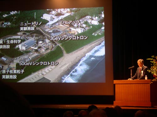
Shoji Nagamiya, director of J-PARC, outlines the accelerator complex

By Michael Banks

Imagine having a world-leading neutron and muon source, a particle accelerator capable of boosting protons to 50 GeV, and a neutrino facility all on one site.

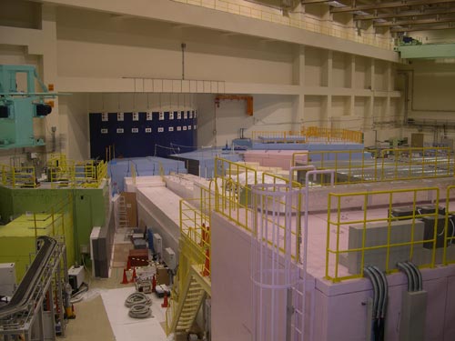
Well, you don’t have to any longer, as this is exactly what the $1.5bn J-PARC accelerator complex in Tokai, 100 km north of Tokyo, has to offer.

Today, physicists from around the world, myself included, met in Tokyo to celebrate the opening of the J-PARC accelerator complex after 12 years of construction.

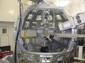
The main aspects of J-PARC revolve around its 3 and 50 GeV synchrotrons. The 3 GeV synchrotron ramps up a beam of protons to smash them into a mercury target producing copious amounts of neutrons and muons that are then used in a range of experiments in biology to condensed-matter physics.

Meanwhile, the 50 GeV synchrotron, which is currently only operating at 30 GeV, accelerates protons before smashing them into a graphite target to produce kaons and neutrinos.

The inauguration held at the Kudan Kaikan centre in central Tokyo was attended by around 1000 scientists.

First to speak was Shoji Nagamiya, director of J-PARC, who has been with the project since its inception in 1999.

Unfortunately, during my few days in Tokyo I haven’t yet picked up the language, so I didn’t understand most of his or the other talks as they were given in Japanese.

After Nagamiya came a roster of dignitaries to the stage to offer their congratulations for the completion of J-PARC. These included the Japanese science minister, Ryu Shionoya, as well as Akito Arima, the former Japanese education minister and Masaru Hashimoto, governor of the Ibaraki prefecture, where J-PARC is based.

Next up was Makoto Kobayashi from the KEK lab, who shared the 2008 Nobel Prize with Yoichiro Nambu from the University of Chicago and Toshihide Maskawa from Kyoto University. Kobayashi gave a brief lecture about the new science that J-PARC hopes to unveil.

DSCN1564.jpg
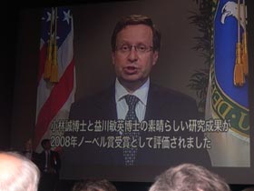
Steve Koonin gives a recorded message of congratulations

Steve Koonin, under secretary for science at the US Department of Energy also give a brief recorded message (in English) of congratulations saying that J-PARC represented “another great venture in Japanese science”.

After the talks had finished, scientists from other countries who helped to build J-PARC were named and invited to the stage.

Then Nagamiya flashed a slide up saying that any foreigner in attendance should come to the stage.

I couldn’t really hide away and was duly encouraged to go on the stage along with some other members from the audience.

I felt somewhat embarrassed to be applauded by over 500 Japanese scientists, but it was a nice touch to the event.

However, it was not all about talks and being red-faced on stage and after the talks a lavish banquet was put on with sushi and sashimi.

After the inauguration I caught up with Nagamiya, who said it was not always easy to build such a big lab that was a partnership between the KEK lab and the Japan Atomic Energy Agency. “Now I feel confident about the project,” says Nagamiya, “but when I started I was less confident. What is especially pleasing is that the world is leaning towards new neutron and neutrino facilities, so we are setting the trends.”

Lasers look inside human bones

Scientists in the UK are set to carry out hospital tests on a new laser technique that could ultimately lead to rapid and reliable detection of bone disorders such as brittle bone disease. The method includes a novel version of Raman spectroscopy — routinely used by physicists and chemists — and could give medics the most detailed characterization yet of bones inside the human body. That is according to the researchers based at the Central Laser Facility in the UK.

The strength of bones comes from two main components: the mineral content, such as calcium and crystalline mineral salts; and a protein-based substance known as collagen. There are various medical conditions linked with these substances including “brittle bone disease” where defective bones result from a deficiency of “type I” collagen.

To detect these diseases and to monitor a patient’s response to treatment, doctors require non-invasive ways of seeing inside bones in the body. At present the two favoured approaches are X-ray spectroscopy and ultrasound but both of these techniques struggle to see the collagen. As a result many cases of bone disease can go undetected.

Elusive proteins

In recent years, a different approach has been suggested that could detect both mineral content and collagen. Raman spectroscopy relies on the fact that light is scattered inelastically as it interacts with matter. In the classic experimental set-up, a laser is shone on a sample, which then scatters the photons at a slightly lower frequency. Measuring this change in colour of photons can determine the identity of the chemical under study.

Already, medical physicists have successfully applied Raman spectroscopy to tissues extracted from the body, but a problem arises when you attempt to observe bones that are still in vivo. Raman signals from surface layers tend to dominate those signals from under the skin of a patient, which are weakened significantly as they scatter sideways. These bone signals are then drowned out even further by the strong fluorescence in the melanin content of skin.

To overcome this problem, Pavel Matousek at the Central Laser Facility and his colleagues have bedecked Raman spectroscopy with an innovative new geometry. These scientists have reduced the dominance of the skins’ signal by creating a distance between the illumination and collection points of the Raman process. They also employ a ringed detector to ensure that all signals are captured effectively.

Souped-up spectroscopy

This technique, known as Spatially Offset Raman Spectroscopy (SORS), has already been used by an independent group in the US to determine the ratio of phosphate to carbonate — a potential indicator of osteoporosis — in a chicken’s shin bone. Matousek and his team now plan to take the next step by testing out SORS on human subjects at the Royal National Orthopaedic Hospital (RNOH) in Middlesex, UK.

Michael Morris, a spectroscopy researcher at the University of Michigan is optimistic about the new development. “Matousek is well-known for his excellent pioneering work in developing spatially offset Raman spectroscopy.” However, Morris also anticipates one of the main challenges ahead. “Confirmation by independent investigators is needed before any new finding can be accepted by clinicians.”

Matousek and his team will initially use their technique to test for brittle bone disease but they hope this will lead on to the investigation of other bone conditions such as osteoporosis, which presently goes undetected in three to four out of ten cases. “This project has the potential to provide a means of detection and confirmation of some of the rare bone disorders that are still difficult to diagnose,” said Richard Keen, a consultant rheumatologist at RNOH.

This work was published in Analyst.

Visiting Asia's oldest observatory

DSCN1476.jpg
The Cheomseongdae observatory

By Michael Banks

Being the International Year of Astronomy, what better time than to go and see Asia’s oldest surviving observatory.

The Cheomseongdae (star-gazing tower) observatory in Gyeongyu, South Korea, dates back to the seventh century and was built during the reign of Queen Seondoek of Silla.

The Silla dynasty began in 57 BC and reigned for almost 1000 years. It was one of the three kingdoms that ruled in Korea, but by 660 AD it had occupied the other two kingdoms – Baekje and Goguryeo — to rule most of the Korean peninsula.

The observatory is around 9m high and is built by 356 stones representing each day in the year. Seemingly everything about the construction of the observatory has some meaning. The observatory has 27 layers of stones as it is thought that Queen Seondoek was Silla’s 27th ruler. Then, above and below the opening is 12 layers of stone for every month in the year.

The Queen’s astronomer would climb to the top of the observatory every day to take a view the sky. Using a ladder, he would clamber through the opening in the observatory and then climb to the top. There he would give information to the Queen about weather patterns and the timings of any solar eclipse.

Copyright © 2026 by IOP Publishing Ltd and individual contributors