Plug me in: the physics of brain–computer interfaces

The human brain is an astounding and complex piece of machinery. With more than 80 billion neurons in the human cerebral cortex, each with a thousand synapses, our brains process some 100 megabits of information per second. Imagine then, attempting to measure, extract and interpret all the signals in our brain in real time, at the speed of thought. Tapping into the brain might once have been solely in the realms of science fiction – from X-Men to The Matrix – but today, it’s actually possible to link your brain to a computer and control a robotic arm, say, or translate your thoughts into text.

A brain–computer interface (BCI) functions as a bridge between your brain and an external device, typically a computer. BCIs collect, analyse and translate electrical signals from your brain into commands that can be understood and executed by a computer. They can also apply external signals to modulate the brain. Thanks to a combination of neuroscience, biomedicine, physics and technology, BCIs can change the lives of people with serious medical conditions. They also have applications across robotics, neuroscience, technology, gaming and computing.

Over the last 25 years, BCIs have allowed paralysed people to operate computers by thought alone. They have restored speech after it has been lost due to a stroke; have allowed those with missing or paralysed limbs to function again or helped them to operate robotic arms and hands. BCIs have diagnosed epilepsy and other neurological conditions, and mitigated them for tens of thousands of people. They’ve even shown promise for restoring sight to the blind.

But most of these examples require brain surgery, in which electrodes are placed on or in the surface layer of the brain (the cortex) and potentially even more deeply, which is risky as it could induce haemorrhages or infections. Another problem is that researchers currently don’t have a clear idea of the impact and potential damage the implanted electrodes could inflict on brain tissue, while also not knowing how long they may last. All this means that electrical implants in their current state cannot safely and reliably help the millions who would benefit from them. In fact, human implantations are carried out only when all other treatment fails, or on an experimental basis – for some 50 individuals worldwide with severe limitations such as paralysis – where the chance to improve a poor quality of life outweighs the dangers.

Fortunately, the solutions to some of these issues may lie in physics principles and methods, which could make these devices safer, more durable and more widely available. Physics could also be used to improve BCI implantation methods and materials. More crucial though, is the need to eliminate or minimize brain surgery by providing ways to interact with the brain via light, magnetic fields or ultrasound. Non-invasive, wireless and portable or wearable BCIs could enhance brain research and medical treatment, and be used in daily life as well.

Tap in with a thought

From antiquity through to the 19th century, physicians and experimentalists, often unwittingly, carried out various rudimentary experiments in which they tried to modify the brain’s electrical activity for medical treatment. In 1924 these efforts became rigorous when German psychiatrist Hans Berger recorded electrical brain activity by using electrodes placed on a patient’s skull, thereby inventing the technique of electroencephalography (EEG). In the 1970s physicist and computer scientist Jacques Vidal demonstrated thought control of an external device, as human subjects fitted with EEG contacts mentally moved a cursor displayed on a computer screen.

EEG remains a valuable non-invasive tool to diagnose conditions such as epilepsy, allowing us to determine the cause and type of seizures a patient might be suffering from, as well as to investigate other conditions such as dementia, brain tumours and concussions. But an EEG samples large groups of neurons, and the signal-to-noise ratio is poor, making it difficult to correlate the signals with specific brain activities.

Implanted electrodes, on the other hand, directly sample selected neurons. This was experimentally demonstrated in 1998, when Atlanta-based neurologist Philip Kennedy placed custom-designed electrodes into the brain of a patient dubbed “JR”, who had been left “locked-in” by a stroke (IEEE Trans. Rehabil. Eng. 8 198). The unlucky patient was in possession of his full cognitive abilities, but unable to move or speak. Eventually, JR learned to communicate by mentally controlling a computer cursor to spell out words.
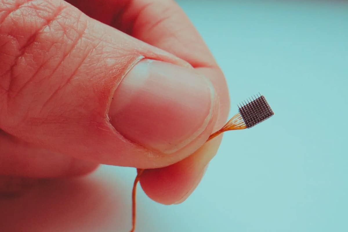
Array of electrodes held in a human hand

Now many researchers and clinicians use an implanted electrode array, known as the “Utah Array” from Blackrock Neurotech. This tailored silicon product is an array of 100 p-type silicon electrodes (in a 10 × 10 configuration), spaced 400 µm apart on a 4 × 4 mm insulating substrate – roughly the size of a peppercorn. The electrodes, 0.5 to 1.5 mm long, are tipped with platinum or iridium oxide. Some 30 people across the world, suffering with different symptoms of paralysis, have been fitted with these devices. For example, in 2015 four arrays were implanted in Nathan Copeland, who was paralysed from the chest down after a car accident in 2004. The implants allow him to control a computer, play video games and control a robotic arm, with his thoughts. At the time of writing, Copeland is the longest-running patient with such an implant, but the truly long-term implications of this invasive technology are not fully understood.

Reducing invasiveness

The trouble with an electrode or any other artificial implant in the brain is that it can trigger an immune response, which inflames and scars nearby tissue. This is aggravated by the mechanical mismatch between a rigid electrode and the brain’s soft tissue, which in turn can also degrade the electrode’s performance.

Finding durable, biocompatible materials with suitable electrical properties for electrodes and substrates is a challenge for physics and materials science

But finding durable, biocompatible materials with suitable electrical properties for electrodes and substrates is a challenge for physics and materials science. Promising candidates include soft and flexible conducting polymers, as well as extremely thin electrical conductors such as carbon nanotubes and silicon nanowires (for another approach, see box below).

Researchers are also working to reduce surgical risks, by adapting existing medical technologies. Stents – tiny hollow cylinders – are commonly used to hold open various types of vessels in the body. In one common use, they keep coronary arteries open and are considered minimally invasive. Neurotech company Synchron has developed “stentrodes” (stent-electrode recording array). They are electrodes mounted on a stent that is permanently implanted into a blood vessel in the brain. They can detect brain signals and wirelessly send them to a computer. In human trials, stentrodes have allowed paralysed subjects to operate computers (J. NeuroIntervent. Surg. 13 102).

Using a different approach, the US firm Neuralink announced in 2019 that it had developed a BCI that would be implanted flush with the skull by a surgical robot, which would also place 1024 or more flexible electrodes into the brain (J. Med. Internet Res. 21 e16194). Neuralink, which was co-founded by Elon Musk, has not published more details since, but after its recent approval from the US Food and Drug Administration (FDA) for human trials, more information may be forthcoming. In whatever forms, electrode-based BCIs will continue to be important because of their high spatial resolution and fast response, but non-invasive approaches are also rapidly developing.

Fibres, nanoparticles and the brain

Polina Anikeeva

Polina Anikeeva is an interdisciplinary scientist at the Massachusetts Institute of Technology (MIT), who works across materials science, brain research, and electronics. Following a BSc in biophysics in Russia, her PhD at MIT focused on organic LEDs and nanoparticles. As a physicist trained in fundamental concepts, Anikeeva realized that she could choose to work on “whatever interested” her that also allowed her to make a difference.

That turned out to be neuroscience working at Stanford University with Karl Deisseroth, who developed optogenetics, a breakthrough technique to control the activity of neurons with light. There for the first time she held a brain in her hands. It wasn’t firm, but soft “like pudding”. This generated an “aha” insight as she realized that physical probes of the brain should match its material properties for minimal invasiveness and maximum stability. Her answer was to fabricate flexible multifunction fibres, tens of microns in diameter, that could stimulate neurons either by using light or by delivering drugs to the patient and electrically recording the responses. After wide use to examine brain function in rodents, Anikeeva and co-workers now report (bioRxiv:2022.10.09.511302) the first use of her fibres to study brain function in non-human primates. This is an initial step toward human trials.

Anikeeva also draws on her materials background to study nanoparticles as sources of brain stimulation. Her results show that magnetic nanoparticles activated by an external field can affect deeper parts of the brain than is possible with “transcranial magnetic stimulation”, where a varying magnetic field applied to the skull induces currents in the neurons below.

Along with her research, Anikeeva and several colleagues have called for developing attitudes toward responsible neurotech within the science and engineering community. Combined with appropriate regulation, she believes this would help individuals and society navigate the ethical questions raised by neurotech and its medical uses such as BCIs.

Photons probe the brain

In the electromagnetic spectrum, near-infrared (NIR) light, which runs from 700 to 1400 nm, can traverse the skull and penetrate the brain centimetres deep, without doing harm, so long as the power density is held to milliwatts per square centimetre. A non-invasive NIR method dubbed “photobiomodulation” has shown that it can stimulate the brain. For instance, in a clinical trial in 2021, patients with dementia were repeatedly exposed to LEDs emitting light at 1060–1080 nm. This group showed notable improvements in cognitive function and subjective mood compared to a control group (Aging Dis. 12 954). It is thought that the light enhances cellular function or reduces inflammation, but more research is needed to establish the exact mechanism.

A second non-invasive method, known as “functional near-infrared spectroscopy” (fNIRS), uses NIR light to measure variations in the light absorbed by haemoglobin in the blood circulating in the brain. The technique can map brain activity because deoxygenated haemoglobin absorbs NIR light differently from the oxygenated form, HbO2. Active neurons need an increased flow of HbO2-enriched blood, making it possible to detect brain function. Two wavelengths are applied to the skull, and a measurement of their different attenuations at specific sites can show which areas are active. fNIRS has been used in the clinic, with US neurotech company Kernel developing a wearable headset version. It covers the skull with 52 modules, each with laser sources emitting at 690 nm and 850 nm and a detector (J. Biomed. Opt. 27 074710). In 2021 the FDA approved the device to test the brain’s response to a psychedelic drug.
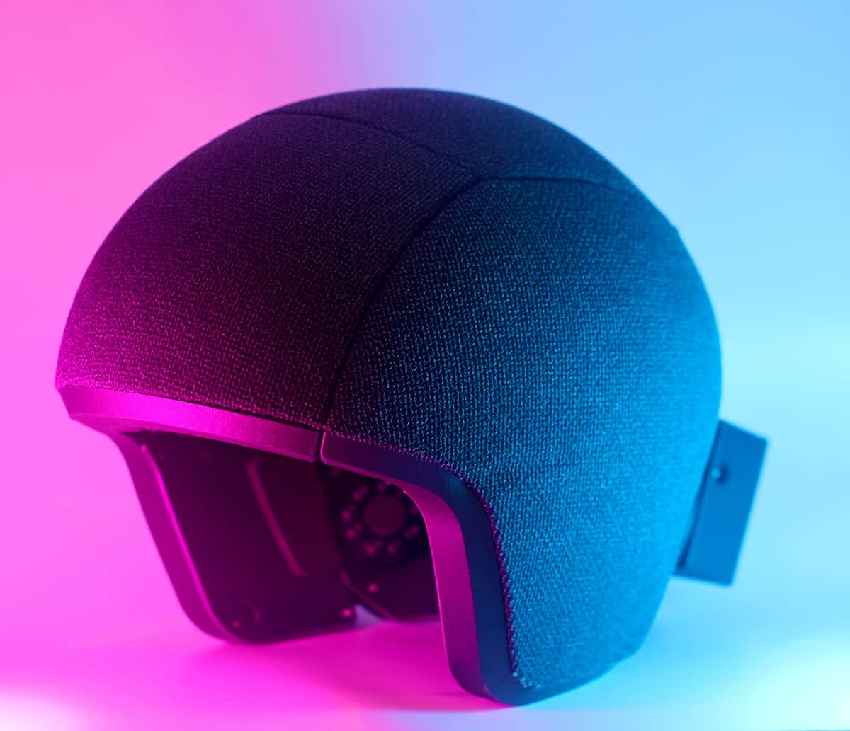
A headset on a pink and blue lit background

Although it takes seconds for the oxygenated blood flow to develop – making fNIRS too slow to control an external device – it does deliver a higher spatial resolution and better signal-to-noise than EEG, meaning it can pinpoint brain activity more accurately. An fNIRS headset could measure brain activity even in a freely moving subject, making it possible to map the brain and diagnose neural conditions under varying conditions.

Faster responses can be obtained with another method – known as “event-related optical signal” (EROS) – that uses infrared light to measure changes in the optical properties of cortical brain tissue. The interaction of light with neural tissue changes when neurons are active because that increases the optical scattering, lengthening the paths of photons traversing the brain and delaying their arrival at a detector.

In early experiments on human subjects, NIR light applied through optical fibres penetrated the skull and was detected a short distance away, delayed by 0.1 s or less after neurons had been excited. Further work has been limited because these measurements are technically demanding, but recent results suggest that EROS combined with fNIRS could form the basis for non-invasive BCIs with good spatial and temporal resolution.

The magnetic brain

Yet another established non-invasive method to trace the brain’s neural activity is “functional magnetic resonance imaging” (fMRI). Standard MRI detects the behaviour of protons in water and fat in the body, within a strong magnetic field, to image bodily structures. fMRI instead detects signals from blood flow in the brain that, as mentioned, depend on the oxygenation level of haemoglobin. Like fNIRS, this allows fMRI to label regions of neural activity but at a spatial resolution of 1 mm rather than 1 cm. The time lag of seconds allows mapping nearly in real time, but it is still too slow for brain control of external devices. fMRI also requires a large, expensive installation with a superconducting magnet.

Faster response times come with non-invasive “magnetoencephalography” (MEG), which tracks neural activity by detecting the femtotesla (10–15 tesla) magnetic fields that are generated as ionic currents flow among active neurons. These fields are measured by sensitive superconducting quantum interference (SQUID) devices placed near the scalp, within a shielded room to prevent magnetic interference. MEG provides a spatial resolution of 1–2 mm and a response time of milliseconds, but requires a bulky device with high operating costs.

Young child wearing a blue helmet

A new type of detector, the “optically pumped magnetometer” (OPM), improves MEG by measuring the brain’s magnetic field at room temperature. OPM uses a small cell filled with an alkali atom vapour. A laser diode tuned to a specific quantum transition optically pumps the vapour, which aligns the atomic magnetic moments. This magnetization interacts with the brain’s magnetic field to change the opacity of the vapour as determined by a detector, which makes it possible to measure the magnetic field.

Earlier this year, UK-based firm Cerca Magnetics won an award in quantum innovation for the development of its OPM-MEG wearable brain scanner. This comprises 50 LEGO block-sized units mounted on a whole-head helmet to cover the brain. The prototype wearable OPM-MEG BCI allows neural diagnosis as a subject moves. With its high spatial and temporal resolutions, it could possibly control external devices.

Listening to the brain

Ultrasound technology is widely used as a portable non-invasive method to image bodily structures, including red blood cells, as they reflect high-frequency sound waves. In the last decade, the technology has developed to the point that “fast functional ultrasound” (fUS) can use Doppler measurements of the brain’s blood flow to identify active neurons. In fUS, probes generate ultrasonic plane waves and gather data over hundreds of channels. A computer then synthetically focuses the waves and analyses the data to rapidly produce high-resolution images of brain function. Studies in non-human primates show that fUS operating through a minimally invasive port in the cranium could support a BCI that tracks the neural impulses representing bodily motion (Neuroscience 474 110).

Ultrasound also serves in transcranial ultrasound stimulation (TUS), a method to modulate neural behaviour that can be targeted to within a few cubic millimetres within the brain. After extensive animal studies, some human trials suggest that TUS can treat neurological or psychiatric problems such as pain and depression.

The future of non-invasive BCIs

Complementing and perhaps someday replacing implants, other physical methods can access the brain with minimal invasiveness, enabling safer, cheaper and wider medical use of BCIs. Andrew Jackson, a physicist-turned-neuroscientist at Newcastle University, UK, says that, when it comes to recording the brain, the most exciting technology at the moment is wearable OPM-MEG. “It’s interesting physics too!” he adds, noting the value of ultrasound for brain stimulation. Jackson warns, though, that none of these non-invasive technologies yet has the spatial resolution that you can get with implants. Much remains to be done for clinical use, and perhaps beyond it.
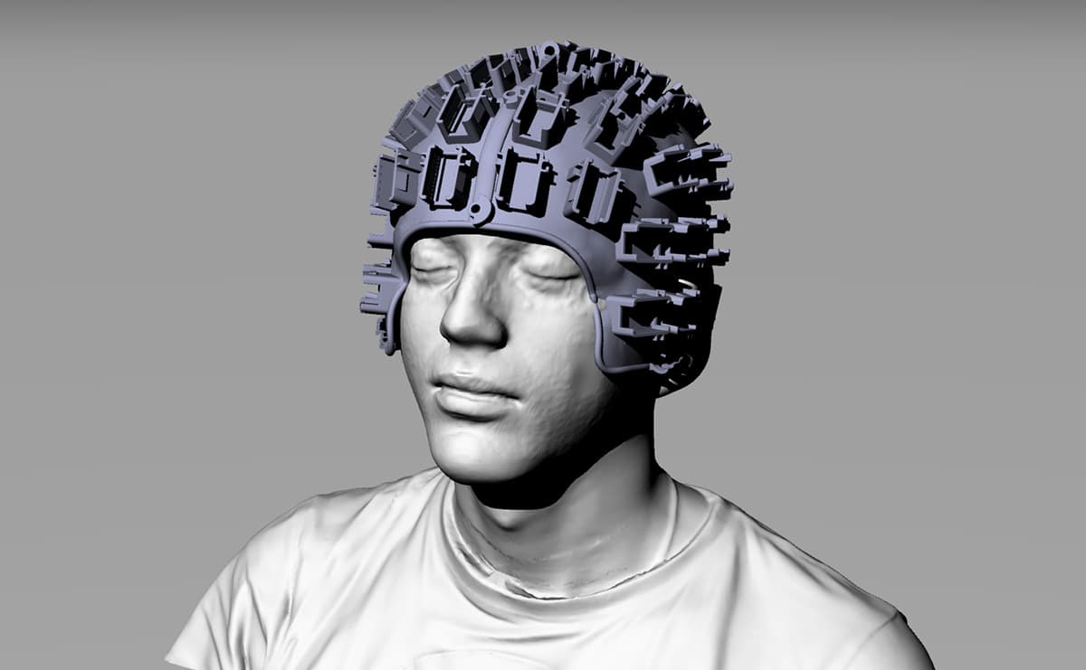
Computer-generated model of a person wearing a helmet

If non-invasive BCIs eliminate surgical risk, healthy individuals could be motivated to use them for real or perceived mental augmentation. Noted neuroscientist Kristof Koch has related how “awesome” it would be to have a safe BCI that links brains to computers so people could download information directly into their brains.

In 2021 San Francisco start-up MindPortal raised $5m to develop a headband for mental control of a virtual-reality game. It uses proprietary technology, perhaps a fast-NIR method. In another application, transcranial direct current stimulation (tDCS) devices are readily available at modest prices. These apply milliamp electric currents to the skull that supposedly improve cognition.

Seeing the rise of consumer neurotech, neuroethicists point to the harm that could come without effective oversight and regulation – which would also need to consider issues such as privacy and mind control. In developing non-invasive BCIs, researchers are hugely advancing brain research and treatment, helping to restore the independence of severely disabled individuals. At the same time, researchers should be aware of the many ethical quandaries that these devices raise, beyond the lab and clinic.

How sound is the model used to establish safe radiation levels?

Ionizing radiation can damage living organisms, that’s clear. But there are big questions over the validity of the linear no-threshold model (LNT), which essentially states that the risk of cancer from radiation and carcinogens always increases linearly with dose. The LNT model implies, in other words, that any amount of radiation is always dangerous and that zero risk is present only at zero dose.

But it is not the only dose-response model that exists. The “threshold model”, in contrast, says there is a dose below which there is no perceptible risk, implying that small exposures to radiation are harmless. Then there is the “hormesis model”, which says a small dose at or slightly above natural background levels can, in fact, trigger beneficial repair mechanisms.

Developed almost 80 years ago, LNT has been integral to the US government policy on radiation protection over the last half century, and many other countries have followed suit. Even if LNT is incorrect in detail, it may appear sensible to err on the side of caution concerning matters of human health. But adopting it in this way, as a kind of “precautionary principle”, can have unforeseen and undesirable consequences.

The LNT can also promote a false sense of security by suggesting that the removal of minute doses guarantees safety

Incorporated into policy the LNT model can require companies and government agencies to clean up tiny and perhaps non-hazardous amounts of radiological material, consuming funds that might be better spent tackling more dangerous sites. The LNT can also promote a false sense of security by suggesting that the removal of minute doses guarantees safety despite the presence of other toxins already in the environment.

The model may even discourage use of radioactive material for beneficial uses, such as X-ray imaging, stress tests, and medical diagnosis and treatment. As a result, the LNT has become a hotly debated topic among historians, public policy makers, toxicologists and medical physicists. One paper in the Journal of Nuclear Medicine (58 1) has even accused the LNT model of leading to “needless public and professional radiophobia”.

Complex beginnings

That radiation can cause genetic mutation in living organisms was discovered by the US geneticist Hermann Muller (1890–1967). In 1927 he published a paper in Science (66 84) entitled “Artificial Transmutation of the Gene”, his title ambitiously and audaciously invoking the artificial transmutation of elements, which had been discovered a few years earlier.

Muller wrote that his discovery in fruit flies that “relatively heavy doses of X-rays induced the occurrence of true ‘gene mutations’” might explain the mechanism for evolution and the ability of X-rays to cause cancer. Over the next few years, he cited his own studies to argue for the proportionality of radiation dose and damage, which soon became known as LNT.

In 1946, Muller was awarded the Nobel Prize in Physiology or Medicine “for the discovery of the production of mutations by means of X-ray irradiation”. Based at the time at Indiana University in Bloomington, Muller proclaimed in his Nobel lecture that there is “no escape from the conclusion that there is no threshold dose”. Individual mutations, he explained, result from individual “hits”, producing genetic effects in their immediate neighbourhood.

Muller’s work made him a celebrity, and opened up the field of “X-ray genetics”. Scientists now began to look in more detail at Muller’s methods and assumptions and to conduct further experiments on fruit flies and also mice. Some researchers questioned whether his X-rays were really inducing mutations or just knocking out pieces of chromosome. Others questioned his key assumption that the genetic damage does not depend on dose rate delivery.

What’s more, after the 1953 discovery by Francis Crick and James Watson of the structure of DNA – which carries genetic information – scientists found that the molecule often repairs itself after being buffeted by chemicals and radiation already present in the environment. Still other scientists pointed out that LNT is not an empirical fact but a hypothesis generated by extrapolation from massive to minute doses.

For a while, US policy for setting levels of radiation protection in the workplace was based on a threshold model. Then, in 1955, the US National Academy of Sciences established the Committees on the Biological Effects of Atomic Radiation (BEAR). Its genetics panel, whose 17 members included Muller, recommended switching from a threshold to a linear dose response model when estimating risk assessment.

Its report, published in 1956, stated that radiation from any source, including natural environmental background radiation and X-rays, is “harmful to life”. The report was highly influential and made the front page of the 13 June 1956 edition of the New York Times, among other newspapers. The report led to changes in public perception and to acceptable radiation-protection levels.

Continued questions about the validity of the use of LNT in radiation protection have led to the Million Person Study

In the 1960s, rising fears of low levels of radiation led the US Congress to set up the Biologic Effects of Ionizing Radiation (BEIR) committee, which in 1972 published a report that essentially endorsed LNT.  In 1975 the US Environmental Protection Agency (EPA) began using the LNT model to fix clean-up levels for radiologically contaminated environments.

In the past decade, however, continued questions about the validity of the use of LNT in radiation protection have led to the Million Person Study (MPS) of US radiation workers and veterans. A joint effort by several US universities and national labs to study the health impacts of exposure to low doses of radiation, the MPS has enrolled a million people to evaluate effects involving different cancers, types of radioactivity and differential impacts on men and women.

The MPS faces huge challenges. Dose response at minute levels can be masked by noise and drowned out by the many other sources of damage such as those posed by genetics, diet, lifestyle, oxyradicals, carcinogens and background sources of radiation from earth and sky. But success would help frame more responsible policies and better protect workers and the public.

The critical point

One of the many reasons for the need to study the validity of LNT is that convictions of its accuracy continue to be used as an argument against nuclear power plants, in connection with their operation as well as their spent fuel rods. Nuclear power may be undesirable for reasons other than this. But the critical need to find a workable alternative to fossil fuels for energy production requires an honest ability to assess the validity of this model.

High-precision measurement of the strong force is made at CERN

In a thorough review of proton collision data from CERN’s Large Hadron Collider (LHC), physicists working on the ATLAS experiment have determined the strength of the strong force to the highest level of precision to date. While the result is in agreement with the Standard Model of particle physics, the approach could be used to search for new physics in future experiments.

As defined by quantum chromodynamics (QCD), the strong force is responsible for the binding of quarks together via exchanges of massless gluons. The strong force also holds protons and neutrons together in nuclei.

The strong force differs from gravity, the electromagnetic interaction and the weak force because it does not diminish as particles move apart. This imbues a complexity to QCD that makes it very difficult to determine the strong coupling constant. As a result, the strong force is the least precisely defined out of the four fundamental forces in the Standard Model.

Through a combination of refined QCD theories and improved experimental techniques, our understanding of the strength of this interaction – defined by its coupling constant – has steadily improved over the past few decades.

Key parameter

“The strength of the strong nuclear force is a key parameter of the Standard Model, yet it is only known with percent-level precision,” explains Stefano Camarda, a CERN-based member of the ATLAS Collaboration. “For comparison, the electromagnetic force, which is 15 times weaker than the strong force at the energy probed by the LHC, is known with a precision better than one part in a billion.”

To boost our understanding of the strong force, the ATLAS team has now revisited data collected by the LHC in 2012, when beams of protons collided head-on inside the ATLAS detector at energies of 8 TeV.

During these collisions, quarks in protons were occasionally annihilated by their antiquark counterparts in the opposing beam. This process is mediated by the weak interaction, leading to the creation of massive, neutral Z-bosons.

During the collisions, gluons radiated off the annihilating quarks boost the momentum of the resulting Z-boson along the collision axis. Crucially, QCD predicts that the magnitude of this momentum kick is tied to the value of the strong coupling constant.

More stable pairs

Shortly after their appearance, the Z-bosons decayed further into more stable pairs of either electrons and positrons, or muons and antimuons – which were subsequently picked up by ATLAS’ detectors. In their latest analysis, the ATLAS collaboration identified more than 15 million of these pairs. These observations allowed them to calculate the momenta of the Z-bosons that created them.

By comparing these values with theoretical predictions calculated by only considering the weak interaction, the researchers could determine the extent of the strong interaction’s influence on the process.

This means that the ATLAS team has measured the strong coupling constant to the highest level of precision achieved in any experiment to date. “That we have now measured the strong force coupling strength at the 0.8% precision level is a spectacular achievement,” Camarda says. “It showcases the power of the LHC and the ATLAS experiment to push the precision frontier and enhance our understanding of nature.”

While this is a great experimental achievement, it does not appear to put us any closer to physics beyond the Standard Model. The team’s value for the strong coupling constant agrees closely both with previous, less precise measurements, and with the latest QCD predictions. So this is yet another case of the Standard Model standing firm against close scrutiny.

However, if the same approach is used to analyse even higher-energy collisions in the future, researchers could be better placed to pick out discrepancies in the Standard Model’s predictions. Perhaps this could get us a step closer to answering some of the most long-standing questions surrounding the fundamental nature of matter.

The research is described in a preprint on arXiv.

NASA launches $1.2bn Psyche asteroid mission

NASA has launched a mission to study an unusually metal-rich asteroid dubbed 16 Psyche. Launched from the Kennedy Space Center in Florida at 10:20 local time today via a SpaceX Falcon Heavy rocket, the $1.2bn mission is expected to reach the asteroid in 2029.

16 Psyche is around 220 km in diameter and consists of almost pure nickel–iron metal. It was discovered in 1852 by Italian astronomer Annibale de Gasparis, who named it after the goddess of the soul in ancient Greek mythology. Located in the asteroid belt between Mars and Jupiter, astronomers speculate that the asteroid is the exposed core of an early planet that lost its rocky outer layers due to a number of violent collisions billions of years ago.

In 2021 astronomers used the Atacama Large Millimeter/Submillimeter Array (ALMA) to gain a more detailed picture of the composition of the asteroid finding that Psyche’s composition is not uniform and its surface is at least 30% metal.

The Psyche mission will help scientists understand how planets and other bodies separated into layers – including cores, mantles and crusts. On it way to the asteroid – a total journey of 3.6 billion kilometres – the craft will conduct a Mars fly-by in 2026. Once Psyche arrives at the asteroid in 2029 it will spend over two years analysing the body from four different orbit that get successively closer.

It will use a gamma-ray and neutron spectrometer to determine the chemical elements that make up the body as well as a multispectral imager to provide information about the mineral composition of Psyche as well as its topography. It will use a magnetometer to look for evidence of an ancient magnetic field. The craft will also test a new laser communication technology, called Deep Space Optical Communication.

Launch delays

The mission was originally expected to launch in 2022 but was delayed due to issues with its navigational software. An independent review board found later that year that the delay was caused by staff shortages at Caltech’s Jet Propulsion Laboratory, which manages the mission.

The craft was then scheduled to launch during a launch window that opened on 5 October, but shortly before then officials discovered that a subcontractor had provided incorrect data concerning the spacecraft’s thrusters. The problem was soon fixed, with a new window opening from 12 October.

Psyche was selected in 2017 from five proposals as part of the space agency’s Discovery Program, which was created in 1992 and launches smaller missions under shorter development times.  Yet while the initial estimated cost of the craft was $450m, at $1.2bn Psyche is one of the most expensive missions to be launched as part of the Discovery Program.

Psyche was chosen in 2017 alongside the Lucy mission, which launched in 2021. It will visit Jupiter’s Trojan asteroids and study the origins of giant planets by looking at the fragments left over from their formation.

Meet the artist behind the Nobel portraits, how to avoid ‘nobelitus’

Since 2012 the first pictures we see of Nobel prize winners are often portraits produced by the Swedish artist Niklas Elmehed. I find the simple drawings iconic and I always enjoy comparing them with the photographs of laureates that emerge later. For me, Elmehed’s portraits often capture aspects of a subjects’ personality that photographs cannot.

Just about everything to do with the process of choosing Nobel winners is shrouded in secrecy, so I had assumed that little information would be available about who did the portraits. After all, the artist would have to know about the winners well in advance, and could be targeted by those trying to discover the names of winners before they are announced.

Undaunted, Elmehed has written about the portraits on his website.

“The graphical concept behind the portraits is to give the portraits the expression of breaking news − a strong and unique visual impression,” he says.

Golden makeover

In 2017 the Nobel prize got a graphical makeover – with gold replacing blue and yellow as the main colours. Elmehed responded in his 2018 portraits by starting to use real gold (see figure).

“I experimented a lot with different gold paints and fell for the gold foil, a super thin metal foil that you can put on the painting with a special glue,” he explains.

In addition to being the subject of a golden portrait, life can get pretty weird for Nobel laureates thanks to all the publicity. In particular, winners are often portrayed in the media as polymaths whose opinions on wide-ranging topics must be listened to – regardless of whether a laureate has any expertise in that field.

This can lead to a condition called “nobelitus”, where this deference gives a laureate a platform for crackpot ideas. Perhaps the most infamous case of nobelitus in physics is William Shockley, who after winning a prize in 1956 became an outspoken proponent of “scientific racism” and eugenics.

Avoiding nobelitus

Writing in the Guardian, the British geneticist Paul Nurse – who shared the 2001 Nobel Prize for Physiology or Medicine – explains how he avoids nobelitus with help from friends, family and colleagues.

Nurse also talks about the doors that his Nobel has opened to him and his projects – most notably the founding of the prestigious Francis Crick Institute in London in 2010. Nurse was a key proponent for the medical research facility and is its first (and current) director and CEO. In 2010 he also began a five-year term as president of the Royal Society, which is the UK’s premier scientific organization.

One thing that a Nobel couldn’t get Nurse automatically was a green card to reside in the US. His initial application was rejected because he did not submit a long-form birth certificate, which includes the names of his parents. When he procured the document, he was shocked to discover that the person he knew as his sister was in fact his mother, that his father was unknown, and that his parents were actually his grandparents. Sadly, such arrangements did occur in an age when births outside of marriage were stigmatized.

Nurse made his application after becoming president of Rockefeller University in 2003. If he had not won a Nobel, perhaps he would not have gone to the US and he may have never discovered the truth about his family. So winning the prize can indeed be life-changing.

Physics for fairness: tackling global sustainability challenges through science

This year, 2023, marks the half-way point to the 2030 deadline for achieving the 17 Sustainable Development Goals (SDGs). Recent global developments, including conflicts and the COVID-19 pandemic, make SDGs such as “Zero Hunger” and “Reduced Inequalities” seem more daunting than ever. The scale of the challenge is clear; but professionals working on the frontline of sustainable development are as committed as ever to enabling positive change.

That includes physicists and engineers, and July 2022 – July 2023 was the International Year of Basic Sciences for Sustainable Development. In this episode of the Physics World Stories podcast you will hear from with two physical scientists whose careers enable them to apply their scientific knowledge to tackling inequities. As always, the episode is hosted by Andrew Glester.

Ruhi Chitre is an intern at UNESCO, Paris, who was previously president of the International Association of Physics Students. Chitre believes that international policymaking can be strengthened by the contribution of more people with backgrounds in fundamental science, not least because they have a nuanced understanding of the concept of risk.

Later in the episode, you will hear from Destenie Nock, a civil & environmental engineer at Carnegie Mellon University in the US. Nock’s research includes looking for patterns in energy usage that can reveal local inequalities, such as correlations between household incomes and use of heating in winter. In her teaching, Nock encourages her students to take engineering back to its fundamental purpose – to think about how innovation can improve livelihoods.

Nock is among the invited speakers at Environmental Research 2023, a series of free-to-attend virtual events on 16 October – 23 November hosted by IOP Publishing (which publishes Physics World).

Ask me anything: Kim Nygård – ‘The better I am at building relationships with these colleagues, the smoother the project progresses’

What skills do you use every day in your job?

For the past six years, I’ve been responsible for the construction and operation of the ForMAX beamline at the MAX IV Laboratory in Lund, Sweden. We provide a state-of-the-art instrument for the structural characterization of materials using X-ray scattering and imaging, with a special focus on the development of advanced materials from renewable forest resources.

My tasks are diverse. I lead a small team that maintains the instrument, further develops it, and supports external users from academia and industry during their experiments. Some of these tasks are technical, such as working together with our automation and motion-control engineers to implement new hardware updates for example, or with our software engineers to enhance our control and data acquisition systems.

Other tasks are scientific in nature – such as developing new X-ray methodologies and carrying out experiments with external scientists or the in-house team. And these are all alongside budgetary responsibilities and outreach with our user communities. During the ForMAX construction phase, my remit also included conceptual design of the instrument, procurement and project management.

If I were to identify one specific skill that I use daily, I guess it would be the ability to learn “on the fly”. I appreciate my physics education – it gives me the technical basis for my current role – but the diversity of tasks at MAX IV is such that I am always encountering new problems and learning new skills.

What do you like best and least about your job?

There are lots of things I really enjoy about my job. For starters, I get to work with, and learn from, colleagues with a wide range of expertise on a daily basis – whether that’s vacuum technicians and mechanical engineers at the laboratory, or external researchers studying, for example, novel cellulose-based materials or degradable bone implants.

On a personal level, I’ve always enjoyed developing new experiments, something I can continue to some extent within my current role. At the same time, I am now involved in R&D that matters at a wider societal and economic level. Earlier in my career, I studied fluids under spatial confinement. This is an interesting soft-matter problem and I think we contributed to significant advances in the field – although the impact of this small-scale basic research was rather limited. If I can now support, say, applied X-ray studies to develop sustainable food packaging, the impact of my contribution is amplified greatly.

Less good, and probably common to a lot of jobs, is the fact that I can rarely set aside a full day to focus exclusively on the challenging task at hand.

What do you know today that you wish you knew when you were starting out in your career?

I’d like to highlight two things. First, the importance of soft skills. For example, more than a hundred colleagues were directly involved in the construction of the ForMAX instrument. Communication is key. The better I am at building relationships with these colleagues, the smoother the project progresses and the more fun it is.

Second, I’ve always been open to new challenges, having worked in physics, chemistry and nanotechnology labs as well as large-scale synchrotron facilities like MAX IV. I’ve been happy with this path, but often wondered if I should have “settled down” earlier. With hindsight, though, I should have worried less and enjoyed the ride more, trusting that things will turn out fine as long as I continue to develop my skills and am ready when an opportunity appears.

Sonobiopsy provides a non-invasive route to brain tumour diagnosis

Diagnosing a brain tumour usually entails neuroimaging with CT and MRI, followed by surgical resection or tissue biopsy. A non-invasive and inexpensive alternative is blood-based liquid biopsy, which analyses circulating biomarkers in the blood to obtain molecular and genetic information about the tumour and guide treatment decisions. Unfortunately, brain tumour-derived biomarkers are detected only in scarce amounts, as the blood–brain barrier (BBB) prevents the transfer of such biomarkers into the peripheral circulation.

To address this problem, researchers at Washington University in St. Louis are using focused ultrasound (FUS) and microbubbles to temporarily disrupt the BBB and release large amounts of biomarkers into the bloodstream for analysis. In a first-in-human prospective trial, they found that FUS-induced release of biomarkers into the bloodstream – a method they call sonobiopsy – is feasible and safe for use.

“With this technique, we can obtain a blood sample that reflects the gene expression and the molecular features at the site of a lesion in the brain. It’s like doing a brain biopsy without the dangers of brain surgery,” explains co-senior author Eric Leuthardt in a press statement.

Transcranial low-intensity FUS, used in combination with intravenously injected microbubbles, provides temporal and reversible opening of the BBB and can target lesions in the brain with millimetre accuracy. The microbubbles, which are traditionally used as ultrasound contrast agents, undergo cavitation upon exposure to FUS and amplify its mechanical effects.

To perform sonobiopsy, a technique pioneered by Leuthardt and co-senior author Hong Chen, the team developed a compact FUS device that can be directly attached to a clinical neuronavigation probe, enabling precise positioning of the FUS transducer. This design enables easy integration of sonobiopsy into existing clinical workflows without requiring neurosurgeons to undertake additional training.

To assess the feasibility and safety of sonobiopsy with the neuronavigation-guided FUS transducer, Leuthardt, Chen and colleagues carried out a pilot single-arm trial of five patients with high-grade glioma (four had glioblastoma, one had a diffuse high-grade glioma).

The researchers performed sonobiopsy on anaesthetized patients prior to planned surgical brain tumour removal. Using MRI and CT images acquired beforehand to register the patient’s head position, they positioned the FUS transducer to align its focus at the tumour location. After intravenous injection of microbubbles, they applied FUS sonication for 3 min.

Analysis of blood samples collected before and 5, 10 and 30 min after sonication revealed that sonobiopsy increased the concentration of circulating tumour DNA (ctDNA). This included maximum increases of 1.6-fold for mononucleosome cell-free DNA (cfDNA) fragments, 1.9-fold for patient-specific tumour variant ctDNA, and 5.6-fold for ctDNA with TERT mutations (which are present in more than half of glioblastoma patients and associated with poor treatment outcomes).

The study also verified that the procedure was safe and did not damage brain tissue. During FUS sonication, patients did not exhibit any significant fluctuations in vital signs and there were no adverse events. Tumour samples collected during surgery showed no microhaemorrhage or structural changes between sonicated and non-sonicated regions.

The researchers conclude that their work “marks a crucial initial milestone in demonstrating the feasibility and safety of sonobiopsy in patients with high-grade glioma”. They point out that while this study was performed in an operating room prior to surgery, operative environments and anaesthesia are not essential, and sonobiopsy could be used in a clinic or at a patient’s hospital bedside.

“With this capability to non-invasively, non-destructively access every part of the brain, we can now obtain genetic information from tumours at every stage of patient care, ranging from tumour diagnosis to treatment monitoring and detection of recurrence,” says Chen. “We can now start to interrogate diseases that traditionally don’t undergo surgical biopsies, such as neurodevelopmental, neurodegenerative and psychiatric disorders.”

The study is described in npj Precision Oncology.

Fusion industry has ambitious plans for 2035, rounding out this year’s Nobel prizes

Nuclear fusion is what powers the Sun, and if we could harness it here on Earth it would be a significant source of clean, carbon free energy. Fusion power plants were first proposed in the 1940s and since then physicists and engineers have struggled to overcome a range of technological challenges that have gotten in the way of practical fusion energy.

In the past, most fusion R&D was done in universities and government labs and involved large-scale facilities. Today, there is also a growing number of companies that are developing alternative routes to practical fusion technologies – and some of these companies say that fusion could be delivering electricity to the grid by 2035.

The US-based Fusion Industry Association represents this private sector and it has released a report called The Global Fusion Industry in 2023. In this episode of the Physics World Weekly podcast, the association’s CEO Andrew Holland talks about what the report reveals about the global fusion industry and what companies are doing to try to meet the ambitious goal of fusion power in just 12 years.

Five physicists win Nobel prizes this year

Last week, six Nobel prizes were awarded to 11 people. Five of the new laureates have backgrounds in physics – including chemistry and peace laureates.  Four out the 11 laureates this year are women – which is certainly progress over previous years. However, Anne L’Huillier is just the fifth female physics laureate since 1901, so much more work must be done on diversity and inclusion in physics.

To chat about this year’s Nobel prizes, I am joined by the physics and philosophy student Hannah Schmalstich, who has written a series of blogs for Physics World about historical and societal aspects of previous prizes. One article explored why the physicist Lise Meitner did not win a Nobel prize for her important work on nuclear fission. Our discussion explores connections between the shunning of Meitner and some of the prizes awarded in 2023.

Pairs of rogue planets found wandering in the Orion Nebula

More than 500 free-floating planetary-mass objects have been discovered wandering through the Orion Nebula thanks to new observations by the James Webb Space Telescope (JWST). Most bizarrely, about 40 of these newfound objects in the nebula’s Trapezium Cluster exist in wide binary pairs, confounding expectations about how these so-called “rogue planets” form.

Free-floating planetary-mass objects that do not orbit a star have been discovered in star-forming regions before, but never in the numbers seen by the JWST’s Near-Infrared Camera (NIRCam). These objects are visible because they are still young and glowing with the heat of their formation. Over time, they will cool and fade from view.

There are two hypotheses for how such free-floating planets form. One is that they originate like stars, collapsing and condensing directly out of a star-forming molecular cloud. The other is that they form through core accretion in orbit around young stars, only to be expelled later by gravitational tidal forces and resonances.

A JuMBO mystery

For most of the 540 planetary-mass objects found in the Orion Nebula, “it’s almost certainly going to be a combination of both [processes],” says Samuel Pearson, a research fellow at the European Space Agency (ESA) and lead author of a not-yet-peer-reviewed pre-print on the new observations.

However, the 40 or so wide binaries – 9% of the total – do not fit this picture. Pearson calls them JuMBOs, for Jupiter Mass Binary Objects, and they are a mystery.

The main problem, Pearson explains, is the JuMBOs’ mass. Models predict that the minimum mass that can form directly from a molecular cloud is three times that of Jupiter. This minimum limit arises because smaller objects are better at retaining heat, which prevents them from contracting further. One might therefore expect smaller planetary-mass bodies (including the smallest object in the JWST observations, which is about 0.6 Jupiter masses) to instead have formed through core accretion – that is, from the bottom up, like Earth – and to have been ejected from their system of origin.

“But then the problem now is that we are finding them in pairs,” Pearson tells Physics World. “How on Earth does that happen?”

Planet-formation scenarios

So far, there are two options on the table. One is that the initial mass function, which describes the distribution of masses that stars are born with, extends to much smaller masses than anyone realized. The other is that these JuMBOs are somehow being ejected from planetary systems.

Though there are no firm models of how a binary pair could be ejected, one possible explanation comes from Rosalba Perna of Stony Brook University, together with Yihan Wang and Zhaohuan Zhu of the University of Nevada in Las Vegas, both in the US. In a separate pre-print, they propose a model in which two giant planets orbiting at great distance from their star can both be ejected if they happen to move into alignment at the same time as the system experiences a close encounter with a nearby star.

Perna and Wang point out that in models of planet formation, giant planets can coalesce directly out of the planet-forming disc at distances of over 50 astronomical units (that is, 50 times the Earth-Sun distance) from their star.

“In our scenario, JuMBOs prefer to form from planetary systems with two giant planets with semi-major axes close to each other,” Wang and Perna tell Physics World. “For example, if the planetary system is wide, the most likely configuration is a giant planet at ~100AU and another giant planet at 70AU.”

Pushing into a new domain

In a paper published in 2022, Aleks Scholz of the University of St Andrews, UK and colleagues predicted that the JWST would be able to detect significant numbers of low-mass rogue planets in young star clusters. Scholz says the new findings in the Orion Nebula justify this conclusion.

“These newly discovered planetary-mass objects are really exciting – they are surprisingly plentiful, and the large number of binaries is completely unexpected,” he tells Physics World. “This is what happens when you push into a new domain: you find unexpected stuff.”

For now, the onus is on getting more data. Pearson and his co-author Mark McCaughrean, who is ESA’s senior advisor for science and exploration, want to take a second look at the JuMBOs in a future study, this time using the JWST’s Near-Infrared Spectrometer (NIRSpec). These observations should confirm that they are indeed of planetary mass by providing details of their temperature, surface gravity and atmospheric composition. Beyond that, Pearson and McCaughrean plan to look for planetary-mass objects in a young star cluster, NGC 2244, which is born from the Rosette Nebula and located 5 000 light years away in the constellation Monoceros. The stars in NGC 2244 are packed less densely than those in Trapezium, Pearson says, meaning the opportunities for stellar encounters plucking planets from their orbits are slimmer.

“It’s way less likely to happen in really sparse regions, so if we suddenly stop seeing JuMBOs, that would be a smoking gun for how these are forming,” he explains.

Though little is known of these rogue planetary-mass objects, Pearson says there could be more of them in our own Milky Way galaxy than there are stars. This raises questions about how to define them: are they planets, sub-brown dwarfs or something else? Given the already blurred lines between these categories, it might not matter, and Pearson is steering clear of such discussions.

“How we try and define them is a great way to cause an argument,” he says, laughing.

Copyright © 2026 by IOP Publishing Ltd and individual contributors