Wouldn’t it be amazing to have a tool like Google Earth but for the human body, where you could zoom in from a full organ down to its cellular structures? That’s now becoming a reality thanks to the Human Organ Atlas project, and physics is key to this innovation. This short videos explains how the Human Organ Atlas project emerged from the COVID-19 pandemic and how it could help medical scientists in a range of clinical contexts.
Discover more about the Human Organ Atlas in this feature, originally published in the March issue of Physics World.
In this age of information, we expect to have knowledge at our fingertips. If we’re looking to obtain a first impression of someone, many of us head straight to their social-media pages. If we want to understand a new topic, we don’t buy a textbook – most of the basics are waiting for us on Wikipedia. And if we want to explore a new city, we can do much of it by moving around in Google Earth. Information that was once costly or exclusive is now free to all.
But what about medical images? Suppose you want to explore what a real human heart looks like, from the entire organ down to the smallest blood vessels. Currently, for most of us, that’s impossible. True, a heart surgeon could obtain radiological images of a patient’s heart, and order biopsies of specific volumes. But even then, the doctor will be easily frustrated by the limitations of individual imaging methods.
Clinical computed tomography (CT), which uses X-rays to build up 3D images slice by slice, is restricted to millimetre resolution. So too is magnetic resonance imaging (MRI), which peers inside the body using magnetic fields and radio waves. Microscopy of biopsies, meanwhile, is usually limited to millimetre-sized volumes. The dream of seeing an organ – or the entire human body – with micron or near-micron resolution has simply been out of the question, whether you are a specialist or not.
Not any more. For the last two years, dozens of scientists in Europe have been busy compiling the most detailed 3D views of real organs ever seen. Like a Google Earth of the human body, the Human Organ Atlas, as the team’s project is known, is both simple and astonishing. Its goal is to create a freely accessible, online image bank of highly “zoomable” human organs, revealing everything from their biggest features (on the scale of centimetres and metres) all the way down to micro-scale structures.
The project has already led to the creation of 3D images of lungs, a brain, a heart, a kidney, a spleen and a liver. By 2025 the Human Organ Atlas team wants to have imaged an entire human torso and, not too far beyond that, an entire human body. The work is impressive for scientists and non-scientists alike – so much so that the project is being bankrolled by some high-profile funding agencies in the UK, EU and US. Even Google has taken an interest.
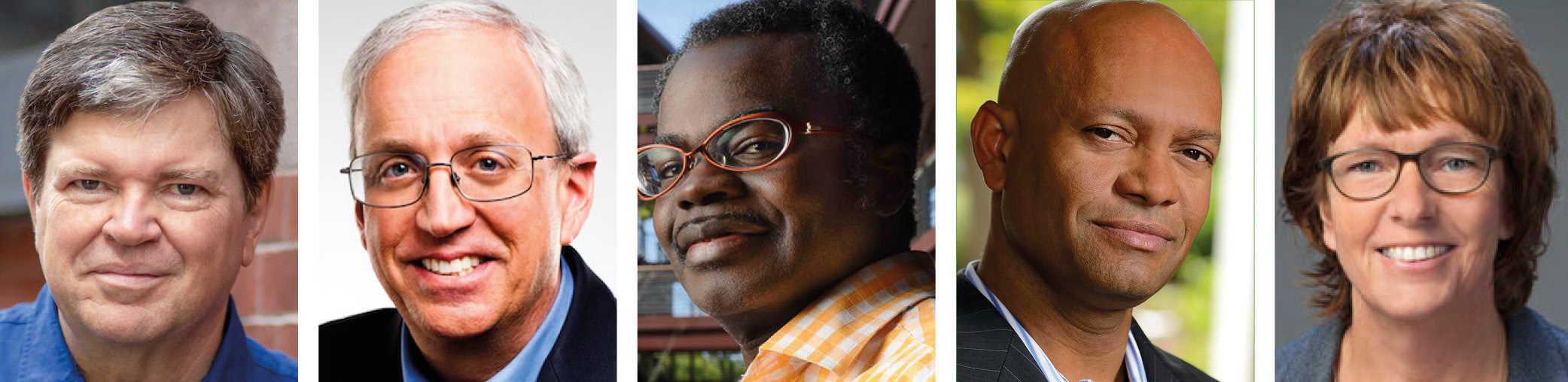
One scientist who has been collaborating on the project is Danny Jonigk, a lung pathologist at Hannover Medical School in Germany. He feels as if he has spent his entire career doing research under candlelight, only for someone “to suddenly switch the lights on”. Then there’s Daniyal Jafree, a medical student at University College London (UCL) in the UK, who’s doing a PhD in kidney imaging. When he heard what was being developed elsewhere at UCL, Jafree couldn’t quite believe it. “I thought that sounds ambitious,” he says. “Then I saw the images.”
X-rays at your service
The Human Organ Atlas project wouldn’t be possible without physics. It began at the European Synchrotron Radiation Facility (ESRF) in Grenoble, France, which has been one of the world’s foremost X-ray light sources since it opened more than 30 years ago. Unlike the X-rays delivered by a clinical CT scanner, synchrotron X-rays have high energy and a high spatial coherence. That means their waveforms remain very much in phase with one another as they propagate, allowing researchers to exploit minute changes in X-ray phase to produce tomographic (section-by-section) 3D images of very high detail and contrast (see box below).
For many years, this phase-contrast X-ray technique has delivered incredible reconstructions of biological specimens. In 2011, for example, ESRF beamline scientist Paul Tafforeau helped produce what is still the most detailed scan ever of the inside of a skull of an early human ancestor, Australopithecus sediba. More recently, he has produced scans of small dinosaur fossils, ancient human teeth and even mummified crocodiles.
Seeing more deeply Claire Walsh from University College London (left) and Paul Tafforeau from the European Synchrotron Radiation Facility in Grenoble, France, are among the scientists to have developed the new imaging technique of Hierarchical Phase-Contrast Tomography (HiP-CT). Originally used to scan donated human organs, including lungs from a patient who died from COVID-19, the technique is central to plans for a zoomable Human Organ Atlas. It will provide 3D reconstructions of entire intact organs that can then be explored anywhere down to the cellular level. (Courtesy: ESRF/Stef Candé)
Then, in 2020, two things happened. The first was that the ESRF finished commissioning a new, “fourth-generation” source, making it the world’s brightest synchrotron lab. More than a decade in planning and construction, the Extremely Brilliant Source (EBS) delivers X-rays that are 100 times brighter than before, and 100 times more coherent in the transverse (horizontal) plane, making them almost laser-like at low energies. The EBS has done wonders for tomographic imaging, enabling users to scan bigger objects, in more detail and at a greater range of scales.
The second big event of 2020 was, of course, the COVID-19 pandemic. For many scientists, the pandemic brought research to a full stop. Not for Tafforeau. Unexpectedly, he received a call from Peter Lee, a regular ESRF tomography user at UCL, who in turn had been approached by Jonigk. Could the ESRF be of help, Lee wondered, in reconstructing lung tissue samples from people who had died after catching COVID-19? It was a great question and almost overnight Tafforeau switched from study-ing ancient fossils to human organs.
“The COVID-19 pandemic changed a lot of things for many people,” Tafforeau recalls. “I realized that several imaging techniques that we originally developed for palaeontology could open access to a new level of imaging precision on complete human organs. Then, while developing the techniques further, we realized that it may be a game-changer for biological imaging in general.”
Swiftly, Lee composed an international, multidisciplinary team to see what could be done: synchrotron imaging scientists at UCL and the ESRF; mathematicians and computer scientists at UCL; medical scientists at Hannover Biobank, as well as the universities of Mainz and Heidelberg in Germany. As the apparent potential of the new tomographic imaging grew, so did the breadth of the collaboration: it now includes more than 50 people.
The scientists called the technique hierarchical phase-contrast tomography (HiP-CT), thanks to its ability to provide 3D reconstructions of entire intact organs that can then be explored anywhere down to the cellular level. As a result, the technique bridges the gap in scales between clinical CT and MRI, and the microscopy of biopsies. In November 2021 the project was formalized as the Human Organ Atlas, with a goal to provide a reference database of organ imagery that is accessible to all.
Hierarchical Phase-Contrast Tomography (HiP-CT) in a nutshell
Most simple imaging methods – including conventional computed tomography (CT) – involve measuring the loss of intensity (the attenuation) of an electromagnetic wave as it passes through a sample. In 1953, however, the Dutch physicist Frits Zernike won the Nobel Prize for Physics for developing an alternative – and potentially more illuminating – imaging method that involves measuring shifts in the phase of electromagnetic rays.
Zernike’s “phase-contrast” microscopy was initially fit only for visible light. But in 1965 it started being extended to X-rays too thanks to the work of Ulrich Bonse and Michael Hart – two physicists at Cornell University in the US – who used a crystal interferometer to convert phase changes into interference patterns.
Limitations with interferometers meant that phase-contrast X-ray imaging of biological samples had to wait until the 1990s through the efforts of Atsushi Momose at Hitachi and Tohoru Takeda at the University of Tsukuba, Japan, and others. At roughly the same time, Anatoliy Snigirev and others at the European Synchrotron Radiation Facility in Grenoble, France, realized they could deduce phase changes without an interferometer, simply from the interference of highly coherent synchrotron X-rays in free space. By combining many propagation phase-contrast 2D images in CT mode, they were able to produce 3D reconstructions of small biological samples with far more detail than that available from clinical CT scanners.
With the upgrade of the ESRF to a “fourth-generation” X-ray source in 2020, “hierarchical” phase-contrast CT (HiP-CT) became possible. The lab’s ultra-coherent X-rays provide information on phase changes over very long propagation distances up to 40 m, allowing samples of up to 2.5 m × 1.5 m in size – including human organs, torsos, even entire bodies – to be imaged in 3D at micron resolution.
The atlas in action
A video of a human brain, as imaged by HiP-CT, gives an impression of the technique’s capabilities (figure 1). It starts off conventionally enough, moving through cross sections of the entire organ. Here the brain looks like it does with a clinical CT scan, albeit at some 50 times the resolution. The various lobes are clearly visible, as are some of the external blood vessels. Then the “camera” zooms in to the back of the brain, the cerebellum, perfectly transitioning from big to small.
At 5 μm resolution, the smallest features of white and grey matter come into view; at 2.5 μm resolution, the tiniest blood vessels can be discerned. Even pyramid-shaped cells can be seen, known as Purkinje neurons, which are largely responsible for human motor function. Finally, the view retreats and the reconstruction morphs to depict blood vessels only. Now the incredible density and complexity of the brain’s “vasculature” become apparent. As the system that delivers and receives the oxygen, glucose and metabolic waste, it keeps every one of us alive and thinking.
The HiP-CT video looks like the cutting-edge CGI you see in sci-fi blockbusters – yet it is perfectly real. What’s more, as all the raw data have been collected and post-processed, it’s possible for scientists to explore different parts of the brain at will. In fact, the sheer wealth of information in the imagery is so great that interpreting it is a major problem in itself. The team divides the work, with Tafforeau in charge of reconstructing the images and the UCL team trying to make sense of them.
1 The Human Organ Atlas in action Stills from a video of a human brain imaged using Hierarchical Phase-Contrast Tomography (HiP-CT) shows what the technique can do as you zoom in and back out. a A cross-section of the entire organ reveals lobes and some external blood vessels. b A closer image of the back of the brain, the cerebellum. c At 5 mm resolution, white and grey matter come into view. d At 2.5 mm resolution, you can see the tiniest blood vessels. e As the view retreats, the reconstruction depicts the blood vessels only, revealing the full complexity of the brain’s ‘vasculature’. f Zooming back out again, the blood vessels in situ. See videos on YouTube. (Courtesy: ESRF/HiP-CT: C L Walsh, P Tafforeau, W L Wagner et al.)
“It’s a little bit overwhelming, like being a kid in a sweetie shop,” admits Claire Walsh, a biophysicist in UCL’s computational analysis team. “Medics and histologists can tell us when something looks weird, but we have to quantify that: exactly how weird?” One example is the size of alveoli, which has in the past been used to indicate the seriousness of lung disease. Previously, says Walsh, the alveoli were assumed to be roughly spherical, like grapes on a vine. But the new technique reveals them to be more irregular.
As a result, the researchers have had to define new parameters, with input from their medical collaborators, to capture the potential of the new information. Joseph Jacob, a chest radiologist who joined the UCL team early in the pandemic, stresses the scale of the interpretation challenge. “When I first saw the images, I felt amazement and apprehension – probably apprehension more than amazement,” he says. “It was definitely what I wanted to work on, but the complexity of labelling it – obviously it would only be possible with computer science.”
Fortunately, Jacob knows how vital the image-processing of X-ray data is, having developed algorithms to stitch together hundreds of CT images to see the lung in detail. He believes the reward now is well worth the labour. “[This new technique] is going to show us things we never knew existed,” he says, which could be vital given how medicine is a very “organ-centric” discipline. “As a chest specialist, I just look at the lungs – I don’t look at the heart, for instance. But disease doesn’t necessarily work that way. If you could image a whole torso, you could understand how disease is affecting other organs; it would be a much more rounded approach.”
The way ahead
As things stand, almost all the organs in the atlas have been imaged at the ESRF’s long-serving BM05 beamline. In December 2021, however, the team acquired its first HiP-CT images at BM18 – a new ESRF beamline that has been designed to maximize the benefits of the EBS for microtomographic images of large objects. Although the beamline won’t be fully operational until the end of 2022, it will eventually be able to image a torso – and even an entire human body.
Imagine one day being able to explore, in virtual reality, human bodies of all ages, backgrounds, states of health and disease. As Lee points out, the damage wrought by new diseases could then be easily compared with that of existing conditions, to indicate possible known methods of treatment. People could see what sort of processes might be going on inside themselves. Medics could entertain pure curiosity, without having to resort to the knife.
We are not there yet, but preliminary images have already given some indication of the benefits of the large-scale, detailed view of HiP-CT. Reconstructions of several lungs from COVID-19 victims have revealed heterogeneous damage that appeared on previous clinical CT scans merely as a fuzzy, ground-glass texture (Nature Methods18 1532). The result is helping to determine whether it is the connectedness of lung damage, or the sheer amount of it, that is the cause of death by the virus.
Meanwhile, Jafree is keen to find out if HiP-CT can help us to give us a better understanding of the kidneys, the organs he specializes in. We know that the number of blood-vessel networks, or glomeruli, is a proxy for general kidney function. But no-one knows how losing some of these networks affects those that remain, or whether their volume or shape affects kidney health. “HiP-CT allows us to look at things in a different way,” says Jafree. “It also encourages [students] like me to learn some of the image-analysis techniques. We need that expertise to generate something meaningful for biology and medicine – and we have an incentive now.”
Vital signs These HiP-CT images of human kidneys could reveal if the volume or shape of blood-vessel networks affects the onset of kidney disease. (Courtesy: ESRF/HiP-CT: C L Walsh, P Tafforeau, W L Wagner et al.)
Sarah Teichmann, a cellular geneticist at the Wellcome Sanger Institute in Hinxton, UK, says she was “blown away” by the first HiP-CT images she saw, letting her view the cellular structures inside organs in exquisite detail, before zooming out to see the whole tissue. “Not only do these images and videos give a new appreciation of the beautiful complexity of the human body,” she says, “they are also stocked full of information about how our bodies work.”
Teichmann believes that the whole-organ or whole-body approach could benefit our understanding of diseases such as cancer. She also reckons that the Human Organ Atlas project ties in well with the Human Cell Atlas – an international consortium that she co-founded to create a comprehensive reference map of all human cells. “[It] could help us to see where these cell types – which we characterize at the molecular level – fit into the bigger picture of the organ. This will help to bridge the gap between cells and systems, painting a more holistic picture of the human body.”
Beautiful times
Alongside the huge scientific impact of this imaging technique, there is also an inherent beauty to the images taken by the Human Organ Atlas. In December 2021 National Geographic magazine picked a HiP-CT image of a lung as one of its favourite science images of the year. Francesco Sette, the physicist who has been director-general of the ESRF since 2009, has even compared the advancement of the technique with Leonardo da Vinci’s anatomical drawings of the early 16th century.
Those drawings gave unprecedented insights into the workings of the human body, especially its biomechanics. It is not yet clear what the ramifications of the Human Organ Atlas will be, although the concept is proving popular. The collaboration’s Nature Methods paper has been downloaded more than 50,000 times, and is in the top 1% of Nature articles in terms of its Altmetric score, or reach.
The project is also gaining some serious backers, not least a $2.75m donation from the Chan-Zuckerberg Initiative (CZI), which was set up by Facebook founder Mark Zuckerberg and his wife Priscilla Chan. The CZI is independent of Facebook – which may be a good thing, as the Atlas team is just beginning a collaboration with Google to make its database available to the public. According to Lee, the plan is to create something like an anatomical version of Google Earth, with 3D “satellite” resolution of 40 μm resolution for a whole organ, and a 3D “street view” resolution down to 1 μm to expose individual cells.
After Google Earth, Google Maps and Google Sky, perhaps it is fitting that one day we will have a Google Body search tool too.
Placing large-scale solar farms on the Arabian Red Sea coastal plain could dramatically increase rainfall in this arid part of the world, a new modelling study claims. According to the researchers, simulations show that such installations could change the reflectiveness of the land enough to unsettle coastal air circulation. The resulting changes in local weather patterns and rainfall could potentially produce enough water to meet the annual needs of five million people. Although idealized, the team says its research points to the feasibility of freshwater recovery from sea breezes by land surface geoengineering.
As the world warms, water security is becoming a major concern. This is a particular issue in hot, arid parts of the world like the Middle East. Many countries in this area, such as Saudi Arabia, are in the middle of a water crisis. They have little rainfall and are exhausting underground aquifers. Water desalination is already widely used to increase freshwater supplies, but current methods are unsustainable due their high energy use. Desalination also unlikely to be able to meet future water demands.
There is increasing interest in the idea of artificially increasing rainfall over the Arabian Peninsula, using techniques such as cloud seeding. There is a lot of water in the air in the region because the Red Sea loses 0.7 teratonne of water every year through evaporation. This is equivalent to nearly 8% of the mass of all water vapour in Earth’s atmosphere. On the Arabian Red Sea coast, sea breezes blow this water across the land, but little of it falls as rain. Instead, it is transported south towards the equator and the middle of the Indian ocean.
Changing albedo
Georgiy Stenchikov, an expert in climate and atmospheric modelling at King Abdullah University of Science and Technology in Saudi Arabia, and his colleagues wondered how changing surface albedo – the reflectiveness of the land – over the region would impact water transport and change rainfall patterns. The idea, Stenchikov told Physics World, is to try to utilize this vast natural freshwater resource by increasing precipitation.
In their latest work, published in the Journal of Hydrometeorology, Stenchikov and colleagues ran a series of numerical simulations in a regional weather research and forecasting model. They focused on three scenarios: extensive forest planting, increased albedo and decreased albedo.
The simulations show that afforestation and increased surface albedo along the Arabian Red Sea coastal plain would reduce rainfall. Sea breezes in the region are driven by the horizontal thermal contrast between land and sea. The warmer land and cooler sea create a pressure gradient that pushes moist sea air towards the land. The models show that afforestation and a more reflective surface cools the land, which dampens sea breezes and reduces the movement of water vapour from sea to land.
Strengthening sea breezes
Conversely, decreasing surface albedo would increase rainfall. It would warm the coastal region, strengthening sea breezes and increasing water vapour transport to the shore, the researchers found. The increase in convection currents over the land increases vertical mixing in the lower atmosphere, particularly of water vapour, the models show. This increases humidity, cloud formation, and instability and turbulence. “The basis for all this is increased water surface flux,” Stenchikov says.
Solar panels are known to alter the surface energy balance by absorbing solar energy and heating up. The team found that a reduction in albedo that corresponds with the large-scale installation of solar panel plants would increase rainfall in the region by around 1.5 gigatonne in the dry summer season. This is linked to an almost doubling of wet days from July to September, compared with current conditions. Smaller reductions in albedo would also have significant impacts on the number of wet days and rainfall volumes.
Stenchikov says that this proof-of-concept study shows that the idea could work, but more analysis is needed. “We have to work further to try to optimize this scheme, looking at the sizes [of solar installations needed] and optimal distribution for this specific area,” he explains.
Stenchikov says that such schemes could in theory work in any coastal region with sea breezes. They will work best, he explains, in areas with strong sea breezes, high levels of evaporation from the sea and large water vapour fluxes.
Read it now: as a service to the community you can read the March 2022 issue for free
As a service to the physics community, we’re offering you complimentary access to the March 2022 issue ofPhysics Worldmagazine.
As usual, there’s a great mix of in-depth features, comprehensive news and analysis as well as incisive opinion pieces, careers articles and book reviews.
The cover feature of this free sample issue looks at how physicists are using X-rays to create a zoomable “Google Earth” of the human body.
There’s a great feature about the life of the pioneering astronomer Cecilia Payne-Gaposchkin, who battled sexism and discrimination to succeed.
You can find out how researchers on big-physics experiments are lowering the “carbon footprint” of their supercomputing calculations.
And don’t miss our take on the cultural impact of the Netflix movieDon’t Look Up, see why physics awards need to be as fair as possible, and explore how firms are trying to build commercial fusion reactors.
• KATRIN sets neutrino mass limit – Björn Lehnert from the Lawrence Berkeley National Laboratory talks to Richard Blaustein about what a new measurement of the upper mass of the neutrino means for particle physics
• JET smashes fusion energy record – The Joint European Torus has achieved 59 megajoules of fusion energy in a single fusion “shot”, more than doubling the previous record. Michael Banks reports
• We need to rethink scientific awards – Jess Wade and Maryam Zaringhalam say that prize processes must be reformed to avoid discrimination
• Fusion: it’s hotting up – James McKenzie applauds recent record investments in commercial fusion power plants, which could help us to create a net-zero economy
• Disaster signalling – Robert P Crease wonders what lessons we can learn from movies about comets and asteroids heading towards Earth
• The body, exposed – A new synchrotron-imaging technique is letting researchers create a “Google Earth” of the human body. Jon Cartwright zooms in
• The woman who found hydrogen in the stars – Sidney Perkowitz delves into the work and life of Cecilia Payne-Gaposchkin, from her stellar astronomical findings to a career-long struggle with bias against women in the early 20th century
• The huge carbon footprint of large-scale computing – Researchers have been able to cut their carbon footprint by jetting off to fewer international conferences, but physicists working on large-scale experiments may also have to consider the significant environmental impact of the computer power they require. Michael Allen investigates
• Technology with characters – Andrew Robinson reviews Kingdom of Characters: a Tale of Language, Obsession and Genius in Modern China by Jing Tsu
• Putting the physics into science fiction – Kate Gardner reviews The EXODUS Incident by Peter Schattschneider
• Industry or academia? – How to choose your path – After doing a PhD and postdoc in
quantum technology, in the UK, Joanna Zajac spent three years in industry before returning to fundamental research. She now works as a quantum scientist at Brookhaven National Laboratory. So how do academia and industry differ and which one is right for you?
• Ask me anything – Careers advice from Maksym Sich, the co-founder and chief executive of quantum-photonics spinout Aegiq
• Conceptual juggling – Laura Hiscott finds out how circus skills can help students to learn physics
Scientists and engineers continue to push the boundaries of what can be achieved with artificial intelligence (AI), with the last few years seeing impressive gains in areas such as speech recognition and natural language processing. But experts agree that the current state-of-the-art still falls some way short of the thinking machines that are widely depicted in science fiction.
“AI is very good for solving very specific problems, as long as there is enough data to train the system,” says Yann LeCun, chief AI scientist at Meta and a Turing Award Laureate for his research on deep learning at New York University. “But current systems do not understand how the world works, and that’s what is needed to realize transformative applications such as domestic robots, virtual assistants and fully autonomous self-driving cars.”
Scientists at the forefront of AI research are still figuring out how to make the paradigm shift from data-driven number crunching to more intuitive human-like thinking. Most researchers believe there is a role for computer algorithms that mimic the biological brain, with artificial neural networks becoming a mainstream approach for solving problems through trial and error rather than rules-based programming.
“Over the last few years there has been a lot of progress in self-supervised learning, where a system can learn to represent the data for a specific task without being trained,” says LeCun. “Once self-supervised learning can work more generally, machines will be able learn how the world works by watching videos – opening the door to solving problems much more simply than we can today.”
While LeCun believes that neural networks running on standard computer chips offer the best route to building next-generation AI systems, others contend that building processors that mimic the functionality of the biological brain would yield more powerful AI systems that also consume less energy.
Such “neuromorphic” processing systems typically exploit analogue circuits to create artificial silicon neurons, and mixed-signal analogue/digital circuits to implement spiking neural networks. These neuromorphic circuits are designed to replicate the way that synapses and neurons in the brain light up during neurological activity.
“In our brain there is no distinction between an abstract algorithm and the computing substrate,” explains Giacomo Indiveri, director of the Institute of Neuroinformatics at the University of Zurich and ETH Zurich, and editor-in-chief of a new open-access journal, Neuromorphic Computing and Engineering. “We separate the software and hardware in computer science, but in neuromorphic computing there is a merging of the two. To achieve the optimal solution we need to co-design the architecture and the computing substrate.”
Advocates for neuromorphic computing believe that in certain situations these bio-inspired systems have the potential to outperform standard digital technologies. “Neuromorphic systems can be implemented as massively parallel architectures in which artificial neurons are in different states and ready to fire within a few microseconds, providing a quicker response that can be typically achieved with conventional digital computation,” says Indiveri. “Analogue/digital spiking neuromorphic architectures have also been shown to consume power in the microwatt range, orders of magnitude lower than conventional digital processors.”
Indiveri believes that the sweet spot for neuromorphic systems lies in applications that require low latency and low power, such as localized processing of sensor data. “It’s more suited to processing continuous streaming data in real time,” he says. “That might be a vision system for gesture recognition, or detecting whether someone has fallen in the home. For biomedical signal monitoring, a low-power neuromorphic system implemented on a ‘wear-and-forget’ wristband could detect any anomalies and raise an alert, without needing to be connected to a mobile phone.”
But LeCun is not convinced that neural computing needs to be neuromorphic to be effective. “I am interested, but I am sceptical,” he says. “The question is whether you are better off exploiting the progress of digital technology or try to follow the neuromorphic philosophy.”
While LeCun agrees that neuromorphic systems could play a role in processing sensor data at the edge of the network, he believes that current analogue technologies have fundamental disadvantages for building larger neural nets. “There may be a good reason for the brain to produce spikes, but it might not translate to electronics and software,” he says.
These differing points of view will be aired in a virtual debate entitled “The future of high-performance computing: are neuromorphic systems the answer?”, which can be watched live on Monday 7 March 2022 at 4.00 pm GMT and then subsequently on-demand. LeCun will be in the sceptics’ corner, along with Bill Dally, a professor at Stanford University and chief scientist at NVIDIA – a company that designs the graphical computer processors that underpin many AI systems.
On the other side of the debate will be Kwabena Boahen, founder and director of the Brains in Silicon laboratory at Stanford University. Boahen and his team are developing silicon-based artificial neurons to emulate the way the brain works, and have demonstrated Neurogrid – a circuit board composed of 16 chips that each includes analogue circuitry for more than 65,000 artificial neurons.
“We’ve built hardware models of neural systems to learn about how the brain functions, which lets us test ideas about how cognition could come from the properties of neurons,” Boahen explained in an article for Stanford University. “Informed by what these models have taught us, we’re now working on developing a computer that works more efficiently, like the brain does.”
Taking sides: speakers at the debate will be Kwabena Boahen (centre) and Ralph Etienne-Cummings (second right), who will be attempting to convince Yann LeCun (left) and Bill Dally (second left) of the need of neuromorphic approaches for more efficient computation. Regina Dittmann (right) will be the chair.
He will be joined by Ralph Etienne-Cummings, director of the Computational Sensory-Motor Systems Laboratory at Johns Hopkins University. Etienne-Cummings has studied bio-inspired vision sensors and their use in robots, and more recently brain–machine interfaces and neural prosthetics that are designed to restore function after injury or to overcome disease. His wide-ranging research has convinced him of the need for neuromorphic computing to “perform recognition tasks as effortlessly living organisms, create legged robots that are as efficient and elegant as humans, and design prosthetics than can seamlessly interface with the body.”
The discussion will be chaired by Regina Dittmann, an expert in memristive devices who is currently at the Peter Grünberg Institute of the Forschungszentrum Jülich in Germany. With Boahen and Etienne-Cummings attempting to convince LeCun and Dally of the benefits of neuromorphic computing over mainstream neural approaches, the session promises to be a friendly, but fiercely contested, debate. You can register for free now.
While the idea of neuromorphic engineering is rooted in concepts first proposed in the 1980s by, among others, microelectronics pioneer Carver Mead, the field has expanded over the years as new approaches have emerged. The term “neuromorphic” now describes any type of analogue, digital or mixed-mode implementation of a neural system, which includes physical devices that are designed to replicate the synapses in the brain, as well as digital chips – such as those demonstrated by Intel and IBM – that provide an electronic implementation of a spiking neural network.
The new journal, Neuromorphic Computing and Engineering, aims to represent this diversity of thought. “It’s the first journal that has tried to encompass all the different aspects of the field, from basic research about the brain to nanoscale technologies and high-performance computing that can hopefully exploit some of the concepts from spiking neural networks,” says Indiveri.
“We have received a high level of submissions and the articles published so far have had a good number of citations. I am happy to see that there is a lot of interest in the area in general and in bringing the different communities together through the journal.”
Register now for the online debate, which will be live on Monday 7 March at 16.00–17.30 GMT and subsequently available to view on-demand.
Pulse oximeters, which measure the percentage of oxygen in the blood, can be used as part of clinical decision-making to triage patients, adjust supplemental oxygen levels, and more. In 2020, researchers analysed tens of thousands of blood oxygenation measurements collected from thousands of patients. The study, though not received without controversy and discussions about study design, found that Black patients had approximately three times the frequency of hypoxemia – an abnormal decrease in the amount of oxygen in the blood – as white patients, and that this condition wasn’t being detected by pulse oximetry in many Black patients. In a letter to the editor in TheNew England Journal of Medicine, the researchers said that missed hypoxemia diagnoses could result in poorer health outcomes in Black patients.
The NEJM letter grabbed the interest of UC San Diego researchers Jesse Jokerst and Yash Mantri. Jokerst, a professor of nanoengineering, materials science and radiology, and Mantri, a bioengineering graduate student, are working to improve human health using a hybrid light and acoustic imaging technique called photoacoustic (PA) imaging. Soon after the NEJM letter was published, they decided to investigate the impact of skin tone on blood oxygenation measurements obtained using a different tool, PA oximetry.
“[This study] ties into some of our other work in wound imaging, wound repair and regeneration,” Jokerst says. “As we were doing those studies, we became more interested about the impact of skin tone. We also were motivated by the paper in The New England Journal of Medicine, among others, which referenced the impact of melanin in pulse oximetry.”
While pulse oximetry offers a quick and non-invasive way to measure oxygen saturation, pulse oximeters may not account for some physically relevant factors, such as absorption and scatter of photons by melanin in the skin.
And though biomedical optics devices such as pulse oximeters, cerebral tissue oximeters, wearables such as smart watches and PA imagers play “an increasingly powerful role in human health,” Jokerst and Mantri say that few of these devices account for differences in skin tone between individuals. Their subsequent proof-of-concept study, published in Biomedical Optics Express, suggests that individuals with darker skin tones have lower oxygen saturation as determined by PA oximetry compared with individuals with lighter skin tones.
Measuring blood oxygenation with PA oximetry: the physics
Most of the light that hits your skin is absorbed by haemoglobin and melanosomes. Haemoglobin is a protein found in red blood cells that carries oxygen throughout your body. Melanosomes are subcellular organelles that produce and store melanin, which largely governs skin tone – individuals with higher concentrations of melanin have darker skin. Darker skin tones absorb more photons than lighter skin tones because they contain higher concentrations of melanin.
PA oximetry is based on the absorption of light and the thermal expansion of absorbed light in tissue. When visible and near-infrared light from a laser or similar light source is delivered to tissue, it interacts with absorbers such as melanin and haemoglobin in the skin. This absorption of light produces a small temperature increase in tissue that leads to an initial pressure increase and then relaxation.
When the tissue relaxes, acoustic (sound) waves are emitted. The acoustic waves travel to the body’s surface, where they are detected by a PA transducer. The PA signals are used to create images that show the differences in optical absorption in tissue, a source of image contrast that can be used to create maps of oxygen saturation.
Skin tone and PA imaging: proof-of-concept study
The researchers measured PA signal, penetration depth and oxygenation differences in nine healthy volunteers with three different skin tones. Skin tones were estimated using the Fitzpatrick scale, which is often used in dermatology to estimate the response of different skin tones to ultraviolet light.
Each volunteer’s left arm and hand were imaged in five easily identifiable locations – selected for their relatively uniform distribution of melanin, flat curvature to facilitate scanning and relative hairlessness – using two PA imaging systems. The researchers acquired images using standardized settings that didn’t attempt to compensate for differences in skin tone, and they quantified oxygen saturation using the PA imaging systems’ built-in methods. They also acquired data using a multi-wavelength approach.
Jokerst and Mantri observed that individuals with darker skin tones had higher PA signal intensity at the surface of the skin, with increased acoustic clutter, a phenomenon caused by reverberations or off-axis scatterers. Further studies showed decreased oxygen saturation in the radial arteries of individuals with darker skin tones compared with those with lighter skin tones.
Correcting biases before devices hit the market
For devices on the market, several techniques that try to compensate for measurement biases already exist. PA intensity can be enhanced during image acquisition by adjusting time gain compensation settings. Noise reduction algorithms can also be implemented. Filtered back projection or PA computed tomography could reduce streaking artefacts even when strong absorbers are present. Applying fluence corrections, multi-angle plane wave ultrasound, focused ultrasound, spatial weighting or deep learning could reduce noise and stationary signals that lower the signal-to-noise ratio and mask blood vessels.
Jokerst and Mantri even developed a basic correction factor to adjust PA oximetry measurements. The factor, defined as the ratio between measurements made with these two methods, corrected PA oximetry measurements to within 2% of pulse oximetry.
But Jokerst hopes that their study, rather than creating more band-aid solutions, prompts the medical devices community to design devices that account for physically relevant factors such as skin tone before biomedical optics devices go to market. (The first PA imaging device approved by the US Food and Drug Administration was recently launched and was not tested as part of Jokerst and Mantri’s study.)
“Ideally, these would all be built into the device. To have the end user fixing the measurement doesn’t make much sense,” Jokerst says.
He suggests that skin tone could be read from images obtained using a built-in CCD camera and that oxygen saturation measurements could be automatically adjusted using this information. Future work will evaluate the results of their proof-of-concept study in larger cohorts, against arterial blood gas measurements, and using a wider variety of imaging equipment.
A scalable and flexible system for the remote monitoring of quantum experiments in noisy and unpredictable environments has been created by researchers in the UK. Thomas Barrett and colleagues at the University of Sussex used the latest sensing, machine learning and database technologies to create and maintain multiple quantum-related experiments involving ultracold atoms. Their approach could soon allow users to carry out, monitor and diagnose problems in advanced quantum experiments in hard-to-access environments – including on satellites.
As quantum technologies become more powerful and robust, they are starting to be used by non-specialists for practical sensing applications in a variety of environments. Much focus has recently been directed towards miniaturized, field-based quantum devices, which can be monitored remotely. Such devices are being developed as gravimeters and navigational accelerometers that exploit the unique quantum properties of ultracold atomic clouds.
Quantum systems are inherently sensitive to external factors such as heat, noise and electromagnetic radiation. Therefore, one of the main challenges for people creating practical quantum devices is how to keep them running in unpredictable environments. To mitigate against thermal disturbances and acoustic noise, components such as mirrors, coils and power supplies can be corrected actively. Yet in many cases, this can only be done after errors have occurred.
Active mitigation
Alongside advances in quantum technology, improvements to electronics and database software have all led to increasingly robust, versatile and modular systems for monitoring and manipulating experiments remotely. Using a combination of remote sensing, machine learning and human input, these systems allow users to collect data on key experimental parameters, diagnose unexpected behaviours and then actively mitigate problems before they occur.
In their study, Barrett’s team extended this technology to monitor a variety of parameters necessary for maintaining several ultracold atom experiments using a shared laser system. These parameters included temperature, vacuum chamber pressure, laser beam power and magnetic field strength.
Within the system, sensors were networked together, and the data they collected were recorded in an external database – which could then be accessed through user dashboards. This way, the team showed that users could efficiently control multiple experiments at the same time, allowing them to mitigate any problems quickly and remotely.
Such capabilities are particularly crucial for maintaining quantum devices in harsh and inaccessible environments. This includes space-borne experiments on satellites and interplanetary probes. Since the system is both scalable and highly flexible, the team says it could also be extended to a diverse array of other applications. These include particle accelerators, sensor networks for monitoring glaciers, and remote teaching labs – which could even allow students to access quantum experiments remotely.
Physicists backing Japan to build the ¥600bn ($5bn) International Linear Collider (ILC) must re-evaluate their plans and “shelve” the question of the country hosting the proposed next-generation particle collider. That is the stark message to emerge from a panel of senior Japanese officials who have examined progress made towards realizing the ILC. They conclude that it is too early for Japan to proceed towards construction of the ILC and instead call for further research and international support towards the project.
First mooted well over a decade ago as the successor to the Large Hadron Collider at the CERN particle-physics lab near Geneva, the ILC – if built – would accelerate and smash together electrons with positrons to study the Higgs boson and other particles in precise detail. Collisions using these particles would be much “cleaner” than the proton–proton collisions that are carried out at the LHC.
The ILC’s five-volume technical design report was published in June 2013 and included a 30 km-long linear collider operating at around 500 GeV. The Japanese physics community expressed their desire to host the machine, with a site in the Tōhoku region, about 400 km north of Tokyo, chosen as a potential location.
But with little support for the go-ahead from the Japanese government, in 2017 physicists looked for cost savings and revised the plans to reduce the ILC’s energy to 250 GeV — an energy aimed to study the 125 GeV Higgs boson, which was discovered in 2012 at the LHC. This included shortening the length of the main tunnel to around 20 km, but leaving the option of later upgrading the collider to the original 500 GeV proposal and eventually to energies of around 1 TeV.
Yet in 2019 an independent committee of the Science Council of Japan (SCJ) failed to support the ILC’s construction in Japan, pointing out that the ILC did not yet have enough international backing. The Japanese government then laid out a condition that the ILC should be more widely supported by the Japanese and international scientific communities.
This would likely involve, for example, the ILC being included in the next roadmap of large science projects put together by Japan’s Ministry of Education, Culture, Sports, Science and Technology (MEXT), as well as pledges being made by other countries to support the project. Following that move officials at the KEK particle-physics lab then began negotiations on cost sharing arrangements for a “preparatory laboratory” for the ILC that would carry out the technical development and engineering design needed for the start of ILC construction.
In June 2021 ILC scientists published a 48-page plan for the ILC pre-lab and also submitted a report updating MEXT on progress since 2019. Those moves resulted in MEXT convening a committee in July 2021 that was tasked with evaluating the updated developments.
Our attention should now be more focussed on trying to find a host for a linear collider elsewhere in the world
Brian Foster
On 14 February the panel reported its findings, which were then released on 25 February. While recognizing the “academic significance of particle physics and the importance of the research activities, including that of a Higgs factory”, the panel says it is still “premature” to give the ILC pre-lab phase the go-ahead as they say this would be “coupled with an expression of interest to host the ILC by Japan”.
The panel adds that given the “increasing strain in the financial situation” of some countries that may provide support for the ILC, it recommends that those backing the collider should not only “reflect upon this fact and to re-evaluate the plan” but also “re-examine the approach towards a Higgs factory in a global manner”. This would involve considering progress made in other proposed collider projects such as the Future Circular Collider (FCC) – a 100 km-circumference collider that would be built by CERN.
The panel also calls for more work in key technologies towards a next-generation accelerator by “further strengthening international collaboration” among institutes and labs therefore “shelving the question of hosting the ILC”.
In response to the panel’s findings, KEK notes that it will now “re-examine the path for realizing the ILC as a Higgs factory” and consider the progress made in the FCC feasibility study. “KEK and the Japanese ILC community is committed to further advance important technological and engineering development in the accelerator area and to continue the effort for the realization of the ILC,” KEK noted in a statement.
Looking elsewhere
John Ellis from King’s College London says that the explicit reference to the FCC in the panel’s recommendations “sounds like a tacit judgement” that FCC studies have been making progress while the ILC has been stagnating. Ellis adds that the panel’s recommendations are in line with previous reviews that stated the ILC community had not done enough to line up broad scientific support within Japan or have substantial international commitments on board. “It sounds like they suggest the ILC is interesting, but advise to find another way of doing the physics by collaborating with the FCC,” adds Ellis.
Yet Hitoshi Murayama from the Kavli Institute for the Mathematics and Physics of the Universe in Tokyo told Physics World that while MEXT’s conclusion towards the pre-lab is “certainly unfortunate”, it is not all bad news. “KEK management is optimistic to acquire funds from MEXT to carry out key development work,” he says.
“The problem MEXT has is that they don’t want to fund the ILC pre-lab which may be interpreted as their tacit approval of the ILC as a whole by some proponents,” adds Murayama. “As long as the technology development does not carry the name ILC or specify the site in Japan, they may support the activity.”
Some remain unconvinced. Particle physicist Brian Foster from the University of Oxford, who was European regional director for the ILC’s Global Design Effort, raises the prospect that the ILC may never be built in Japan. He says the MEXT review statement is “very disappointing” not only because it means delaying the ILC but also due to the way the decision was reached.
“It is apparent that some members of the committee did not approach their task with the openness of mind and belief in evidence-based conclusions that one would expect from senior members of the scientific community in an independent and unbiased review,” says Foster. “While we should continue to work with our Japanese colleagues to progress work on the ILC, our attention should now be more focused on trying to find a host for a linear collider elsewhere in the world.”
That view is backed by Phil Burrows, from the University of Oxford, who is the UK lead scientist in the ILC International Development Team. “Despite the best efforts of the particle physics community, not least via the International Committee for Future Accelerators, the Japanese government has yet to embrace the prospect of hosting ILC as a forefront global science facility,” he says. “Perhaps the time has indeed come to explore options elsewhere.” He points out that CERN, for example, has the “know-how and technical capability” to build a linear collider, although he admits that this “is not currently its stated top priority for a future project”.
International Linear Collider Q&A
What is the International Linear Collider (ILC)?
The ILC is a 20 km-long particle collider that will accelerate electrons and positrons to energies around 250 GeV. To do so, it will consist of thousands of superconducting radio-frequency accelerator cavities made of niobium. The ILC will then smash these beams together roughly 7000 times a second at an “interaction point”. The ILC will have two all-purpose detectors based around the interaction point — SiD and ILD — that would take turns being in the beam. Interchanging the detectors is estimated to take around a day to complete.
How is this different from CERN’s Large Hadron Collider?
The 27 km-circumference LHC is a circular collider made up of more conventional technology such as radio-frequency cavities that accelerate the beam, dipole magnets that bend the particles along a circular path, and quadrupole magnets that focus the beam. The ILC instead uses superconducting cavities to accelerate the beam along a straight path before being focused by quadrupole magnets. This linear acceleration has the advantage that the electrons do not lose energy via X-rays when travelling along a circular path. The benefit of a circular machine is that it allows for more integration points — four in the LHC’s case, with no need to swap detectors.
Has this accelerator technology been tested before?
Yes. The European X-ray Free Electron Laser (E-XFEL) facility near Hamburg, Germany, uses 768 superconducting niobium cavities to accelerate electrons to 17.5 GeV over 1.7 km. Rather than collide the particles, however, the E-XFEL makes them produce X-rays that are then used for a range of experiments from biophysics to condensed-matter physics. The E-XFEL is considered to some extent as an ILC prototype.
What would the ILC study?
Its main aim would be precision studies of the Higgs boson, which was discovered in 2012 at the LHC. The LHC has managed to measure the properties of the Higgs – notably how it couples to other particles – with a precision of around 20%. Yet the LHC’s proton–proton collisions suffer from a large amount of “debris” that affects the precision of the measurements. As electrons and positrons are fundamental particles, their collisions are much “cleaner” meaning that the ILC would improve this precision to 1% or lower. The ILC could also be later upgraded to higher energies to study the top quark.
Why is this exciting?
Physicists hope that the door to “new physics” could be opened through precision studies of particles such as the Higgs. This would come from deviations from those predicated by the Standard Model of particle physics.
Why has Japan dragged its feet for so long?
Japan has balked at the potential cost of building the ILC, which is one reason why physicists proposed a scaled-down version in 2017 that is both cheaper and would not take as long to build.
When could the ILC see the light of day?
Likely to be 2040s. If Japan gave it the go-ahead in the coming years then negotiations and preparations could take around four years to complete, with construction then taking a decade.
What other designs are on the table?
The CERN-led Compact Linear Collider would offer higher collision energies up to around 3 TeV, but the technology is not as mature as the ILC. Two other designs are circular colliders. China’s Circular Electron Positron Collider and the Future Circular Collider would both be 100 km-circumference colliders that would initially be used as a Higgs factory but could later be used for high-energy proton–proton collisions.
The assembly of 2D nanosheets on the surface of emulsion nanodroplets leads to the stabilization of the emulsion and requires only a tiny amount of material – report physicists in the UK. Furthermore, the team has developed a model that allows fine-tuning of the solvent composition so the droplets could be used to create components for a range of applications from strain sensors to batteries. The emulsions also raise the possibility of printing large-scale devices in a matter of minutes, which could have an important impact on electronics fabrication.
Wouldn’t it be great to print with 2D materials such as graphene? The answer is yes for many reasons, but how to do so is a puzzle that has confounded scientists working with 2D materials since Konstantin Novoselov and Andre Geim first isolated 2D sheets of carbon (graphene) in 2004. A thousand papers have been published on this subject, but the question regarding printing remains open. One important challenge is how to overcome the well-known coffee ring effect when using droplets to print thin layers of graphene or other 2D materials. This effect involves solids in a droplet forming a ring when the droplet evaporates on a surface – and results in defects when printing conductive coatings.
Now, researchers at the University of Sussex have come closer to making large-scale printing with 2D materials possible. The inspiration came when physicist Alan Dalton was mixing salad dressing and thought about adding graphene to liquid emulsions. An emulsion is a combination of two liquids that normally do not mix – like oil and water. In an emulsion, one of the liquids exists as droplets within the other liquid – like droplets of oil in a vinegar-based salad dressing.
Thin nanosheets are known to stabilize emulsions by covering the surface of droplets. The result is a Pickering or armoured emulsion, which is also found in some foods.
Armoured droplets
Dalton and colleagues armoured tiny droplets in an emulsion with thin layers of graphene, forming a shell that is just a few nanometres thick. This microscopic, but functional structure requires only 0.001 vol% of graphene in solution, which is the lowest value ever reported.
Dalton comments, “In bringing the graphene coatings of the liquid droplets down to atomically-thin layers, and in opening wide the potential for real-world applications by being able to do so with any liquid material, this research development will significantly advance the emerging and scientifically exciting field of liquid electronics”.
The armoured droplets offer a solution to the coffee ring problem because a continuous conductive pathway is formed upon drying. As a result, electronic devices could be printed from emulsion droplets.
Fine-tuned for different applications
Moreover, armoured droplets can be produced in any liquid, so they could be fine-tuned for different applications. Conductive droplets could be used for wrapping soft polymers such as silicone, for example, to create wearable strain sensors. Such devices have increased sensitivity at low graphene loading.
An important potential application is the development of better batteries for electric vehicles. Such batteries would be cheaper and more sustainable because they would require less graphene or other 2D nanosheets to coat the droplets used in their manufacture.
“We are also investigating emulsion assembly of battery electrode materials to enhance the robustness of these energy storage devices,” says Sussex’s Sean Ogilvie, who is lead author of a paper in ACS Nano describing the research.
Experimental evidence of long-range attractive forces between cellular proteins has been obtained by researchers in France more than 50 years after the idea was first proposed. The forces are mediated by electromagnetic radiation, and they could explain how molecules find their targets inside the crowded interiors of living cells.
At any given time, around 130,000 pairwise interactions may be occurring between proteins in a living cell. These are mediated by a range of phenomena including van der Waals forces and hydrogen bonding. Biochemical reactions work on a “lock and key” setup, whereby molecules must find and bind to receptors to trigger the processes of life. For this to occur efficiently inside a cell, molecules must find their cognate partners (keys must find the right lock) much more quickly than is possible by simple Brownian motion. However, how this happens so quickly and efficiently is a mystery. One possible explanation is electrostatic attraction, but mobile ions in the cell cytoplasm would screen out static electric fields over ranges longer than 100 nm.
Electrodynamic interactions
In the 1960s, the theoretical physicist Herbert Fröhlich suggested that the resonant exchange of radiation between macromolecules and their cognate partners could be the answer. “Electrostatic interactions are shielded, but electrodynamic interactions are not, in principle, provided these take place at sufficiently high frequency,” explains Marco Pettini of Aix-Marseille University – the work’s principal theorist. Fröhlich reasoned, therefore, that if molecules underwent collective oscillations in an excited state, they might behave like antennas and emit terahertz radiation. If the cognate partner resonantly absorbed this radiation, the two would attract as a result.
Pettini explains, “Biomolecules behave as special kinds of antennas called Hertzian antennas, whose size is much smaller than the wavelength of the radiation, and it’s a consequence of Maxwell’s equations that we can excite these attractive interactions”. However, there was no evidence at the time that proteins could show this collective resonance, so Fröhlich’s suggestion gathered dust.
The principal problem, explains Jérémie Torres of the University of Montpellier, was that the main constituent of a cell’s cytoplasm – water – strongly absorbs terahertz radiation, making terahertz spectrosopy extremely challenging.
Terahertz-frequency fluctuations
In 2018, Torres and colleagues labelled molecules of the protein bovine serum albumin (BSA) with a fluorophore that absorbs 488 nm light. They then dispersed this protein in water and irradiated droplets of the solution with a 488 nm laser. Using a nanowire, they detected terahertz-frequency fluctuations of the electric field around the droplet that matched theoretical predictions from Pettini and colleagues. This showed that proteins could indeed produce collective modes, but not how strong the resulting forces were. Pettini explains, “In principle these forces could be very weak and thus of no interest in biology,” says.
Now in a new paper published inScience Advances, the researchers demonstrate that electrodynamic forces can indeed excite phase transitions in biomolecules. They first use a refined version of their nanowire-based technique to study both the labelled BSA and a second, algal protein that naturally absorbs light. This time, instead of looking simply for collective oscillations in the proteins, the researchers looked at how these oscillations changed when they varied the laser power.
They found that, on increasing the laser power, the frequency of the oscillations shifted, showing that the interaction between the proteins was becoming stronger, as predicted by theory. When they reduced the power, the process was reversed. Moreover, for the algal protein, they used a new, independent spectroscopic technique to show that at high laser powers, the proteins form large clusters: “This is the combination of a very sensitive experimental set-up with a physical mechanism that gives you an absorption that’s even higher than water due to this collective dipole moment,” explains Torres, who led the new experimental work.
The researchers now intend to investigate the effects of these phenomena. Torres points to a 2014 optogenetic study in which two proteins aggregated mysteriously when irradiated by light. “We produce quite similar results but with deeper insight from the physical point of view,” he says. More generally, they want to know how the forces operate in cells, where there are no lasers. “Our work opens a new and broad research topic,” he explains, “During the evolution of life on the planet, nature has exploited all physical phenomena. It would be hard to understand why nature hasn’t resorted to these.”
“It’s a really interesting experiment,” says biophysicist Cécile Fradin of Canada’s McMaster University. “Whether it applies in cell I think remains to be proved and that will determine the ultimate significance, but it’s the first step – well, the second step.” Among several ideas for further work, Fradin suggests that whether proteins and other molecules are resonantly attracted to their cognate partners rather than simply themselves needs investigation. “I would see the next step to be finding out whether you can have two different types of molecules reacting together against a background of all different types of molecules that you don’t want to interact”.