Elastic polymers can be stretched and released repeatedly without tearing and are widely employed in applications from disposable gloves to heart valves. Their main drawback is that they can generally be made either stiff or tough, but not both at the same time. A team at the Harvard John A Paulson School of Engineering and Applied Sciences (SEAS) has now developed an elastic polymer, or elastomer, that has both properties. The new material could find use in applications such as tissue regeneration, bioadhesives, bioprinting, wearable electronics and soft robots, and might also help reduce plastic pollution by increasing the durability of products made with elastomers.
Polymers are fabricated by linking together building blocks of monomers into chains. To make elastic polymers, these chains are crosslinked by covalent chemical bonds. These crosslinks connect the polymer chains so that when a force is applied to stretch the elastomer, the material recovers its shape when the force is removed. The more crosslinks, the shorter the polymer chain and the stiffer the material. This is because the energy stored in the materials decreases, making it brittle.
Entanglement bonds
Led by Zhigang Suo, the researchers made their new polymer simultaneously stiff and tough by using physical rather than chemical bonds. These types of bonds are known as entanglements, and have been known about for a long time, but until now they were only thought to affect polymer stiffness, not toughness.
In their work, Suo and colleagues fabricated a polymer in which all chains are long and cross-links are outnumbered by entanglements. The latter act as slip-links and stiffen the polymer. Unlike cross-links, though, they do not make the polymer brittle. This means that when an entangled polymer is stretched, the applied tension is transmitted along one chain and then to many other chains before a chain breaks.
“Crowded” monomers
The researchers studied two polyacrylamide hydrogels – 3D polymer networks that can hold a large amount of water. They found that they could introduce many entanglements into the material using a precursor made of a concentrated monomer solution that contained 10 times less water than usual. The monomers are thus “crowded” into the solution, which forces them to become entwined like a tangled string of yarn, explain Junsoo Kim and Guogao Zhang, the study’s co-first authors. “Just like with knitted fabrics, the polymers maintain their connection with one another by being physically intertwined,” they say.
By adding enough entanglement into the polymer chains, the team found that the hydrogels they studied could become tough without losing any of their stiffness. Indeed, with hundreds of these entanglements, only a few chemical crosslinks are required to keep the polymer stable and prevent the polymer chains from disentangling, they say. The hydrogels also have low friction and high wear resistance, which increase the durability and lifespan of the polymer. This last point is important since it could help decrease the consumption of plastics, Kim and Zhang suggest.
Looking forward, Suo and colleagues tell Physics World that they are now busy developing highly entangled hydrogels using polymers suitable for biomedical applications.
This year’s Fall Meeting of the Materials Research Society will, like many other scientific conferences at the moment, be held as a hybrid event. Delegates can choose between attending the live conference in Boston, Massachusetts, or catching up with all the symposia and featured events through a dedicated virtual meeting.
The live conference will take place from 29 November through to 2 December, and with networking and social events will enable attendees to reconnect with friends and colleagues who they may not have seen for some time. The virtual meeting will follow a few days later, on 6–8 December, allowing registrants to access all the symposia and presentations on demand. Registrants for the virtual meeting will also be able to tune into a live stream of featured talks during the live event.
Whether attending in person or online, the MRS Fall meeting will provide a forum for discussing breakthrough materials research. The Symposium X sessions, which are designed to highlight work at the frontiers of materials science, will include talks on superconducting qubits for quantum computers, the internal structure of biominerals, and novel materials for next-generation lithium-ion batteries.
The scientific programme will also feature two Nobel laureates: Sir Kostya Novoselov of the University of Manchester in the UK will deliver the distinguished keynote lecture on Wednesday 8 December, while in the plenary session on Tuesday 9 December Sir Fraser Stoddart of Northwestern University in the US will highlight recent advances in artificial molecular machines.
Registrants to both the live and virtual meetings also have the opportunity to engage with scientific equipment vendors, either in the exhibit hall at the Hynes Convention Center or via online exhibit booths. Some of the latest innovations that will be presented are highlighted below.
Single instrument combines synchronous source and measure capabilities
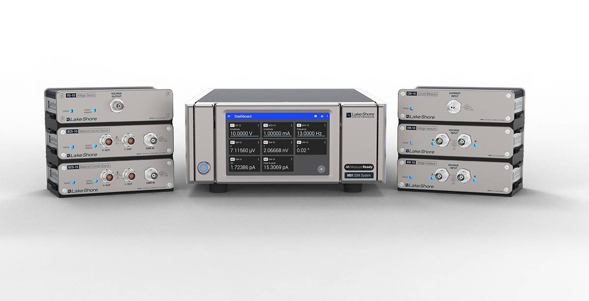
The M81-SSM from Lake Shore Cryotronics has the potential to simplify complex experimental configurations (Courtesy: Lake Shore Cryotronics)
The new, modular M81-SSM (Synchronous Source and Measure) System from Lake Shore Cryotronics provides a flexible and powerful solution for a wide range of material characterization applications. The M81-SMM combines highly synchronized DC, 100 kHz AC, and mixed DC + AC sourcing and measuring in a single instrument, and also offers both voltage and current lock-in measurement capabilities.
The multichannel system supports up to three remote mountable source and three measurement modules, while its unique modular architecture enables signal and source amplifiers to be located as close as possible to the sample being characterized. Minimizing the signal wiring to the sample reduces noise and increases sensitivity, resulting in more accurate and repeatable measurements.
The amplifier modules feature 100% linear circuitry and leverage the M81’s patent-pending MeasureSync™ real-time sampling technology, which ensures synchronous sourcing and measuring across all channels. Multiple samples can be characterized under simultaneous sampling conditions to enable users to obtain consistent data. Combining both DC and AC sourcing and measurement in a single instrument reduces the need for different pieces of equipment, simplifying the set-up and operation of complex characterization configurations.
The M81-SSM system also uses the standard SCPI programming interface and is supported by Lake Shore’s MeasureLINK™ application software, which provides easy control of temperature and field set-up. The software also supports general electrical characterization measurements, with enhanced versions available for custom applications.
Visit Lake Shore Cryotronics at booth #417
Automated imaging speeds up AFM experiments
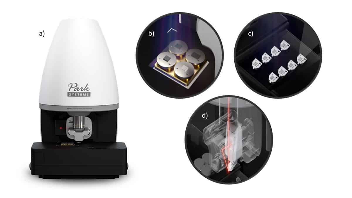
The FX40 atomic force microscope from Park Systems automates the entire imaging process to eliminate the manual procedures that are typically needed to set up an AFM (Courtesy: Park Systems)
The latest atomic force microscope from Park Systems, the Park FX40, integrates robotics and machine learning throughout the design to produce the first AFM that fully automates the imaging process for research users. The FX40 exploits a robotic system to automatically change and replace its own tips, and also features an optical design that enables automatic beam alignment.
“The Park FX40 takes care of all the set up before and during scanning,” says Ryan Yoo, the company’s vice president for product development. “All the tedious and time-consuming manual processes are now a thing of the past. The Park FX40 performs them all autonomously.”
The FX40 also features a unique dual-camera design that makes it much simpler to locate the region of interest. The first camera offers a view of the whole sample stage, which can accommodate up to four samples at a time, to allow the user to quickly find and select the target location. The stage then automatically moves to the chosen position, at which point the system switches to a high-resolution camera for a closer view and any detailed adjustment.
Once in the correct location, the system automatically brings the probe close enough to the sample’s surface for measurements to be taken. With the FX40, Park Systems has also updated its true non-contact mode to boost the data quality and the precision of measurements.
Visit Park Systems at booth #613
Materials support TEM nanocharacterization
TEM grids from MilliporeSigma are available in a range of materials and patterns, and with a choice of films to support the specimen, to suit the needs of different applications (Courtesy: MilliporeSigma)
MilliporeSigma supplies a variety of specialized materials for characterizing nanomaterials and nanodevices using transmission electron microscopy (TEM). The most detailed information can be obtained from TEM studies when the samples for analysis are mounted onto a TEM grid that matches the type of material to be studied.
TEM grids are available from MilliporeSigma in different materials, including copper, nickel and molybdenum, as well as with a range of mesh patterns and sizes. Grids are also available with a choice of films for supporting the specimen, such as a continuous amorphous carbon film with various thicknesses or a lacy carbon film with different hole sizes.
Locator and finder grids, which are used to quickly and easily check a particular feature, or to examine another TEM, are also available in a variety of designs and materials. In addition, MilliporeSigma offers a comprehensive range of supporting components for characterizing nanomaterials, including tweezers, TEM window grids with various thicknesses, a magnetic pick-up tool, cryo-capsules, and lift-out grids in copper and molybdenum. Substrates for atomic force microscopy are also available in various dimensions.
Breakthrough analyser delivers precision ARPES measurements for small samples
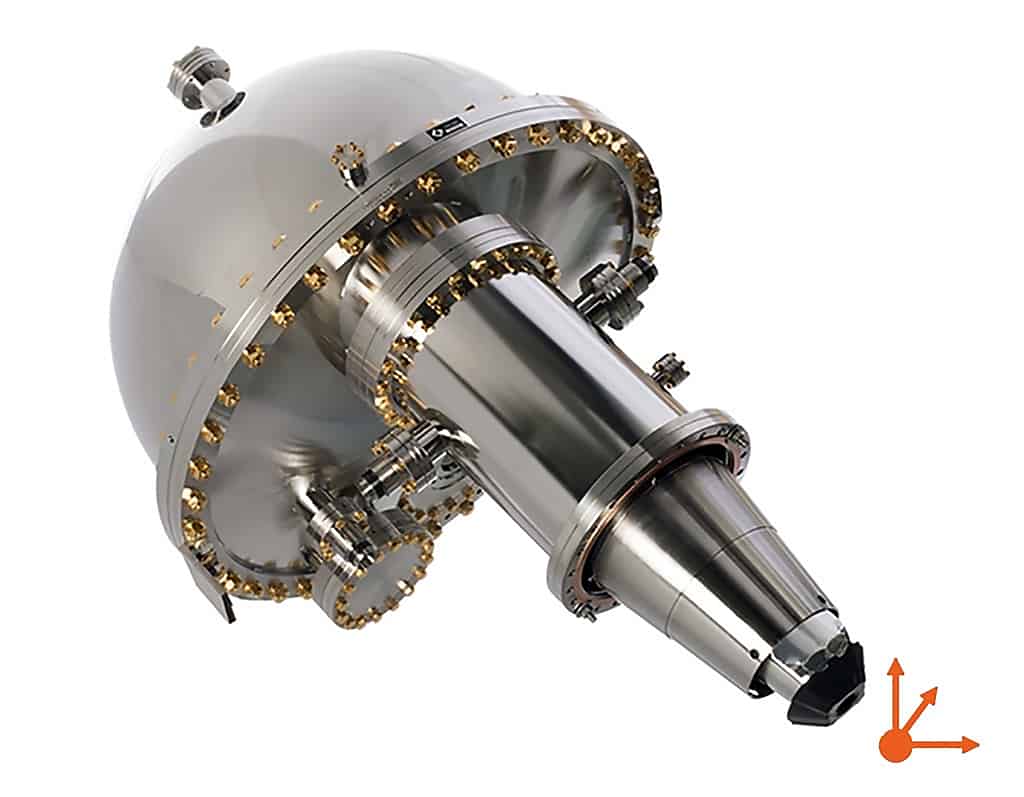
The DFS30 analyser is equipped with electrostatic 3D focus adjustment to match the analyser focal point to the emission spot on the sample, and is ideal for small-spot angle-resolved photoelectron measurements. (Courtesy: Scienta Omicron)
New from Scienta Omicron is the DFS30, a next-generation analyser for angular-resolved photoemission spectroscopy (ARPES). The DFS30 – which is now available as part of the company’s ARPES Lab system – is optimized for measuring the small-spot band structure of novel materials and micron-sized samples, and is perfect for measuring the electronic band structure of 2D materials.
High-quality ARPES measurements, particularly on small samples, require accurate alignment of the photon source, the sample, and the focal point of the analyser. The DFS30 exploits electrostatics to adjust the focal point of the analyser, for the first time replacing imprecise mechanical movements. This speeds up the workflow and improves reproducibility when optimizing experimental conditions.
The DFS30 extends Scienta Omicron’s wide range of analytical and turnkey solutions for surface science and nanotechnology. The company’s field-proven technologies provide configurable solutions for both standalone systems and for full Materials Innovation Platforms (MIPs) that offer full control over the materials development process.
One of these MIPs might include an epitaxial growth system for creating a material structure, an XPS Lab to evaluate the sample’s surface composition, or a bulk-sensitive 9.25 kV HAXPES Lab to probe interfaces below the surface. Other options include the HiPP Lab for studying gas or liquid surface interfaces under ambient pressure using XPS, and the LT STM for studying surfaces at ultralow temperatures with STM, spectroscopy, and Scienta Omicron’s QPlus® AFM.
In 1545 the English warship Mary Rose sank in a battle off the south coast of England and was raised more than 400 years later. The ship and some of its contents are now on display at the Mary Rose Museum in Portsmouth.
In this episode of the Physics World Weekly podcast, the materials scientist and deputy chief executive of the Mary Rose Trust, Eleanor Schofield, explains the science behind conserving objects that have spent centuries underwater.
The LIGO–Virgo observatories are one of the success stories of 21st century physics. The LIGO detectors were the first ever to detect a gravitational-wave signal – from the merger of two black holes – back in 2015. Since then, LIGO and Virgo have detected a total of 90 gravitational-wave signals. Cardiff University physicists and LIGO–Virgo members Katherine Dooley, Stephen Fairhurst and Fabio Antonini are on hand to talk about the latest haul of observations and what we can expect in the future.
For a relatively simple chemical compound, water – especially its frozen varieties – is surprisingly poorly understood. An international team of researchers has now chipped away at the mystery by measuring the structure and properties of a form of ice known as ice XVIII and another superionic ice phase, ice XX. The team’s work could shed light on the formation of the unusual magnetic fields of the planets Uranus and Neptune, which are thought to contain these two phases of ice deep in their interiors.
Although most of the ice found on Earth has a hexagonal structure (think of snowflakes), ice can take on at least 20 different crystalline and amorphous forms. This curious behaviour comes in part from the weak intermolecular bonds between its two hydrogen atoms, which are separated by a single atom of oxygen.
Hot ice and other icy phases
One of the most-studied high-pressure forms of ice is ice VII, also known as “hot ice”. This exotic body-centred cubic (bcc) crystal phase forms at ambient temperatures at pressures above 2.3 GPa (about 23,000 times atmospheric pressure at sea level) and it has been theorized to exist in cold subduction zones within the Earth’s crust as well as on Saturn’s icy moon Titan.
Another novel ice phase, superionic ice or ice XVIII, is predicted to exist at higher temperatures and pressures, around 1000 K and 40 GPa. This form of frozen water contains liquid-like hydrogen ions – that is, protons – that quickly diffuse through a solid lattice of oxygen atoms. Superionic ice could make up a large fraction of the interiors of Uranus and Neptune, with the fast-diffusing protons helping to generate the strong and complex magnetic fields characteristic of these “ice giant” planets.
Mimicking the conditions inside the ice giants
“The superionic phase of ice was predicted two decades ago, but when we have started our experiments 10 years ago there were no direct experimental evidence for the existence of this superionic phase,” co-team leader Vitali Prakapenka, a beamline scientist at the University of Chicago, US, tells Physics World. “And theoretical estimations of the pressure and temperature stability range for this phase were extremely controversial.”
In the past, Prakapenka explains, various research groups had collected experimental data on these phases using X-ray and optical techniques. These data hinted that water behaves in unusual ways under high pressure-temperature conditions, but the scattered nature of the results meant that the overall picture was unclear. “We have now solved this puzzle,” he says.
Together with colleagues Alexander Goncharov from the Carnegie Institution of Washington and Sergey Lobanov of the German Research Center for Geosciences GFZ Potsdam, Prakapenka measured the structure and properties of ice XVIII and another superionic ice phase, ice XX. To do this, the researchers began by placing a room-temperature sample of water in a diamond anvil cell and increasing the pressure to 150 GPa, producing ice VII or ice X. They then increased the water’s temperature to 6500 K by heating it with high-power laser light. These are the conditions that prevail several thousand kilometres inside Uranus and Neptune, which likely contain more than 60% water.
Next, the researchers used X-ray diffraction techniques to observe how the crystalline ice structure changed and became superionic. They carried out these experiments using the extremely bright synchrotron rays at the Advanced Photon Source (APS) of the Argonne National Laboratory, backed up by optical spectroscopy measurements at Carnegie’s Earth and Planets Laboratory that aimed to determine the ice’s optical conductivity. They observed two phases – ice XVII and ice XX – and determined the pressure and temperature at which these phases suddenly undergo a change in their optical properties.
Conductive water-rich liquid
The researchers say their results could help explain Uranus’ and Neptune’s magnetic fields, which are unusual in that they do not run quasi-parallel and symmetrically to the axis of the planets’ rotation (as Earth’s magnetic field does). Instead, the magnetic fields of the ice giants are skewed and off-centre. Models of how they formed therefore hypothesize that they are generated not by the motion of molten iron in the core (as on Earth) but by conductive water-rich liquid in the planets’ outer third.
“We can draw the pressure and temperature in the interiors of Uranus and Neptune,” Lobanov explains. “Here, the pressure can roughly be taken as a measure of the depth inside. Based on the refined phase boundaries we have measured, we see that about the upper third of both planets is liquid, but deeper interiors contain solid superionic ices. This confirms the predictions about the origin of the observed magnetic fields.”
“Our study is only a first step in characterizing the structure of superionic phases, their density, optical properties, and pressure-temperature boundaries,” Prakapenka adds. Future studies, he says, will focus on pinning down the nature of this unique state of matter by combining theoretical simulations with experimental measurements of the electrical conductivity of superionic phases, their viscosity, chemical reactivity and composition effects.
Since it was first successfully isolated in 2004, the “wonder material” graphene, with its honeycomb-like 2D structure and its wide gamut of interesting properties, has been keenly studied by material scientists. In recent years, however, the focus has shifted from simply exploring its physical properties to using graphene as a platform for testing and realizing a plethora of novel ideas. This was especially apparent with the “From graphene physics to graphene for physics” theme of the 2017 Solvay workshop on graphene. And, like a relentless spring of fresh water from different sources, graphene is still unveiling surprises.
Today, there are numerous areas of research in the field where lively discussions are being had. Superconductivity in rotated graphene layers, for example, is of interest to many researchers, as it may open the door to a better understanding of superconductivity as a whole. But a key puzzle in graphene is the material’s perplexing non-local response, which has seeded a series of studies addressing the use of graphene’s “valley degree of freedom”.
Electrons have fundamental properties – such as “charge” and “spin” – which allow for a number of emergent phenomena such as magnetism. Certain semiconductors and 2D materials possess a similar property known as the “valley” degree of freedom. In these materials, the states at the Fermi surface are distributed in pockets, in the crystal momentum space. Each of these pockets is called a valley, which corresponds to a specific electronic state. Graphene is a zero-gap semiconductor with two valleys (usually called K and K’); its conduction and valence bands meet at discrete points, known as Dirac points.
In the valley
Just as “spintronics” – which makes use of the internal degree of freedom of spin to store and manipulate bits of information – this valley degree of freedom is at the centre of a stream of research aiming to exploit it for “valleytronics”, where the information would be stored as discrete values of the crystal momentum. One of the challenges in this field is that transport measurements sense charge not valley, making it difficult to detect valley currents. Furthermore, a high crystalline quality of graphene is required, as atomic defects would normally produce intervalley scattering.
However, in 2007 Di Xiao and colleagues, then at the University of Texas, Austin, found that if inversion symmetry is broken and an electric field driving a current is present, then the charge carriers would bend their trajectories, as in the presence of a magnetic field. Their direction of travel is then determined not by the sign of their charge, but by their valley (Phys. Rev. Lett.99 236809). Essentially, graphene can be coaxed into exhibiting a “valley Hall effect” – similar to the spin Hall effect – by creating an asymmetry between the two valleys, such that electrons move across the sample in opposite directions when a current is run across it.
A puzzling non-local response
In 2014, nearly a decade after the isolation of graphene in electrical devices, condensed-matter physicist Roman Gorbachev at the University of Manchester, UK, and collaborators including graphene Nobel laureates Andre Geim and Konstantin Novoselov, characterized multi-terminal graphene samples, by looking for topological currents in graphene superlattices (Science346 448). Inspired by experiments on quantum and spin Hall systems, their experiment was designed to probe the non-local response, and led to the observation of a remarkable effect that has puzzled scientists ever since.
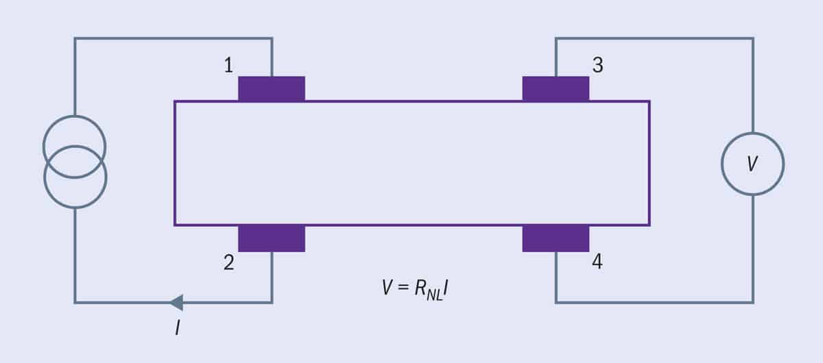
1 A non-local surprise in graphene Schematic of the basic set-up used by the team at the University of Manchester to measure non-local resistance, RNL. The current, I, is applied between terminals 1 and 2, and the voltage, V, is measured at a distance of several microns, between terminals 3 and 4, where no current is expected to flow.
In Gorbachev’s measurements a current, I, is applied between two terminals while the voltage, V, is measured far (several microns) away, between two different terminals (figure 1), where no current flows and an ohmic contribution is not expected. In these experiments graphene was placed on a special substrate of hexagonal boron nitride (hBN), which, when carefully aligned, breaks the graphene lattice inversion symmetry. Furthermore, the Fermi level could be tuned thanks to a back-gate voltage. The team’s findings showed sharp peaks in the non-local resistance V/I versus back-gate voltage, only when inversion symmetry was broken. The peaks appeared near Dirac points in the absence of a magnetic field and implied that a voltage developed microns away from the current probes.
To rationalize these unusual findings, Gorbachev and colleagues relied on the valley Hall effect, which they connected to the non-local response. More details on the theory were published almost a year later (PNAS112 10879) by Justin Song at the California Institute of Technology, and colleagues. The researchers modelled a system made of graphene stacked atop hBN, and wrote that the “Berry curvature manifests itself in transport via the valley Hall effect and long-range chargeless valley currents. The non-local electrical response mediated by such currents provides diagnostics for band topology.” The experiments were then explained in terms of such charge-less valley currents flowing far away close to the voltage probes (figure 2a).
Strikingly, and unlike other cases where topological effects are tied to robust states at the edges (through a bulk-boundary correspondence), the charge-less valley currents predicted by Xiao’s research are not due to states appearing at the boundary but rather states in the Fermi sea beneath the gap, and are signalled to have a consequence in the non-local response.
Further findings, and seeds for a controversy
The story continues with a number of subsequent experiments (Nature Phys.11 1027; Nature Phys. 11 1032; Appl. Phys. Lett.114 243105), all of which looked at topological valley currents, and confirm the observation of a large anomalous non-local resistance. This time, the experiments were not carried out on graphene stacked on hBN. Instead, they looked at bilayer graphene, where the strength of the inversion symmetry breaking term could be tuned by means of a perpendicular electric field. Other related experiments attempted to map the current flow leading to the non-local signal (Nature Comms8 14552), and even explored a ballistic version of these phenomena (Comms Phys. 3 224).
But at the same time, a 2015 paper by theorist George Kirczenow of the Simon Fraser University in Canada triggered a debate on the interpretation of the results (Phys. Rev. B 92 125425). By using scattering theory, Kirczenow pointed out that the non-local resistance peaks cannot be taken as a signature of the anomalous velocities produced by an electric field and the non-vanishing Berry curvature. A crucial point leading to this conclusion was the fact that the use of a semiclassical theory is unclear because, in the experiments by Gorbachev and colleagues, the Fermi level is tuned within an electronic gap.
Kirczenow argued that a more suitable approach is provided by the Landauer–Büttiker theory – one of the most widely used quantum-transport theories, where voltage and current sources appear on equal footing. Current flow is expressed in terms of transmission probabilities, whereas in other views (including the semiclassical one) it is seen as the response to an external field. All quantities can then be computed from the scattering matrix obtained for a Hamiltonian in absence of the electric field, thus depriving the non-local resistance of the crucial ingredient associated with the anomalous velocity.
Fermi sea versus surface
Considering the disparate views, the scene was set for a clash of interpretations and methodologies and many colleagues wondered whether we had hit a so-called “valley of death”. Indeed, a 2018 study by Juan Marmolejo-Tejada of Montana State University and colleagues aimed at deciphering the non-local resistance in graphene on hBN using an ab initio or “first principles” method of quantum transport. They showed that graphene with zigzag edges on hBN hosts dispersive edge states near the Dirac point (J. Phys. Mater. 1 015006). The team suggested that these peculiar edge states, absent in simplified descriptions, lead to non-local signals that can persist at long distances, while withstanding edge disorder. Marmolejo-Tejada and collaborators concluded that the non-local signals in earlier experiments may stem from edge states at the Fermi surface, rather than being a topological response from bulk states deep in the Fermi sea (figure 2b).
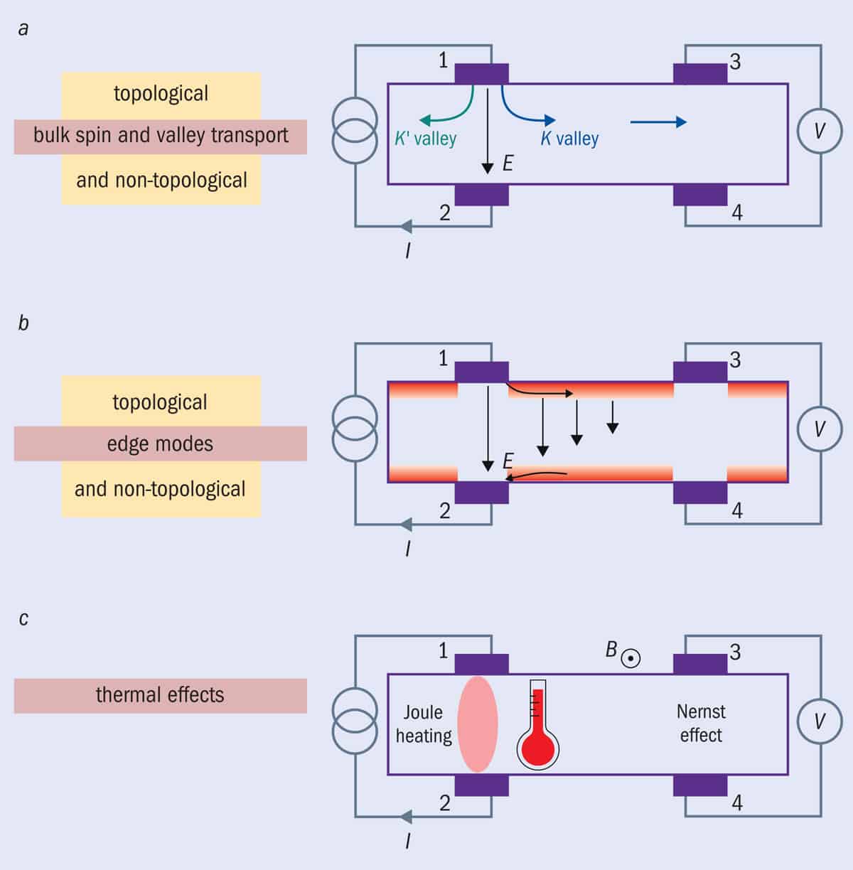
2 Multiple mechanisms Varying mechanisms that may lead to the curious non-local resistance in graphene: (a) bulk spin and valley transport, which might be of topological origin; (b) topological or non-topological edge modes; and (c) thermal effects (Joule, or resistive, heating). Current is passed through contacts 1 and 2 (applied electric field E) while the voltage is measured far away between contacts 3 and 4. While the proposed mechanisms involving the valley Hall effect and edge modes can result in a non-local signal at zero magnetic field, the scenarios based on spin and thermoelectric (Nernst) effects rely on the presence of a significant magnetic field (B), of up to 5 T.
Earlier this year, Thomas Aktor from the Technical University of Denmark and collaborators brought a new twist to this debate by suggesting that the valley Hall effect can indeed enhance the non-local resistance (Phys. Rev. B103 115406). However, they claim that the mechanism would arise not from Berry curvature effects inside a bulk gap, but from the non-uniform gap profile produced in graphene/hBN heterostructures by the Moiré (interference) pattern. The authors propose a way of engineering substrates and tailored defects (through controlled deposition of atomic clusters or drafted molecular patterns) to produce bulk-driven valley Hall effects.
Topological or non-topological response?
New experiments published this year, looking at the long-range non-topological edge currents in charge-neutral graphene, add even more surprising pieces to this puzzle (Nature593 528). As in the original experiments by Gorbachev’s team, Amit Aharon-Steinberg at Weizmann Institute of Science in Israel and colleagues have implemented non-local transport measurements in hBN-encapsulated graphene at low temperatures. Their experiments, however, are carried out under moderate magnetic fields of up to 5 T; and they combine transport experiments with a superconducting interference device on a tip for thermal imaging and scanning gate microscopy.
When it comes to transport experiments, the observations largely reproduced previous investigations, in particular the unexpectedly large non-local resistance at zero magnetic field (figure 2c). This becomes orders of magnitude larger as the magnetic field is increased from 0 to 5 T, a phenomenon that was ascribed to spin and thermo-magnetoelectric effects by Gorbachev’s team in 2011 (Science332 328). However, scanning thermal imaging in the same sample suggested that Joule heating and non-local resistance are highly correlated, even at zero magnetic field. Indeed, when a large non-local resistance develops, the temperature increase spreads across the sample, which casts doubt about the relevance of non-dissipative valley currents.
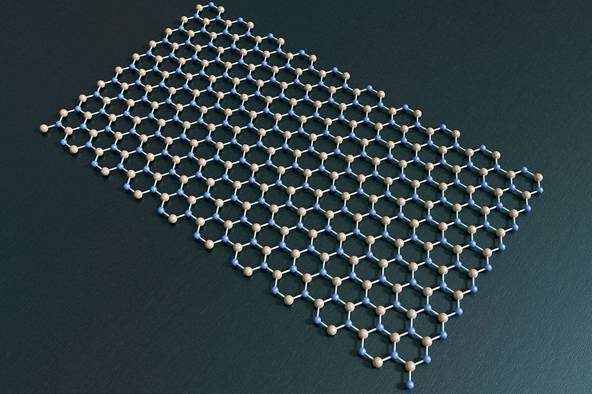
Special substrate A number of the experiments to study graphene’s non-local response include the use of the material being placed on a substrate of hexagonal boron nitride, which, when carefully aligned, breaks the graphene lattice inversion symmetry. (Courtesy: Robert Brook/Science Photo Library)
The temperature distribution and scanning gate microscopy measurements point towards predominant edge transport, driven by the presence of edge charge accumulation resulting from band bending, thereby ruling out the bulk mechanisms invoked in earlier works. As Aharon-Steinberg and colleagues remark in their paper, the edge currents are not protected by topology and are extremely sensitive to edge disorder. They coexist with the bulk conduction and their effect can be felt over long distances if the conductivity of the edge states is relatively large in comparison to the bulk.
Within the scenario they propose, the sharp peak non-local resistance at zero magnetic field would be largely trivial. In regions where the bulk carrier density is suppressed – either by tuning the Fermi level at a Dirac point in graphene or by opening a gap in bilayer graphene – edge transport is bound to become more relevant. However, although the mechanism relies on non-topological edge states just as suggested by Marmolejo-Tejada, there seem to be some pieces that are still missing.
Indeed, it is unclear whether the edge conductance found by Aharon-Steinberg is sufficient to explain the non-local response that Gorbachev observed, at zero magnetic field. In addition, the correlation between broken lattice symmetry discovered by the Manchester team, with the observation of large non-local resistance, remains inconclusive. Aharon-Steinberg’s results correspond to a single sample in which the alignment between graphene and hBN is not addressed.
More than one phenomenon
All of this leaves us with many questions and it is fair to wonder whether the non-local response in the samples Gorbachev and colleagues studied is actually due to crystal alignment, or simply some serendipity, explained by the occurrence of uncontrolled edge impurities and defects. However, this seems unlikely, given that so many samples were used in the study, thus suggesting that there may be more than one phenomenon at play. If there is a contribution to the non-local response at zero magnetic field originating from both edge and valley currents, when inversion symmetry is broken, it will be necessary to determine how important their relative contributions are and whether a unified framework can be used to describe them. This in itself seems difficult due to the clash of methodologies explained earlier.
The comparison between the results at zero and non-zero magnetic fields also requires further attention. Experiments have largely focused on one case or the other, and specific mechanisms have been invoked to explain them. When a magnetic field is applied, the observations have been ascribed to spin and thermo-magnetoelectric effects, or to edge states. However, these observations at varying magnetic field share many features and therefore could be manifestations of the same phenomenon – even though, currently, a combination of multiple phenomena cannot be ruled out.
For the moment, these observations call for a careful re-examination of some of the reported non-local transport phenomena
As Argentine author Julio Cortázar writes in his famous novel Hopscotch“I discover new worlds which are simultaneous and alien, and every time I get the feeling more and more that to agree is the worst of illusions.” In effect, contrasting viewpoints are the fuel that move science forward. For the moment, as Aharon-Steinberg writes, these observations call for a “careful re-examination of some of the reported non-local transport phenomena”. Further experiments may well provide us with more decisive clues. Whatever the outcome of this debate, it seems certain that a valley of opportunities still lies ahead.
For researchers entering a new field of study, or trying to get to grips with a new experimental approach, one of the most valuable resources is an up-to-date and comprehensive reference book. Unlike research papers or a review article, a book offers an expert synthesis of current best practice – allowing scientists to set up their experiments more quickly, obtain more reliable results, and understand how best to analyse and interpret their data.
One important example in structural biology is single-particle cryogenic electron microscopy, or cryo-EM, which since 2013 has enabled researchers to obtain high-resolution 3D structures of biological macromolecules. Pioneered by a small community of like-minded scientists with a background in biophysics, this powerful characterization technique is now becoming a popular tool in research studies ranging from biochemistry to cell biology.
“Many different people are coming into the field who don’t have previous training in single-particle cryo-EM,” comments Robert Glaeser of the Lawrence Berkeley National Lab (LBNL) in the US, who together with two co-editors and multiple expert authors has just published an ebook entitled Single-particle Cryo-EM of Biological Macromolecules. “They have a lot of details to learn about, and there was no single place for them to find the information they need.”
Many different people are coming into the field who don’t have previous training in single-particle cryo-EM.
Robert Glaeser
The big advantage of cryo-EM is that it offers a way to image macromolecules that are difficult or impossible to characterize using more established characterization techniques. For a start, it only requires a small amount of material to yield high-resolution structures, which makes it possible to study macromolecular complexes that are too scarce to provide the quantities needed for spectroscopic studies based on nuclear magnetic resonance. There is also no requirement to crystallize the sample, which can be a major barrier when attempting to characterize large protein complexes using X-ray crystallography.
Inside knowledge: A new ebook on single-particle cryo-EM shares the insights and experiences of multiple expert authors to provide newcomers with an authoritative introduction to the field (Courtesy: IOP Publishing)
What’s more, cryo-EM allows the macromolecule to be contained inside a biochemical buffer, which opens up the possibility of studying its biological function. “Macromolecular complexes have moving parts, they are a type of machine,” explains Glaeser. “They go through a catalytic cycle with many intermediate steps, and taking a picture with cryo-EM reveals all the steps in the process.”
The technique has advanced rapidly since 2013, when a new generation of cameras enabled the first high-resolution images to be captured. Since then the resolution of the technique has increased from 3.5 Å to better than 1.3 Å – enough to distinguish individual atoms in the structure. As a result, tens of thousands of macromolecular complexes have now have been imaged with cryo-EM, and its importance for biochemical studies was recognized by the 2017 Nobel prize for chemistry.
Early pioneers like Glaeser and his co-authors have been immersed in developing the technology for three decades or more, gradually accumulating the knowledge and understanding that comes from testing new ideas and learning from mistakes. But newcomers to the field can easily get confused by the methods used to gather and process the data. “They know the value of having a high-resolution structure, but it can be hard for them to figure out the concepts and approaches used in cryo-EM,” Glaeser comments.
There is still a lot of value in having an authoritative textbook on a subject rather than forcing people to dig back into the literature.
Robert Glaeser
An academic reference book was seen by many people in the biophysics community as the best way to fill this gap in knowledge and skills. The Biophysical Society, as part of its publishing partnership with IOP Publishing, sought authors for an ebook on the topic – which has the additional advantage making all the content easily accessible online. “There is still a lot of value in having an authoritative textbook on a subject rather than forcing people to dig back into the literature,” comments Glaeser. “It makes it easier for people who are coming into the field, particularly for postdocs and early-career scientists.”
The problem, says Glaeser, was finding an author. “Writing a book is a huge amount of work,” he points out. “My academic colleagues are generally too busy to write a chapter, let alone a complete book.”
Fortunately, Glaeser came up with the idea of producing an edited volume that brings together the expert knowledge of multiple authors. Together with two co-editors, Wah Chiu at Stanford University and Eva Nogales at LBNL, he invited a number of leading academics to write individual sections of book, each one about 3000 words long. “We asked people to write about the topic they know best,” he says. “That made it easy for them to compose the text, put the figures together, and select the most relevant references.”
The response was overwhelmingly positive. Just about everyone who was asked to write a section accepted the invitation. “The first ones to say yes were some of the busiest and most prominent people in the field,” says Glaeser. “They are often the toughest people to get to write, but obviously the need for the book was something that resonated with them.”
The big challenge for the three editors was to ensure that a book composed of some 35 individual contributions would come together as a coherent whole. Although there are plenty of research monographs that collect together articles from different authors, their aim was to create a practical reference book with a logical structure and a single voice.
Glaeser, Chiu and Nogales started by mapping out a detailed outline for the chapters, as well as for all the individual sections. From that they decided who would be the best author to write each individual section. “We know most of the people really well, so we had a good idea of what they were likely to write,” says Glaeser. “We might have made suggestions of what should be covered in each section, and provided a few guidelines, but for the most part we didn’t need to direct them. ”
Glaeser also took the time to read and edit each individual contribution. “It sounds like a lot of work, but it really wasn’t,” he says. “It helped to make it a book rather than a collection of articles, plus I learnt as much from my detailed reading of each section as the intended readers.” The result is an ebook that provides a comprehensive, accessible and authoritative introduction to cryo-EM. As well as clear explanations of the underlying concepts, it offers a practical guide to the key steps of sample preparation, data collection and analysis, and the final validation of results.
This multi-author model may be an unconventional approach to producing a book, but it offers several important benefits for both the authors and the readers. For a start, distributing the effort between many different contributors reduces the burden on any individual person – even for the three editors who took responsibility for managing the process and bringing the content together. “It was easy for me and my co-editors, and it was easy for each of the individual contributors,” comments Glaeser.
Distributing the work also speeds up the whole process. This particular ebook took just two years from initial proposal to final publication, while Glaeser says that another book he wrote with just a few co-authors took as long as 16 years. Such rapid publication ensures that all the content is up-to-date and relevant to present-day experiments, which makes the information much more useful to researchers who are learning to use the technique. It certainly seems to have found its mark, with almost 4000 chapter-downloads since the ebook was published in May 2021.
Perhaps most important, though, the book provides direct access to the knowledge and experience of multiple experts in the field. “What really makes the book work is the authoritative content provided by each of the section authors,” says Glaeser. “I don’t know everything, but I know the right authors and they do know everything. I really want to thank them for their selfless and whole-hearted participation. They are the ones who made this a success.”
Artificial intelligence (AI) has potential to play a pivotal role in many areas of medicine. In particular, the use of deep learning to analyse medical images and improve the accuracy of disease diagnosis is a rapidly growing area of interest. But AI is not perfect. A new study has revealed that radiograph labels can confuse AI networks and limit their clinical utility.
The problem arises due to a phenomenon called hidden stratification, in which convolutional neural networks (CNN) trained to analyse medical images learn to classify the image based on diagnostically irrelevant features.
For example, a neural network trained to diagnose malignant skin lesions was discovered to actually be looking for the presence of a ruler, included for scale in images of cancerous lesions. Elsewhere, CNNs trained to detect pneumothorax (collapsed lung) on chest radiographs used the presence of a chest tube as a shortcut to identify such events, resulting in missed diagnoses if no tube was present. Other confounding features include arrows on the image or radiograph labels – commonly used to identify the radiographer or distinguish right from left on an X-ray image.
In this latest study, Paul Yi, director of the University of Maryland Medical Intelligent Imaging (UM2ii) Center, and collaborators assessed how radiograph labels impact CNN training, using images from Stanford’s MURA dataset of musculoskeletal radiographs. They hypothesized that covering up such labels could help direct the CNN’s attention towards relevant anatomic features.
The researchers used 40,561 upper-extremity radiographs to train three DenseNet-121 CNN classifiers to differentiate normal from abnormal images. They assessed three types of input data: original images containing both anatomy and labels; images with the labels covered by a black box; and the extracted labels alone.
For each CNN, a board-certified musculoskeletal radiologist inspected heatmaps of 500 test images to identify which features the CNNs emphasized. The researchers found that CNNs trained on the original images focused on the radiograph labels in 89% of the 500 heatmaps. When the labels were covered, in 91% of the heatmaps, the CNNs shifted the emphasis back towards anatomic features such as bones.
The team also assessed the area under the curve (AUC), a measure of how well the algorithm performs, for the three training regimes. For CNNs trained on original images, the AUC was 0.844; this increased to 0.857 for images with covered labels. A CNN trained on radiograph labels alone diagnosed abnormalities with an AUC of 0.638, a greater-than-chance accuracy. This indicates the presence of hidden stratification, with some labels more associated with abnormalities than others.
The researchers conclude that CNNs are susceptible to confounding image features and should be screened for this limitation prior to clinical deployment. “Because these labels are ubiquitous in radiographs, radiologists developing CNNs should recognize and pre-emptively address this pitfall,” they write. “Covering the labels represents one possible solution. In our study, this resulted in significantly improved model performance and orientation of attention towards the bones.”
“We are now actively working to better understand hidden stratification, both in identifying potential confounding factors for deep-learning algorithms in radiology and in developing methods to mitigate these confounders,” Yi tells Physics World. “The implications of these issues are huge for ensuring the safe and trustworthy implementation of AI and this initial study has only scratched the surface.”
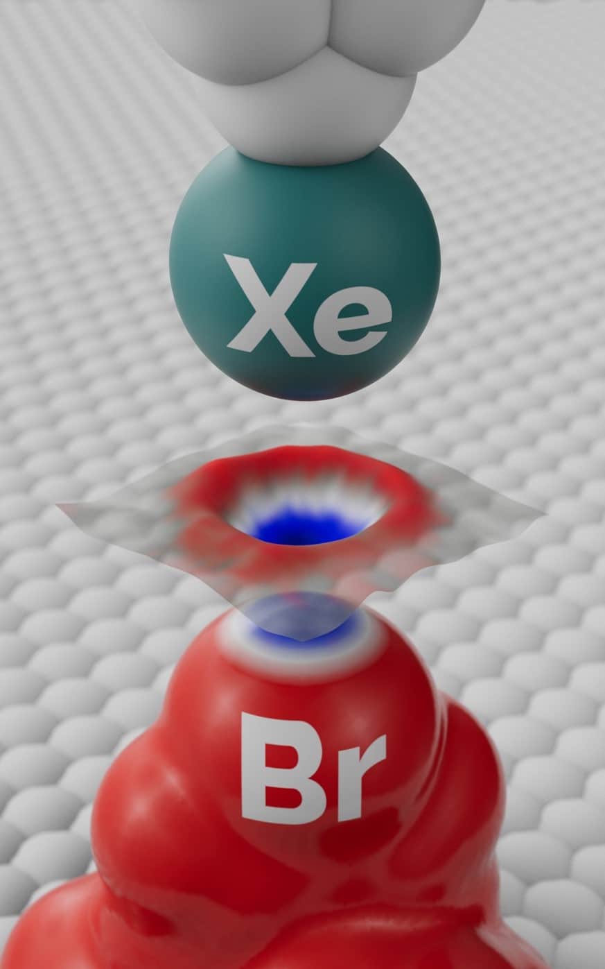
Positive crown: Schematic view of the experiment used to visualize the sigma-hole on a bromine (Br) atom in a molecule using a scanning microscope tip functionalized with a single xenon (Xe) atom. (Courtesy: FZU/DRAWetc)
Scientists have long suspected that bonds between certain negatively charged halogen atoms are made possible by regions of positive charge called sigma-holes, but they lacked experimental proof. Now researchers in Czechia have used a method known as Kelvin probe force microscopy to image these subatomic structures directly – an achievement they claim will lead to a better understanding of molecular crystals and the folding of biomolecules, among other phenomena.
The stability of certain molecular crystals had long been a puzzle to scientists, since the crystals contain pairs of negatively charged atoms that would ordinarily be expected to repel one another. These pairs consist of either two atoms from the halogen group of elements (such as bromine), or one halogen atom and another electronegative atom such as oxygen or nitrogen.
Several groups have proposed that halogen bonds come about through an anisotropy in the atoms’ charge distribution. In other words, rather than having a spatially uniform negative charge, they consist of a belt of negative charge topped off by a positively charged crown – the sigma-hole. However, while quantum-mechanical simulations and observations of crystal structures provided indirect support for this idea, no-one to date was able to image the anisotropy directly.
Kelvin probe force microscopy
A group headed by Pavel Jelínek of the Czech Academy of Sciences in Prague and Palacký University in Olomouc has now achieved this feat by exploiting Kelvin probe force microscopy. Based on work originally carried out by William Thomson (Lord Kelvin) at the end of the 19th century, and adapted more recently to image intramolecular charge distribution, the technique involves suspending a tiny cantilever over a sample and electronically connecting the two so that they form a capacitor. The next step is to set the cantilever vibrating and record how its frequency of vibration shifts as it is brought close to the sample. This shift is measured over a range of voltages and the resulting distribution plotted as a “Kelvin parabola” with a peak at a particular value. This value represents the difference between the work function – a macroscopic quantity describing how much energy is needed to remove an electron from a surface – of the cantilever tip and that of the sample.
Scientists had previously used a Kelvin probe force microscope with a tip comprising a single atom to map the atomic-scale local variation in charge density by measuring shifts in the parabola’s peak. Jelínek and colleagues have now refined the theoretical understanding of this technique and optimized the experimental procedure. This allowed them to enhance the sensitivity of the electrostatic interaction between probe and sample, and thereby push the technique’s resolution beyond the atomic scale.
The researchers carried out their experiment by depositing molecules of a bromine-containing compound (tetrakis(4-bromophenyl) methane) on a silver surface inside an ultrahigh vacuum at cryogenic temperatures. Each molecule had a tripod-like shape, with the bromine atom uppermost and so easily probed. At the cantilever tip the researchers placed a xenon atom, which has a uniform charge distribution and therefore avoids confounding signals. By moving the probe through a grid of points above the bromine atom and plotting a separate Kelvin parabola at each point, they were able to map regions of higher and lower electron density within the halogen atom.
In doing so, the researchers found that the bromine atoms do indeed contain a sigma-hole surrounded by a more negative bulk. They backed up their result with simulations based on density functional theory, and also carried out analogous experimental measurements involving the same molecules but with fluorine replacing the bromine. Although fluorine is also a halogen, it attracts electrons so strongly when forming chemical bonds (it is highly electronegative) that a sigma-hole cannot develop. The team’s measurements revealed, as expected, that the fluorine atoms have a uniform negative charge distribution.
Direct imaging of anisotropic atomic charge
According to Jelínek, the findings not only confirm the existence of the sigma-hole and the concept of halogen bonds but also constitute the first direct imaging of anisotropic atomic charge. “The resolution of the sigma-hole opens up a new way to characterize the electron density of single atoms,” he says. “We can now think about measuring the electron cloud’s response to an external field.”
Among the systems that could be probed in more detail, he adds, are atomic defects in 2D materials. Kelvin probe force microscopy, he says, could be used to establish whether a defect is positively or negatively charged and whether the charge distribution is asymmetric or not.
The open hardware movement advocates the sharing of designs for material objects. For the global science community it means people can access instructions to 3D print increasingly sophisticated tools. Just as importantly, the movement is decentralizing knowledge and giving users the ability to customize scientific equipment then repair it when things go wrong.
In the latest episode of Physics World Stories, Andrew Glester meets researchers at the University of Bath who are part of the open science community.
First, social scientist Julieta Arancio discusses the open hardware movement’s origins and some impactful projects. Among them are: Open Science with Drones; GORGAS tracker for Malaria and Human Mobility in the Peruvian Amazon; and Mboa Lab, a makerspace community in Cameroon.
Later, Richard Bowman and Julian Stirling describe the journey of developing a low-cost, laboratory-grade microscope. The OpenFlexure project, developed with the University of Cambridge and partners in Tanzania, can become an important tool in the fight against malaria.
Wearable robotic systems have great potential for assisting locomotion during clinical rehabilitation, as well as use in recreation and to ease demanding occupational tasks. Walking patterns, however, vary according to a person’s age, height and physiology, may be affected by neural or muscular disorders, and change in different environments. As such, there’s a need for wearable robotics that can customize walking assistance to each user and task.
To address this need, researchers at Harvard University have developed a novel robotic ankle exosuit that uses ultrasound measurements recorded during walking to tune the level of assistance to an individual’s own muscle dynamics and walking task. The team – from Robert Howe’s Harvard Biorobotics Laboratory and the Harvard Biodesign Lab run by Conor Walsh – describes this new muscle-based assistance (MBA) strategy in Science Robotics.
The researchers predict that such personalized assistance should improve exosuit performance and support the adoption of wearable robotics in real-world, dynamic locomotor tasks. “By measuring the muscle directly, we can work more intuitively with the person using the exosuit,” explains co-first author Sangjun Lee in a press statement. “With this approach, the exosuit isn’t overpowering the wearer, it’s working co-operatively with them.”
Personalized assistance profiles
The researchers tested their MBA strategy on nine healthy adults. To calibrate the exosuit assistance level, they first recorded the baseline dynamics of each individual’s soleus muscle (one of the large muscles in the calf) while they walked on a treadmill (without wearing a device) at multiple speeds as well as at a 10% incline.
A portable ultrasound system strapped to participants’ calves measured the muscle dynamics as they walked, capturing continuous B-mode ultrasound images of the soleus muscle. For each participant and task, the researchers used these images to estimate the force produced by the muscle, with the system only needing a few seconds of walking to determine the muscle profile. They then designed the exosuit’s assistance profile to be proportional to that estimated force, generating individualized, task-specific MBA profiles.
After a training session, participants performed walking tasks with the ankle exosuit applying assistive force according to their individual MBA profiles. Compared with not wearing a device, the bilateral ankle exosuit reduced users’ metabolic expenditure by an average of 15.9%, 9.7% and 8.9%, for level walking at 1.25, 1.5 and 1.75 m/s, respectively, and by 7.8% when walking at 1.25 m/s on the incline.
The team notes that these metabolic energy benefits were achieved while applying less assistive force than previously reported assistance strategies. This ability to provide assistance using relatively low forces has implications for future exosuit design, enabling the use of smaller and lighter power sources and actuators, which further decreases loading while increasing the user’s comfort.
Finally, the researchers performed a proof-of-concept real-world demonstration of online adaptive assistance by a mobile ankle exosuit. When tested in variable-speed, outdoor walking situations, the exosuit could quickly adapt to changes in walking speed and incline. In future work, the team aims to improve estimation methods and closed-loop controllers to enable real-time dynamic control for real-world tasks.