A wave-like property previously only seen in beams of light and electrons has been observed for the first time in atoms and molecules. By passing beams of helium and neon through a grid of specially shaped nanoslits, researchers led by Edvardas Narevicius of Israel’s Weizmann Institute of Science succeeded in giving the beams a non-zero orbital angular momentum (OAM). The resulting structures are known vortex beams, and they could be used for fundamental physics studies such as probing the internal structure of protons.
Many natural systems contain vortices – think of tornadoes and ocean eddies on Earth, the red spot on Jupiter and gravitational vortices around black holes. On all scales, such vortices are characterized by the circulation of a flux around an axis. In the quantum world, these swirling structures are found in ensembles of particles that can be described by a wavefunction, including superfluids and Bose-Einstein condensates.
Twisted wavefronts
When the phase of this wavefunction, or wavefront, varies in a corkscrew fashion around an axis as a beam propagates through space, the wavefront is described as being twisted, and the beam carries OAM. Since elementary particles also carry OAM, researchers have, in recent years, succeeded in generating OAM-bearing vortex beams with laser photons and even electrons. Such beams have led to impressive advances in a range of fields, including optical imaging, optical and electron microscopy, communication and quantum optics.
Until now, however, no vortex beams had ever been created from non-elementary particles. One reason for this is that composite particles are heavier than electrons, meaning that the length scale at which their wave-like nature starts to become evident – their de Broglie wavelength – is smaller. This is a challenge because to generate wave-like beams, particles need to be sent through diffraction gratings with slit dimensions that correspond to their de Broglie wavelength. For atoms and molecules, this wavelength is typically on the order of nanometres – too small for slits created using conventional micromachining techniques.
Transmission gratings with very small periods
Recent advances in nanotechnology have, however, made it possible to fabricate transmission gratings with spacings, or periods, as small as tens of nanometres. These nanoscale gratings can be used to diffract matter waves, and that is what Narevicius and colleagues did.
In a study published in Science, the team describe creating vortex beams by passing a supersonic gas of helium atoms through specially nanofabricated diffraction gratings that contain structures known as fork dislocations. These dislocations induce a circular phase in the diffracted beam, and the researchers confirmed that this was the case using a detector placed behind the gratings.
Like classical vortices and photon vortex beams, the newly-created atom vortex beams show up as a row of doughnut-shaped rings, and the phase of their wavefunction revolves around the axis of the dislocation. Each “doughnut” observed corresponds to a beam with a different OAM.
The researchers repeated their experiment with neon atoms and helium dimers, and they say that the technique could also be applied to other atomic and molecular gases. One particularly exciting possibility, they suggest, would be to make vortex beams with protons and use them to probe the internal structure of this subatomic particle.
Edward Mausolf and Erik Johnstone, partners at Innovative Fuel Solutions LLC, frame their vision as one of necessity. Ten years ago, they were in graduate school becoming technetium chemistry experts, and as they studied how isotopes of technetium could be produced and separated, they realized that there were issues inherent in the technetium supply chain that the industry would continue to face unless substantial changes were made.
Technetium-99m (Tc-99m), the single most-used isotope for nuclear diagnostic imaging procedures, for example, is typically produced from molybdenum-99 (Mo-99) using nuclear reactors. Scientists can’t produce a stockpile of Mo-99/Tc-99m due its relatively short physical half-life (the amount of time it takes an isotope to decay to half of its initial activity); instead, they must produce and distribute it continuously. Other issues, such as ageing nuclear reactor infrastructure and the increasing demand for radioisotopes, have contributed to several major interruptions in the Mo-99/Tc-99m supply chain over the years.
Simply put: there’s a need for new methods that can produce radioisotopes of technetium more efficiently and with less waste.
Mausolf and Johnstone have since devised a method that relies on deuterium–deuterium neutron generators to produce radioisotopes of technetium. Their approach could solve supply chain and infrastructure issues by safely bringing the production of Tc-99m and the previously underutilized radioisotope Tc-101 closer to a patient’s bedside, while also generating less waste and using fewer resources. Results of their initial experiments, reported in the journal Pharmaceuticals, also suggest that Tc-101 – often considered to be an impurity – could be useful in medical imaging and therapy applications.
Producing technetium isotopes with a neutron generator
“We realized that there might be some out-of-the-box thinking to find ways to increase yield. Instead of having a larger pile, use different isotopic ratios of technetium, to be able to make up the difference of the inefficiencies of the current supply chain,” says Mausolf. “And a neutron generator makes sense because you don’t have long-lived fission waste.”
Deuterium–deuterium neutron generators create neutron radiation by colliding isotopes of hydrogen called deuterium. When two deuterium atoms collide in a fusion reaction, they create free neutrons that can collide with a molybdenum target to produce isotopes of technetium. When neutrons bombard a molybdenum target, Tc-101 is produced alongside Tc-99m. And Johnstone and Mausolf want to put Tc-101 to work. Though its half-life is only 14.22 min, Tc-101 produces beta rays (487 keV, 90.3%) and gamma rays (307 keV, 89.4%) when it decays. These could be harnessed for both therapeutic and imaging applications.
“We believe that small-scale radioactive isotope production close to the point of use could be the way forward because it avoids the need to ship fast-decaying products with half-lives measured in hours across the country or even internationally,” Johnstone adds. “Another key feature of our research was pinpointing the production or isolation of Tc-101, which we see as a shorter-lived isotope of technetium that’s not discussed in the literature.”
The scientists say that Tc-101’s short physical half-life should not deter others from considering its medical applications because other isotopes with even shorter half-lives, such as oxygen-15, which has a half-life of just over 2 min, are widely used.
Instead, Mausolf and Johnstone say that the biggest challenge they faced was optimizing the production of Tc-101 from neutrons produced using a deuterium–deuterium neutron generator. Compared with deuterium–tritium neutron generators (which are more powerful, but require increased shielding and regulatory burden due to tritium’s radioactivity), a deuterium–deuterium neutron generator often produces fewer neutrons that also have lower energies, which reduces the amount of a radioisotope that can be produced.
With the goal of increasing Tc-101 yield, the scientists partnered with David Williams at Adelphi Technology. Together, they devised and coupled an optimized separation process to Adelphi’s deuterium–deuterium neutron generators, which produce neutron yields exceeding 10 billion neutrons per second. Neutrons produced in the generator then hit molybdenum, which in turn decays to Tc-99m and Tc-101. The resulting material is contacted with activated carbon to isolate the technetium isotopes from the remaining bulk, low specific activity molybdenum.
With the optimized system, “you always have some equilibrium period in production,” says Williams. “You have a half-life that’s not a half-life because it’s dominated by that of the parent [molybdenum].”
Additional considerations
The scientists evaluated the yields of Tc-99m and Tc-101 produced with this method and compared yields to those from a deuterium–tritium system as well as natural and enriched molybdenum targets. And though their purpose was not to pinpoint any specific applications of Tc-101, they also considered the impact of producing the isotope from environmental, financial, regulatory and clinical perspectives.
Natalia Mayordomo, a scientist from the HZDR Institute of Resource Ecology who consulted on the study, notes that because of the short physical and biological half-life of Tc-101 and its stable daughter product, ruthenium-101, the amount of radioactive material entering the environment through human waste will be less than that from radioisotopes with longer-lived ground states or decay products.
In addition, says Williams, neutron generator targets have low specific activity, there are no fission products associated with radioisotope production, and shielding requirements wouldn’t be much different to shielding PET isotopes like fluorine-18.
The team’s next steps include raising funding for a pilot production line and scaling up production so that important studies in mice and testing by regulatory agencies such as the US Food and Drug Administration can begin.
Producing isotopes of technetium using a deuterium–deuterium neutron generator and considering new uses for “impurities” like Tc-101 “present a large dynamic change to the argument of how to make technetium work for the medical community,” says Mausolf. “I think it’s got a lot of traction, I think there’s a lot of runway, and I think there’s a lot more to come.”
A new phase-change memory device that uses much less energy than its predecessors could help meet the world’s increasing demand for digital information storage. The device, which was developed by researchers at Stanford University in the US, is made from a so-called superlattice material placed on a flexible/bendable substrate, and it boasts a switching current density of just 0.1 MA/cm2 – making it 100 times more efficient than other memory types of its kind.
The volume of digital information produced worldwide is currently doubling every two years. By 2025, it could reach a staggering 160 zettabytes (a zettabyte is 1021 bytes, or 1 trillion gigabytes). Phase change memories (PCMs), which rely on the ability of certain materials to switch between a crystalline state that conducts electricity well and an amorphous state that does not, could help address this demand. However, switching between the “1” and “0” states in a PCM tends to require relatively high currents, as the material must be heated before it can change phase. This is true regardless of whether the PCM is placed on a rigid substrate such as silicon or a flexible one such as plastic.
Ultralow programming current density
In the latest work, a team led by Eric Pop succeeded in creating a PCM with an ultralow switching current density. The new PCM’s low power use makes it especially attractive for applications related to the Internet of Things (IoT) and mobile devices, both of which often rely on batteries or energy-harvesting systems.
The researchers made their memory devices directly on a flexible polyimide substrate. The phase change material they used was a superlattice made of alternating thin layers of antimony telluride and germanium telluride. They confined this material stack in a “pore” surrounded by insulating aluminium oxide. They then contacted the top and bottom of the structure with titanium nitride metal electrodes.
More energy-efficient on a flexible substrate
As well as its ultralow current density, Pop and colleagues note that their PCM also has another advantage: each step of its fabrication takes place at temperatures below 200 °C, making it compatible with a variety of flexible plastic substrates. This is important because without a good insulating layer, the electrical pulses used to switch the PCM could cause other electronic components in a device to heat up and become less efficient.
“The superlattice material combined with the confined pore-cell design play a key role in this aspect,” Pop explains, “but we found that the very low thermal conductivity of the flexible substrate also helps. In other words, the memory becomes more energy-efficient on a flexible substrate as compared to a conventional silicon substrate.”
Smart applications
The new PCM might be used in any flexible electronic device that requires low-power, non-volatile data storage, Pop tells Physics World. One example is smart sensors for the IoT, which process and store data locally before sending it to the cloud. Such devices could be useful for environmental monitoring, food packaging analysis and electronic skin for robotics as well as biomedical sensing with on-skin or implantable devices. This type of PCM could also be useful in more general-purpose flexible processors, such as those demonstrated recently by ARM and PragmatIC, or for in-memory computing on flexible substrates.
The Stanford team is now exploring other superlattice materials and new designs to further improve the energy-efficiency and thermal stability of their flexible PCM. They say they might achieve this by optimizing the sidewalls of the structure and by reducing the diameters of the pore-like memory cells. “We are also looking into superlattice materials with an even lower thermal conductivity and higher thermal stability to improve the energy-efficiency of our memories,” Pop says.
Five sites have been shortlisted as a potential home of the UK’s prototype fusion energy plant. Known as the Spherical Tokamak for Energy Production (STEP), it is intended to be a working fusion reactor and have many of the features of a fully operational power station when operational in the 2040s. The five potential sites, announced today, include one in Scotland and four in England, with a final decision on the plant’s location to be made by the end of 2022.
In 2019 the UK government announced £222m towards a conceptual design report for a fusion power plant based on the spherical tokamak design. This would include prototyping components, carrying out materials research and robotics development, as well as computer modelling. The design effort will involve over 300 people and be complete in 2024. Despite the impact of the COVID-19 pandemic, the design work remains on track with the aim that the plant is fully operational by the 2040s.
After an open call for site proposals between December 2020 and March 2021, 15 sites were assessed. Following that process, five have now been picked to be studied further. They include four sites in England – Goole in East Riding of Yorkshire, Moorside in Cumbria, Ratcliffe-on-Soar in Nottinghamshire and Severn Edge in South Gloucestershire – while the fifth potential site is in Scotland in Ardeer, North Ayrshire.
“The shortlist of sites is a significant step for the programme as it helps bring this challenging, long-term endeavour to life [and] also increases our focus as we push on with design and delivery of what we hope is the world’s first fusion power plant prototype,” says Paul Methven, STEP programme director at UKAEA. “Through the next phase of assessment, we look forward to working with the shortlisted sites and local communities to gain a more in-depth understanding of the socio-economic, commercial and technical conditions associated with each site.”
A significant STEP
Earlier this year, researchers were buoyed by the first results on MAST-U. The spherical tokamak used a novel divertor that will likely be employed on STEP in which the new configuration led to a 10-fold reduction in waste heat load on the reactor walls.
The divertor is the heat exhaust system in the tokamak and if the results from MAST-U can be extrapolated to working fusion reactors, then exhaust material and other components would not need to be regularly changed – making such reactors more cost effective by allowing them to keep operational for longer.
An electroretinogram (ERG) is a standard clinical method for measuring the function of the human retina. This procedure typically uses either a contact lens electrode or a fibre electrode to record retinal activity, both of which require physical contact with the eye, and therefore cause discomfort for the patient.
Researchers at Aarhus University in Denmark, led by Britta Westner and Sarang Dalal, have tested a potential replacement for these uncomfortable electrodes by using optically pumped magnetometers (OPMs), publishing their results in NeuroImage. OPMs are quantum sensors that detect magnetic fields that are over one billion times smaller than the earth’s magnetic field. Recent advances in quantum technology have enabled the design and commercial production of lightweight and flexible OPM sensors, such as those being incorporated into magnetoencephalography scanners.
While retinal response is typically detected via electrodes, which measure the electrical activity from the eye surface, the OPM sensors instead record the corresponding magnetic field induced by this electrical activity. The resulting magnetoretinograms circumvent the need for direct contact with the patient’s eye.
At Aarhus University, the researchers placed multiple OPMs close to a participant’s eye while performing routine retinal measurements, using a fibre electrode and a flashing light stimulation. During such a diagnostic test, a flash of light stimulates a negative potential in the retina, the “a-wave”, followed by a positive wave, the “b-wave”, which occurs at known times following the stimulus.
Close match: The OPM sensors measure the signature a- and b-waves of healthy retinal activity without any physical contact with the eye. Here, we see the ERG signal and OPM MRG signals overlaid. (Courtesy: CC BY 4.0/NeuroImage 10.1016/j.neuroimage.2021.118528)
These features were clearly visible in the data recorded by the OPMs and subsequent comparison to the fibre electrode measurements showed a close match. To ensure the OPMs were not simply detecting signals from the electrode, the scientists repeated the experiment without the electrode on the eye and found very similar results.
Interestingly, artefacts due to eye blinks were reduced without the fibre electrode, as the OPMs offered a more comfortable scanning environment. However, the OPMs still suffered from a lower signal-to-noise ratio than the fibre electrode, likely due to a noisy magnetic environment.
As the magnetic fields originating from the retina are much smaller than the residual environmental magnetic field, the researchers operated the OPMs in a magnetically shielded room, to screen out some of this magnetic noise. They note that additional cost-effective active magnetic shielding could reduce this interference, resulting in a comparable signal-to-noise ratio to the ERG.
The team concludes that OPMs have the potential to replace fibre electrodes and contact lens electrodes to provide a contactless and comfortable clinical retinal scanning system. Furthermore, the flexibility of these magnetic field sensors will benefit neuroscientific research into human vision and the pathology of vision impairment.
Signatures of ionized calcium in the upper atmosphere of an ultra-hot, Jupiter-like exoplanet have been found by international team of astronomers led by Emily Deibert at the University of Toronto. The researchers say that the ions could have only formed if the upper atmosphere of the exoplanet WASP-76b is either far hotter or far windier than previously thought. The discovery is the first result of the Exoplanets with Gemini Spectroscopy (ExoGemS) survey. Further insights into the origins of the calcium ions could soon be gained through future observations of the atmospheres of other exoplanets – which are planets orbiting stars other than the Sun.
First discovered in 2016, WASP-76b has about the same mass as Jupiter and has a radius almost twice as large. It completes a full orbit of its star in just 1.8 days and this stellar transit is visible from Earth, making WASP-76b an ideal candidate for atmospheric analysis. Previous studies have revealed that the exoplanet has one of the most exotic atmospheres known to astronomers.
Through several years of close observation, astronomers now widely agree that the planet is tidally locked with its star: leaving one side in permanent daytime, with temperatures reaching 2400 °C. Such an environment would vaporize iron, which would then condense in the upper atmosphere and fall as liquid rain. Meanwhile, the other side exists in permanent night, at temperatures of roughly 1300 °C.
Now, WASP-76b has become the first subject of the ExoGemS survey, which uses the Gemini North Telescope on Mauna Kea in Hawaii. ExoGemS aims to probe the atmospheres of over 40 exoplanets, spanning a wide range of masses and temperatures.
Triplet of peaks
As they analysed the absorption spectrum of light from WASP-76b, Deibert and colleagues found the signatures of several different ionized elements in the exoplanet’s atmosphere: including sodium, lithium, and potassium. The most interesting result was a particular triplet of absorption peaks, which suggests an abundance of ionized calcium in WASP-76b’s upper atmosphere. Intriguingly, these ions can only form in very extreme planetary environments.
Through their analysis, the astronomers narrowed down the possible origins of these ions to two possible causes: either temperatures in the exoplanet’s upper atmosphere are far higher than astronomers previously thought; or the atmosphere harbours extremely strong winds. To gain further insights into these possible origins, Deibert and colleagues will now search for similar signatures of ionized calcium on other exoplanets that will be studied by the ExoGemS survey.
By analysing a large sample of alien worlds, the astronomers aim to learn far more about the chemical compositions of their atmospheres. Other factors including wind and rotation patterns; cloud formation mechanisms; and the rates at which their atmospheric gases escape will also be analysed. By studying these features on planets with widely varying masses and temperatures, the team hopes to gain a better understanding of how exotic planetary atmospheres form and evolve.
Radiotherapy plays an essential role in the management of cancer, with roughly half of all cancer patients receiving radiation as part of their treatment. The majority of such treatments are delivered using external beams of X-rays, targeted at the tumour to damage or kill cancerous cells. Another approach is particle therapy, in which tumours are irradiated with beams of protons or carbon ions. While less prevalent than photon-based radiotherapy, the number of proton-therapy centres worldwide has increased at pace in recent years.
The technologies used to deliver photon-based treatments have progressed a great deal over the last few decades, evolving from 3D conformal radiotherapy to intensity-modulated radiotherapy (IMRT) to volumetric modulated arc therapy (VMAT), in which the linear accelerator delivering the therapeutic X-rays rotates around the patient during treatment (see box below). Each of these steps helped to improve dose coverage of the tumour being targeted, while reducing unwanted radiation being delivered to normal tissues within the body.
The last few decades have also seen proton therapy transition from research laboratories into the clinical setting. During this time, the dose delivery technique has progressed from passive scattering to intensity-modulated proton therapy (IMPT) using scanned proton pencil beams. So will protons follow the lead of photons and introduce rotational delivery – and is proton arc therapy (PAT) the next logical step?
That was the question under consideration at the recent ESTRO 2021 congress, where a dedicated conference session examined the latest developments in PAT and the technique’s potential to improve proton therapy for cancer patients.
Radiation-therapy techniques
Three-dimensional conformal radiotherapy, 3D CRT
3D CRT is a photon-based treatment that uses 3D medical images to precisely define the tumour target. The radiation dose is then shaped to match the shape of the tumour by delivering X-ray beams from many directions.
Intensity-modulated radiotherapy, IMRT
IMRT is a type of conformal radiotherapy in which not only the shape, but also the intensity profile, of each treatment beam is varied to precisely target the tumour.
Volumetric modulated arc therapy, VMAT
With VMAT, the linear accelerator that delivers the radiation beam rotates around the patient during treatment. The shape and intensity of the X-ray beam are continuously controlled as it moves around the body.
Stereotactic body radiotherapy, SBRT
Stereotactic radiotherapy uses high radiation doses to treat tumours in the brain and central nervous system in one or just a few treatments. SBRT is similar but refers to treatment of tumours elsewhere in the body.
Intensity-modulated proton therapy, IMPT
IMPT is a proton therapy technique in which scanned proton pencil beams of variable energy and intensity are used to precisely paint the radiation dose onto a tumour.
Proton arc therapy, PAT
With PAT, the proton beams are delivered continuously as the gantry rotates around the patient. During this rotation, the beam energy and intensity are adjusted to match the dose to the target volume.
The promise of PAT
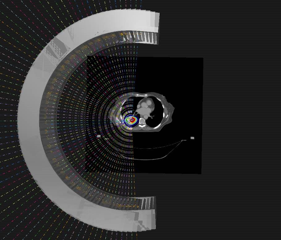
When it comes to PAT, protons are delivered continuously as the gantry – the large circular structure containing the equipment that delivers the protons to the patient – rotates around the patient. First described in the literature back in 1997, the thinking was that PAT could improve dose distribution, increase treatment robustness and reduce delivery time compared with IMPT. Unfortunately, at that time, only 16 proton-therapy centres were in operation, and only one with a gantry. As such, PAT appeared too technically challenging to continue and was soon dropped.
Now, however, there are more than 100 proton-therapy centres worldwide, most with a gantry and using pencil-beam scanning. “The potential for integrating PAT into the clinic is huge right now,” said Laura Toussaint from the Danish Centre for Particle Therapy. Toussaint shared the results of several published treatment-planning studies comparing PAT with other proton- and photon-based treatments. The first example presented a whole-brain treatment, which aimed to avoid irradiating the hippocampus during brain radiotherapy, to spare the patient’s memory function (Acta Oncol.58 483). While VMAT, IMPT and PAT all delivered equivalent target dose distributions, PAT reduced the dose to the hippocampus, as well as to the cochlea.
Toussaint also presented a study of head-and-neck treatments, where robustness can be a particular challenge (Radiat. Oncol.15 21). Here, PAT offered improved target dose conformity and better sparing of nearby vital organs, known as organs-at-risk (OARs), compared with IMPT. The PAT plan was also more robust to changes in patient set-up and anatomy.
She also described a comparison of lung stereotactic body radiotherapy (SBRT) delivered via PAT, IMPT and VMAT. Again, PAT delivered the lowest dose to OARs, and reduced the integral dose. PAT could also mitigate interplay effects better than IMPT. Studies of brain, breast, prostate and spine cases all reported similar findings.
“Overall, from these treatment planning studies, when comparing proton arc to IMPT, we saw no strong evidence of improved target dose conformity or homogeneity,” Toussaint explained. “However, there was a large reduction in the body integral dose and consistently better OAR sparing. There was also potential for improved plan robustness by mitigating range uncertainty and interplay effects.”
PAT could also reduce treatment delivery time, Toussaint explained, citing a publication showing that PAT was 50% faster than VMAT for a spine SBRT case. She noted that the field is moving quickly, and has already seen a paper published on spot-scanning hadron arc therapy using carbon and helium ions (Advances Radiat. Oncol.6 100661).
“Are the demonstrated gains in physical dose and the estimated clinical benefits enough to motivate the technological and engineering efforts needed for implementation of proton arc in clinical practice?” Toussaint asked. “This is up for debate.”
Creating a PAT prototype
Xuanfeng Ding from William Beaumont Hospital in Michigan certainly thinks that PAT offers plenty of clinical potential, and is part of a team working on an ongoing project to create a clinical PAT system.
Currently, there are three main challenges when it comes to proton therapy: range and set-up uncertainties; large spot size; and prolonged treatment times. These can be addressed using approaches such as robust plan optimization, dynamic beam collimation and increased dose delivery efficiency, respectively. “But PAT can address these three together,” said Ding.
PAT delivery is complex, however, with treatment plans comprising numerous energy layers (as the energy of a proton beam defines the precise depth at which it deposits a dose within the target). Ten years ago, the energy-layer switching time of a first-generation pencil-beam scanning systems was up to 6 s, making PAT delivery significantly lengthier than IMPT and limiting its clinical application.
Introducing rotation Proton arc therapy is an advanced cancer-treatment technique in which beams of protons are delivered continuously as the gantry rotates around the patient. (Courtesy: Xuanfeng Ding)
“But things change. The majority of new-generation cyclotron-based scanning systems can reach an energy-layer switching time of 0.5 s, which makes proton arc comparable or even faster than two-field IMPT,” said Ding. “This is good timing to look seriously at how to implement this in a clinical setting.”
Ding described the Beaumont project, which is developing spot-scanning PAT, also known as SPArc. It involves moving the patient couch and gantry, while continuously delivering the proton beam; as well as switching energy layers and scanning the beam during the gantry rotation.
“The goal is to make proton therapy more efficient, more robust and also with better dose conformality. That’s the final clinical endpoints that we’re looking at that will benefit society,” said Ding. “The challenge is it’s a huge gantry, so how can we rotate this gantry precisely while delivering the proton beam with millimetre accuracy?”
The team, now working with Belgian medical technology company IBA, has already demonstrated SPArc delivery on a clinical ProteusOne system (see photo at top of article). The researchers confirmed that during gantry rotation, the central spot maintains a constant size and 1 mm position accuracy, at energies from 70 to 227.7 MeV (Radiother. Oncol.137 130). The collaboration has also developed an iterative treatment planning optimization algorithm, which incorporates machine-specific dose delivery sequences and gantry motion. Indeed, their work established the first dynamic proton arc model that combines mechanical and irradiation sequence models.
Ding also demonstrated that SPArc can be more efficient than IMPT. He compared a SPArc plan with a three-field IMPT plan for a brain tumour case. Both exhibited the same plan quality, but the SPArc treatment took just 4 minutes, while IMPT required 11 minutes. The gain is disease-site dependent, he noted, and treatment delivery time is just one part of the process. But Ding estimates that, when patient set-up time is included, SPArc could provide an average 20% increase in patient throughput for a single-room proton system, and potentially more for a multi-room proton-therapy centre.
Looking ahead, Ding’s group is collaborating with Université catholique de Louvain to develop a linear energy transfer (LET)-optimized SPArc algorithm. The team also plans to investigate hypofractionated treatments, in which the prescribed dose is delivered in one or just a few fractions, as well as investigate ways to mitigate interplay effects between the motion of a tumour and motion of the proton beam.
“I think we’re on the right track to proton arc therapy, eventually clinical users will adopt this technology for a variety of disease sites,” Ding concluded. “More and more vendors and clinical users will play a key effect because different people will invent different innovations for this technology, moving together to implement proton arc into the clinical setting to benefit our patients.”
Biological optimization
In the final talk of the session, Alejandro Carabe from Hampton University Proton Therapy Institute, Virginia, took a look at the third piece of the puzzle. With PAT seemingly technically feasible and dosimetrically advantageous, could it also allow us to control the biological effectiveness of a proton beam and increase the therapeutic index?
The goal of any radiation treatment is to maximize the probability of destroying tumour cells (tumour control probability, TCP), while minimizing the risk of damage to non-cancerous tissue (normal tissue complication probability, NTCP). The therapeutic index represents the ratio between TCP and NTCP, and the higher its value, the more beneficial a radiation treatment.
Looking first at a single photon beam, the high entrance dose will inevitably damage normal tissue more. To reduce NTCP, and thus increase the therapeutic index, rotational therapy can be used to deliver many fields and conform the dose to the target volume. With a single proton beam, however, the entrance dose is already inherently low, which should result in a decreased NTCP with respect to TCP, and a higher therapeutic index than that of the photon beam.
Carabe described a case study comparing VMAT with two- and three-field IMPT plans for oesophageal cancer treatments. While the mean dose to the target was not significantly different for the three approaches, the IMPT plans clearly delivered less mean dose to the OARs.
The study demonstrated that – in addition to a single proton field offering a higher therapeutic index than a single photon field – a low number of proton fields can outperform VMAT, the most conformal photon therapy. With this in mind, is there even any need to incorporate rotation into proton therapy to increase the therapeutic index?
A review of published studies comparing physical dose distributions between PAT and IMPT for lung, brain, head-and-neck and prostate cancer treatments showed no evidence that PAT improved conformity or uniformity compared with IMPT – although it did reduce normal tissue integral dose. “Therefore, we cannot categorically say that PAT significantly improves therapeutic index over IMPT, at least when just looking at dose distribution,” said Carabe.
But, he explained, there’s more to consider than purely dosimetric parameters. Even when a uniform dose is delivered to the target, the linear energy transfer increases towards the distal edge of beam, causing that dose to be more harmful. If proton plans are only optimized for physical dose, not LET, they often place high-LET dose in the normal tissue next to the target. PAT could provide the control to deposit high-LET dose in the target volume.
Carabe and colleagues performed an in vitro experiment to examine whether PAT could be used to control the LET distribution in cultured cells (Phys. Med. Biol. 65 165002). They found that PAT plans generated almost double the LET than three-field IMPT, and killed more cells than IMPT with the same dose. They also analysed a prostate cancer case and found that using PAT to concentrate high LET in the target volume enabled reduction of delivered dose, without reducing the clinical effect.
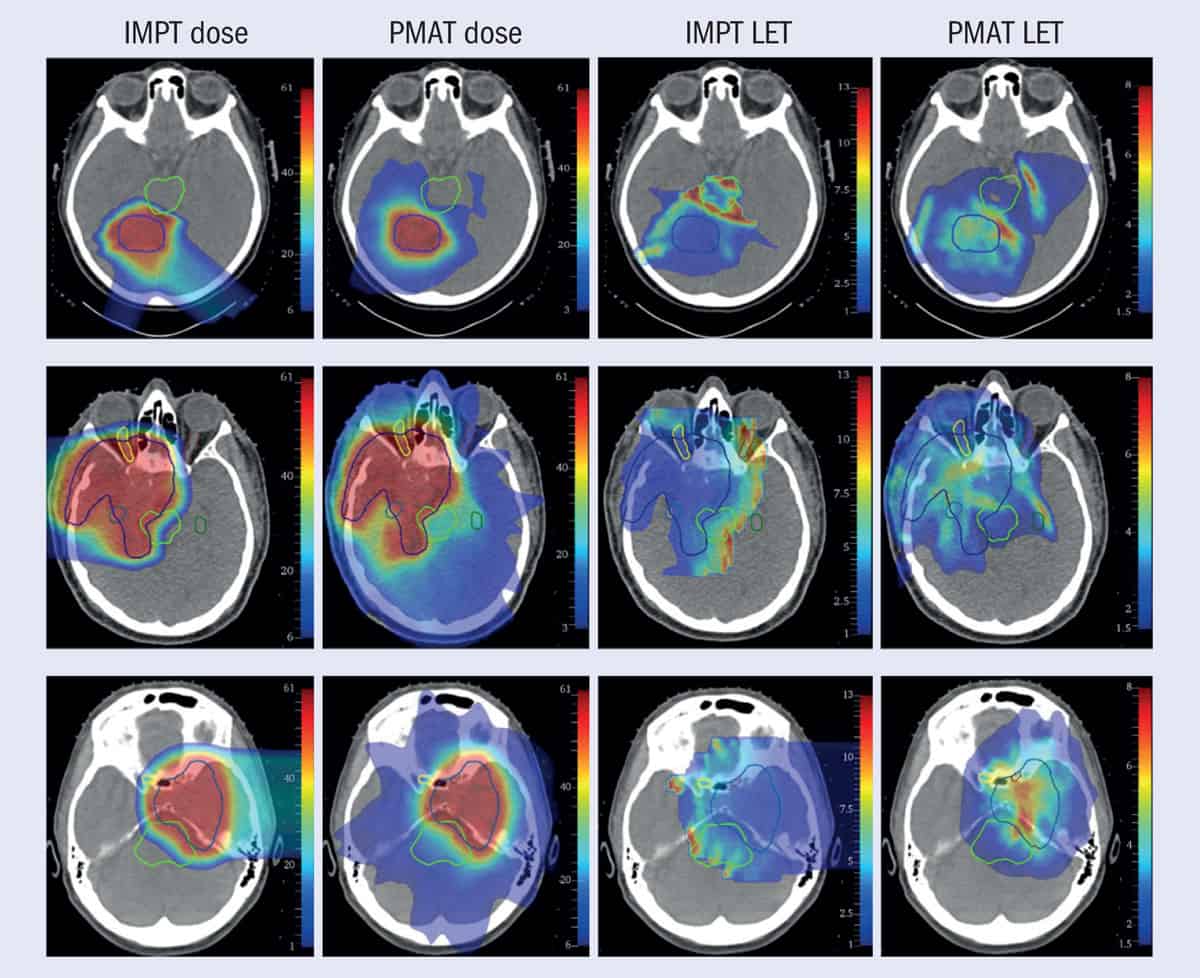
1 LET targeting Dose and linear energy transfer (LET) distributions for intensity-modulated proton therapy (IMPT) and proton monoenergetic arc therapy (PMAT) plans for three brain cancer cases (top to bottom rows). The dark-blue contours depicts the target volume. (Courtesy: A Bertolet and A Carabe Phys. Med. Biol.65 165006/IOP Publishing)
In another piece of research, Carabe compared IMPT with monoenergetic PAT (figure 1) to treat a clinical brain tumour case (Phys. Med. Biol. 65 165006). “With monoenergetic PAT, we were able to avoid critical areas such as the brain stem and produce very conformal plans compared to IMPT plans,” he said, adding that the technique “could concentrate the LET in the area that we wanted and take it away from the areas where it could be very damaging”.
“PAT allows LET painting, which can be utilized to either decrease the risk of radiation-induced toxicity, or increase the biological effectiveness of the treatment, or even reduce the prescribed dose or number of fractions,” said Carabe. “All of this will allow us to have much better control of the effects of a treatment and increase the therapeutic index.”
He emphasized that introduction of this new modality should not just rely on a pure physics argument, but on its biological impact. “The most important thing to remember is that delivery of PAT should not be justified based on increased conformity,” Carabe concluded. “It should be justified based on enhanced biological impact.”
The future of bioelectronics – including wearables, implantable devices and smart technologies – hinges on the ability to sustainably power devices. A number of approaches for converting biomechanical energy into electricity have been introduced, including piezoelectrics and triboelectrics, which function by deriving charge from compressing or contacting materials. Unfortunately, these techniques’ suboptimal electronic properties and vulnerability to ambient humidity limit their effectiveness.
The answer could lie in magnetoelasticity, in which a material’s magnetic properties change under mechanical stress. This effect is usually observed in rigid metal alloys, which have mechanical moduli significantly higher than that of human tissue (they are very stiff). As a result, such materials are unsuitable for biomechanical energy generation. Researchers led by Jun Chen at the University of California, Los Angeles’ Samueli School of Engineering have overcome this difficulty by formulating a new soft magnetoelastic polymer blend. They share their results in Nature Materials.
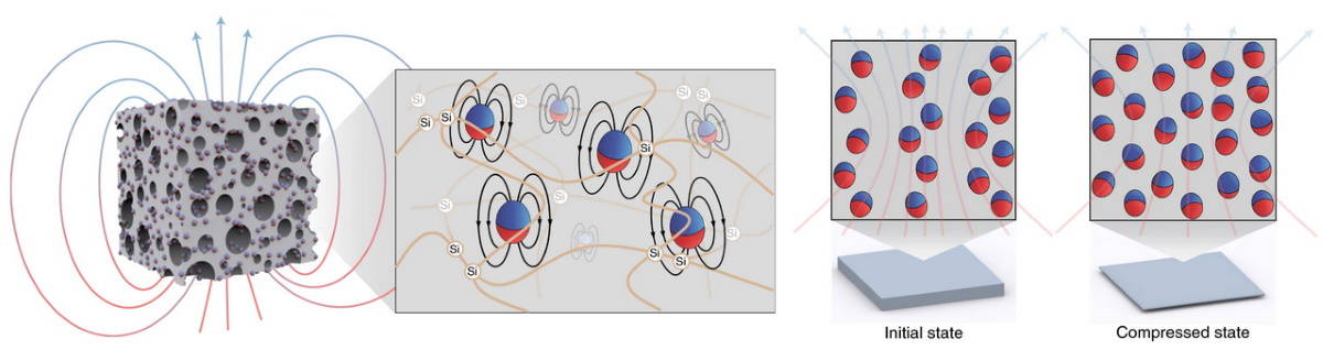
Magnetic control: Micromagnets dispersed in a soft porous system make it magnetoelastic. Compresssing the system shifts the magnets’ positions, changing the overall magnetism. (Courtesy: Nat. Mater. 10.1038/s41563-021-01093-1)
The team created the porous, soft magnetoelastic material by dispersing micromagnets in a silicone polymer matrix. These micromagnets induce the inherent magnetic behaviour of the system so that when the elastomer is compressed, the magnetic field changes. The authors hypothesize that this change in magnetic flux density is caused by a realignment of “chains” of micromagnets under mechanical deformation. They were also able to model this effect theoretically.
In order to harness the energy induced by the magnetoelastic effect, the researchers positioned a magnetic inductor on top of the silicone layer. This inductor converts changes in magnetic fields into an electrical current. Therefore, magnetic deformations can be turned into electrical energy to be used according to the desired application. The authors call this electrically responsive system a “soft magnetoelastic generator”.
Monitoring human health
Many applications for wearable power generation could exploit the electrical coupling provided by a magnetic inductor. For example, with gentle hand tapping, the authors were able to charge capacitors of varying size within seconds. Another magnetoelastic generator was able to drive a commercial wearable thermometer to monitor body temperature.
In addition to powering wearables, the generator could also be used to sustainably power implanted bioelectronic devices, which remains difficult with current technology. Acoustic waves and ultrasound can be utilized to transfer energy to medical implants through tissue. To demonstrate this effect, the researchers implanted a magnetoelastic generator in porcine tissue and excited the tissue using ultrasound. The generator was able to output a power of over 30 µW, a value comparable to that used by bioelectronics such as pacemakers and neurostimulators.
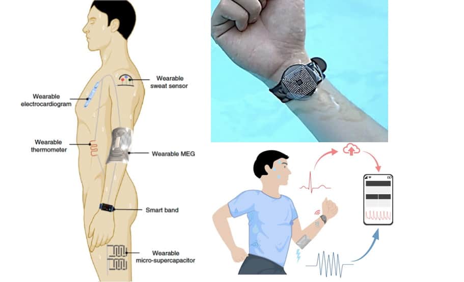
Multiple applications: The magnetoelastic generator can be used to power health monitoring devices, as well as act as a cardiovascular monitor. (Courtesy: Nat. Mater. 10.1038/s41563-021-01093-1)
Lastly, the researchers tested the device’s capacity to act as a cardiovascular monitor. They found that a generator worn on the wrist could detect a human pulse, even when wet. In this scenario, the natural arterial pulse deformed the magnetoelastic generator, inducing a current in the inductor. This sensor was even able to function underwater and through sweat, as the material is inherently waterproof.
Moving forward, Chen’s team aims to further augment the electrical output of the generators by optimizing the device’s design. This work opens a new avenue for practical human-body-centric energy, sensing and therapeutic applications.
A member of NASA’s Astrophysics Advisory Committee has resigned over the agency’s handling of an investigation into whether the James Webb Space Telescope (JWST) should be renamed. The probe was instigated in the wake of concerns that Webb – a former NASA administrator – had been involved in mistreating gay and lesbian people in the 1950s and 1960s. NASA announced in September, however, that it would not be changing the name of the JWST, revealing the news via a single-sentence statement that was released only to certain media outlets.
That decision angered some astronomers particularly because the agency had said it would be fully transparent in releasing the results of the investigation. In response, Lucianne Walkowicz from the Adler Planetarium in Chicago, who is also a co-founder of the JustSpace Alliance, has now announced they are resigning with immediate effect from the committee over its handling of the affair. The JWST is due to be launched in December.
Regarded as a successor to NASA’s Hubble telescope, the JWST was originally known as the Next Generation Space Telescope. In 2002 the then NASA boss Sean O’Keefe renamed the telescope in honour of Webb, who had served as NASA administrator during the Apollo era. A bureaucrat rather than a scientist, Webb had served in various US government roles since the 1940s.
Earlier this year, however, more than 1200 people signed an open letter calling on NASA to rename the JWST, claiming that Webb was involved in anti-LGBT+ activities before taking up the role at NASA. The letter was initiated by Walkowicz as well as Chanda Prescod-Weinstein from the University of New Hampshire, Brian Nord from Fermilab and the University of Chicago as well as Sarah Tuttle from the University of Washington.
“Webb served as the undersecretary of state during the purge of queer people from government service known as the ‘Lavender Scare’,” the authors stated. They added that archival evidence “clearly indicated that Webb was in high-level conversations regarding the creation of this policy and resulting actions”.
The authors also noted that Webb was in charge of NASA when Clifford Norton – a budget analyst at the agency – was sacked in 1963 on suspicion of homosexuality. “We, the future users of NASA’s next-generation space telescope and those who will inherit its legacy, demand that this telescope be given a name worthy of its remarkable discoveries, a name that stands for a future in which we are all free,” the authors wrote.
Lack of transparency
In June, NASA said it would begin an internal investigation, which would examine historical documents and interview historians who had studied Webb. While officials at NASA said the agency would be “transparent” with the decision, on 27 September NASA administrator Bill Nelson issued a single-sentence statement to selected journalists stating: “We have found no evidence at this time that warrants changing the name of the James Webb Space Telescope.”
The news angered astronomers. “What I hear as a queer scientist and a member of multiple NASA collaborations is, ‘The homophobic terror that Clifford L Norton was subjected to doesn’t matter’,” noted Prescod-Weinstein on Twitter. “I find NASA’s single sentence statement about the evidence to be gaslighting, constituted by the sin of omission, and most troublingly, unsupported and thus unscientific. They do not make the case for their claim in light of the publicly available evidence.”
The news also surprised many who sit on NASA advisory committees, who said they only learned of the news from press reports. In an open letter announcing their resignation from the 12-strong committee, Walkowicz criticized NASA’s lack of transparency and called NASA’s response “flippant” and “pathetic”.
“After the past year and a half we’ve had with not only the pandemic, but also national grappling with issues of racism and human rights, it boggles the mind that NASA has so little insight into its own participation in systematic oppression,” Walkowicz writes. “I’m not the first and won’t be the last driven out of a NASA space, where evidently straight people’s opinions are valued and taken more seriously than queer people’s experiences.”
Prescod-Weinstein adds that the logic behind naming the telescope after Webb is that he is responsible for NASA’s successes during the Apollo era. “At the same time, NASA says he is not responsible for the homophobia that occurred at NASA,” says Prescod-Weinstein. “How is he responsible for all of NASA’s successes during his time as administrator but none of its failures? Real people were harmed by those failures. That matters.”