A new universal theory and three basic rules for how defects in semiconductors behave in response to strain could lead to improvements in the electronic properties of a wide range of semiconducting materials. The work, which began with the discovery of a key physical quantity that describes how a semiconductor’s volume changes in the presence of impurities, could help researchers determine the “right” amount of strain to apply, and thus optimize the effects of these impurities.
Defect-induced volume changes
Most materials contain impurities, or dopants, that are either intentionally or unintentionally introduced into the system – for example, during the growth of a crystal. These impurities induce volume changes in their immediate vicinity, thus producing a strain in the material.
In the latest work, researchers led by Bing Huang of Beijing Normal University’s Department of Physics and the Beijing Computational Science Research Center demonstrate that the extent of these volume changes, Δ𝑉, depends on whether the defect is positively- or negatively-charged. More precisely, Huang and colleagues show that volume increases for more negatively-charged defects and decreases for more positively-charged ones; in other words, Δ𝑉 is positive when an electron is added to the defect site and negative when an electron is removed from it.
Three basic rules
To further understand (and therefore predict) the different strain-dependent doping behaviours of semiconductors, Huang and colleagues developed three basic rules to describe how strain affects the properties of semiconductor defects.
The first rule describes how a defect’s formation energy changes in response to strain. In a material under strain, the researchers found that the total energy difference between a neutral semiconductor and a negatively (or positively) charged semiconductor that accommodates extra electrons (or holes) in the material’s valence band will depend on the sign and size of Δ𝑉. This energy difference will either increase or decrease superlinearly as a function of the strain, and the rate at which it does so will be proportional to Δ𝑉. If Δ𝑉 is close to zero, the energy difference will be a parabolic function of strain.
The second rule describes how strain changes the Fermi energy level (a hypothetical energy level that has a 50% probability of containing an electron) when the defect charge state changes. According to this rule, a compressive strain will shift this transition energy level up, while a tensile strain will shift it down.
The third rule describes how strain changes the position of the “pinning” Fermi level. This is an intrinsic effect occurring in semiconductor systems in which the Fermi level is far from the electronic band edge. This effect can significantly limit the doping-induced electron and hole densities, and dramatically degrade the performance of devices like solar cells and transistors. Huang and colleagues’ third rule describes how compressive strain will shift the absolute pinning Fermi level up in energy and a tensile strain will shift it down in energy.
Together, Huang says that these rules could help researchers estimate the “right” strain to apply to a semiconductor to optimize the doping effect of impurities or defects. “We have known for some time that strain can be used to tune doping effects in semiconductors, but a fundamental and general theory to understand the diverse strain-induced changes of different point defects in semiconductors was lacking until now,” he tells Physics World.
A new way of controlling the expansion of matter in a freely falling Bose–Einstein condensate (BEC) has produced the coldest effective temperature ever measured: 38 pK (10–12 K) above absolute zero. The method, which allowed researchers in Germany and France to image the condensate’s evolution for more than two seconds, opens the door to enhanced measurements of the gravitational constant g and photon recoil, and could even offer an alternative means of detecting gravitational waves.
BECs are clusters of particles in the same quantum ground state. Since they were first created experimentally in 1995, they have become a testbed for research on the quantum nature of matter. One example is matter–wave interferometry, which is a type of interferometer that uses the wave character of atoms. When this is done with free-falling BECs, the resulting interference pattern will depend partly on gravitational effects, making such experiments sensitive tests of fundamental physical processes. However, when a BEC is released from the magnetic trap in which it is created, repulsive interactions between its constituent particles quickly get converted into kinetic energy. This causes the BEC to expand until it becomes too dilute to detect via standard absorption imaging, in which a laser beam is sent through the condensate and a camera measures how much light the particles absorbed.
Because the interferometer’s resolution increases with the square of the free-fall time, limiting the condensate’s expansion is crucial. To do this, physicists use magnetic, optical or electrostatic forces to focus the condensate, similar to the way that lenses focus light. These so-called matter–wave lensing methods can cool the BEC’s effective temperature to as low as 50 pK. However, they only affect the temperature along the condensate’s radii, not in the axial direction of its fall. Hence, even with matter–wave lensing, a BEC in free-fall still expands rapidly.
3D matter-wave-lensing
The new method, which is described in Physical Review Letters, addresses this shortcoming by changing the magnetic field used to trap the condensate in a way that causes its shape to oscillate, transforming it from a ball into a thin ellipse. The condensate then enters free-fall when its axial dimension reaches its thinnest point, which keeps its axial expansion rate as low as possible. Magnetic lensing further controls the condensate’s expansion in the radial directions.
To test their technique, researchers led by Ernst Rasel of Leibniz University Hannover used the 110-metre drop tower in Bremen, Germany. The team began by creating a BEC from a cloud of about 100,000 rubidium atoms. These atoms then underwent a 4.74-second free fall, during which absorption images were taken at various points. When the team imaged their free-falling BEC without any matter-wave-lensing techniques, they found that the condensate degraded within 160 ms of release. By applying their technique, they achieved a record-low effective temperature of 38 pK and imaged the BEC for more than 2 seconds. “Our methods enable new experiments or greatly improve existing ones,” says Ernst Rasel, who led the research at IQO.
“A significant step for atom interferometry”
The team say that more complex magnetic lens configurations could reduce some limitations of the current setup, bringing about even tighter control over the BEC’s expansion. Lowering the number of atoms inside the BEC might also lower the expansion rate, reducing the effective temperature to as low as 14 pK. However, it could also reduce the total imaging time as a smaller BEC would more rapidly become too dilute to image.
Florian Schreck, a physicist at the University of Amsterdam in the Netherlands who was not involved in the research, calls the new technique “a significant step for atom interferometry with BECs in situations that allow long free-fall time (e.g. in space or in a drop tower)”. Compared to previous methods, Schreck notes that IQO team’s technique reduces the internal kinetic energy of the gas in all three dimensions, and its simplicity means that it should find wide adoption in rubidium BEC atom interferometry. Translating such a scheme to strontium would, he adds, be “especially interesting”, as strontium is the species of choice for plans to use atom interferometers to detect gravitational waves space.
In the medium term, Rasel and his colleagues plan to demonstrate even longer interferometry times using the Bremen drop tower’s catapult system, after simulations showed that the total imaging time with their technique could be more than 17 seconds. In the future, they also hope to employ their method in the Bose-Einstein Condensate and Cold Atom Laboratory (BECCAL), a planned space-based facility.
A mechanism that causes droplets of boiling water to propel themselves rapidly across hot oil films has been identified by Victor Leon and Kripa Varanasi at the Massachusetts Institute of Technology. The duo used high-speed photography to determine the relationship between the fleeting timescales of bubble formation inside the droplet, and their motions over long timescales.
When water droplet is placed on a hot metal surface it will boil and create a cushion of vapour upon which the droplet will “float”. This well-known phenomenon is called the Leidenfrost effect, and the low friction created by the cushion allows droplets to move at speeds of millimetres per second over hot surfaces.
If instead, the surface is coated in a film of hot oil, it may seem intuitive that a droplet placed on top of it would experience far more friction. Yet when doing just that in a new experiment, Leon and Varanasi observed the exact opposite effect: with droplets moving up to 100 times faster than levitating Leidenfrost droplets. To investigate this strange effect, they used high-speed photography — at 100,000 frames per second — to investigate the mechanisms propelling the droplets.
Trapped bubbles
In their images, the duo identified bubbles of vapour forming at the droplet-oil interface on microsecond timescales, each some distance away from the centre of the droplet. In a Leidenfrost droplet, these bubbles would escape almost immediately – and since they are random, would emerge almost uniformly across the base of the droplet.
In contrast, the oil interface prevents vapour from escaping in this case. As a result, bubbles accumulate inside the droplet, allowing asymmetries to develop over time. Since this vapour is far more insulating than the liquid inside the droplet, it produces thermal disturbances in the oil film. This causes the droplet to vibrate – reducing its friction, while also encouraging additional bubble formation.
Momentum transfer
When the bubbles finally burst through the droplet surface, Leon and Varanasi determined that they transfer momentum to one side of the droplet, propelling it in one direction. Altogether, this process produces a coupling between random, microsecond-scale fluctuations inside a droplet, and its motion over far longer timescales.
When analysing their results using established equations of fluid mechanics, the duo found that their motions were remarkably similar to those of droplets moving across the surfaces of infinitely deep pools of oil – despite the films they used being no more than 100 micron thick. This would greatly reduce the friction experienced by the droplet, allowing it to skim rapidly across the surface.
Through their future research, the duo hopes that the effect could be harnessed to trigger droplet propulsion in controlled directions. If achieved, this could allow for advanced microfluidic devices, which could smartly propel fluids in specific direction. Such devices could include pumps that can operate in microgravity environments; and new breakthroughs in applications such as targeted drug delivery.
Paul Davies has been exploring the esoteric nature of physics in his popular science books since the 1970s. The Arizona State University physicist talks to me about his latest book What’s Eating the Universe and Other Cosmic Questions and also gives some top tips for aspiring science writers.
Tandem solar cells show great promise for boosting the efficiency of solar energy systems. Physics World’s Margaret Harris speaks with Laura Miranda Pérez and Chris Case of UK-based Oxford PV, who explain why combining traditional silicon solar-cell technology with newer perovskite-based devices delivers a significant increase in performance.
Margaret Harris has written an extensive blog about tandem solar cells that explains why the combination of silicon and perovskite technologies offers a way of meeting future targets for boosting global photovoltaic generating capacity.
Paul Davies will be debating the fluid nature of physical laws at the How the Light Gets In festival in London later this month.
A black hole or a neutron star may have merged with a normal massive star and caused it to explode in a supernova, according to Caltech’s Dillon Dong and colleagues. Dong says that such explosions could occur at minimum rate of “one explosion per 10 million years in a galaxy like the Milky Way”.
Many stars are born in pairs, and two stars massive enough to explode as supernovae can be close companions. In such binary systems, the more massive of the two stars will explode first and in most cases a compact object – a neutron star or a black hole – will be left behind.
Astronomers have speculated that if the compact object is close to its surviving companion star, it can spiral into the star’s atmosphere, eventually sinking to the companion’s core where it disrupts the star, causing it to go supernova prematurely. Now, Dong’s team says that it has discovered evidence for such an event in a search of archive data from the Very Large Array Sky Survey (VSS), covering the period between September 2017 and February 2018.
Anomalous sources
The team looked for anomalous but luminous radio sources. They then compared their candidate sources to equivalent observations of the same part of the sky from the Faint Images of the Radio Sky at Twenty Centimetres (FIRST) survey, which was also conducted by the Very Large Array between 1994 and 2005.
One radio source in particular, catalogued as VT 1210+4956, stood out as not being present in the FIRST data. This means that it appeared sometime between 2005 and 2017. Further investigation found that it is situated in a dwarf galaxy about 500 million light–years away.
Following up with the Low Resolution Imaging Spectrometer on the Keck I telescope on Mauna Kea, Hawaii, Dong’s team identified a massive outflow of ionized gas from the source. The Monitor of All Sky X-ray Image (MAXI) experiment on the International Space Station had also detected a soft X-ray burst from the same location on 14 August 2014.
Compact object
With all these observations to hand, Dong’s team were able to piece together what happened. They suggest that long ago, a massive star in a binary system exploded, leaving behind a compact object. When the other star in the system started nearing the latter stages of its life, it ran out of hydrogen in its core and turned to fusing helium instead. This raised the temperature of the star so hydrogen fusion could occur in the star’s envelope, prompting the star to expand. Gradually, the compact object came to be within the star’s atmosphere.
The gravitational tidal interaction between the compact object and the star’s atmosphere saw the star begin to eject huge amounts of material at a rate of 0.04 solar masses per year. This process is estimated to have begun only about 400 years ago, whereas other stars that experience mass loss before going supernova do so for tens of thousands of years before exploding.
This outflow of material formed a dense torus of circumstellar material around the star, which is what Keck observed. Then, in 2014, the compact object reached the star’s core and started ripping it apart from the inside out. An accretion disc of matter around the compact object formed inside the star, and the compact object’s powerful magnetic fields whipped up some of this matter and blasted it away in relativistic jets that emitted the X-ray burst detected by MAXI, and which prompted the star to explode. When the supernova ejecta slammed into circumstellar material, radio emission was released in the form of synchrotron radiation as charged particles in the debris spiralled around magnetic field lines.
As for the star that exploded, if the compact object that merged with it was a neutron star, then “it’s likely it would’ve accreted enough mass to collapse into a black hole,” Dong tells Physics World. “If it was a black hole, it would’ve just become a more massive black hole.”
Type IIn supernovae
Among those astronomers who predicted the existence of such supernovae is Roger Chevalier, of the University of Virginia. In a 2012 paper, he described what such a merger-induced supernova might look like, suggesting that their existence could explain a type IIn supernovae. This poorly understood class of supernova stays brighter for longer and has narrow hydrogen-emission lines. This suggests that the exploding star is surrounded by material ejected by the star that is blocking much of the hydrogen emission, and which is produces a more sustained afterglow as the supernova shock slams into it.
Although Dong does not mention type IIn supernovae by name, Chevalier thinks that VT 1210+4956 looks like a type IIn with its narrow hydrogen line width.
“I think they make a good case,” Chevalier says. “The radio and X-ray [emission] both indicate a central engine. It’s gratifying to see support for a speculative scenario that aimed to explain why the dense mass loss and supernova occurred at about the same time.”
Now that astronomers know that such supernovae occur, the race is on to find one in the process of exploding. “Much remains unknown because we don’t have early time optical and infrared spectra of the explosion,” says Dong. His next steps will include reassessing the rate at which these supernovae occur. If other, more normal looking supernovae can also be triggered by this mechanism, he speculates that they could be much more common than we realize.
Late in 2020, scientists in Germany and Lithuania announced a new milestone in so-called “tandem” solar cells – that is, cells made from two different types of photovoltaic material. Writing in Science, the Helmholtz-Zentrum Berlin/Kaunas University team reported that its perovskite/silicon tandem cell had a photovoltaic conversion efficiency (PCE) of 29.15%, beating out the previous maximum of 26.2% for a tandem cell. The researchers also suggested that their device’s record-breaking efficiency was only the beginning, with plenty of room for tandem cells to improve before running up against theoretical and practical limits.
As it turns out, that prediction was accurate – so accurate, in fact, that the efficiency record lasted only a few months. The jump in PCE wasn’t as big this time – the number to beat is now 29.52%, according to a paper published in Applied Physics Letters – but intriguingly, the new record-holder is not a laboratory prototype. Instead, it’s a commercial product, developed by researchers at a UK-based start-up called Oxford PV and scheduled for installation on residential rooftops sometime in 2022, once the company’s factory in Germany is up and running.
At this point, I should declare an interest. For the past few weeks, I’ve been thinking about getting some solar panels myself, and I was intrigued by the prospect of sticking half-a-dozen record-breakers onto my roof. So I e-mailed Chris Case, the chief technology officer (CTO) at Oxford PV and a co-author of the recent paper, to ask if I should wait until his company’s product becomes available.
His answer was quick and to the point. “Get those PV panels installed!” he wrote back. “You don’t have to wait for ours.” Electricity generated from rooftop photovoltaic (PV) panels is, he explained, already cheaper than the UK’s standard grid price, even at the lower efficiencies typical for monocrystalline silicon cells. “You can’t lose and it’s a step towards saving the planet,” he concluded.
Pragmatic approach
Coming from the company’s CTO, that might sound like a less-than-ringing product endorsement. Yet it fits in well with the pragmatic approach that Case and his co-authors outline in their paper. As of 2020, they note, the world produced only 0.7 TW of solar electricity per year – far below the 14 TW required to meet the International Renewable Energy Agency (IRENA)’s 2050 targets. More efficient solar cells will certainly help close that gap, and Case is keen to emphasize that 29.52% is “nowhere near” the maximum PCE for silicon/perovskite tandem cells. However, the time available for installation is short enough, and the required number of panels large enough, that other considerations also play a role.
One such consideration is the mass per solar cell area of each element in a PV panel’s absorber layers. By plotting this “areal mass density” against the element’s average concentration in the Earth’s crust, it becomes possible to estimate the feasibility of manufacturing large quantities of PV panels from that element. For example, the areal mass density of silicon is the highest of any PV material because silicon isn’t very good at absorbing visible light. Silicon-only cells therefore need a relatively thick absorber layer (at least 150 microns) to function well. However, because silicon is extremely abundant, making up some 27.7% of Earth’s crust by mass, its high areal mass density isn’t an issue for solar PV manufacturers.
Perovskites, in contrast, are complex structures with the chemical formula ABX3 (where A is typically caesium, methylammonium or formamidinium; B is lead or tin; and X is iodine, bromine or chlorine). Their limiting element is iodine, which is vastly less abundant than silicon, making up just 0.000049% of the Earth’s crust. However, Case and colleagues note that tandem cells require half as much iodine as their perovskite-only counterparts. This is partly because the total thickness of the perovskite layer is less, but also because the tandem design allows some of the iodine to be replaced with bromine, which is around six times more abundant. All told, the Oxford PV team argue that while “producers of silicon and perovskite-based technologies would be able to source all the required elements well in advance” of IRENA’s 2050 target, “this would not be achievable at current production levels for any of the other technologies”.
So is it worth holding out for slightly-more-efficient solar panels? For me, the answer will probably depend on a bunch of near-term, non-physics-related factors (including semiconductor supply-chain issues stemming from the coronavirus crisis). Over the next few years, though, it seems likely that the advantages of tandem cells will eat into silicon PV’s current 95% market share, thereby bringing greater efficiency – and an exciting new material – to a rooftop near you.
Female academics are more likely to have authorship disagreements when publishing their research than their male colleagues. That is according to a global survey of more than 5000 scientists, which also finds that women often feel that they receive less recognition for their work than they deserve.
Almost half of the respondents to the survey – conducted by researchers led by Cassidy Sugimoto, an information scientist at Georgia Institute of Technology – were from the natural sciences and engineering (which included physics). About a third, meanwhile, were biomedical scientists and a fifth came from the social sciences.
The authors of the study found that just over half of respondents (53.2%) have had a disagreement over who to list as authors on a paper as well as the order in which those names appear. Women, however, were 1.38 times more likely than men to have experienced a naming disagreement and 1.25 times more likely to have had a dispute about author order.
Natural sciences and engineering had the lowest proportion of female researchers but the largest difference in disputes, with women 1.5 times as likely as men to report a naming disagreement.
Authorship disputes are often rooted in perceptions of whether contributions have been recognised fairly. Female respondents were more likely to state that they distributed authorship fairly but that their colleagues were unfair in their practices. When asked about which authors – first, last or all – receive the most recognition, women were also more likely to report a gap between who is recognised and who should be.
This, the authors write, suggests dissatisfaction with the current status quo. “Disagreements may be more prevalent for women because they perceive the system as not recognizing those it should,” the authors write. Overall, women felt that they received less credit than they deserved while men were more likely to claim that they received more credit than they deserved.
Author hostility
Limiting further collaboration was the most common outcome of authorship disputes for both men and women. There are, however, differences in behaviour following disagreements. Women are more likely to have observed hostility, while men, according to the findings, are more likely to produce fraudulent research “to compete with or undermine the results of a colleague”. In natural sciences and engineering, men are twice as likely as women to undermine colleagues’ work during meetings or talks as payback for such disputes.
Highlighting the importance of communication, respondents who said they discussed authorship issues during collaborative work had fewer disagreements. But the researchers found that men have a more “authoritarian” style when deciding authorship. Women are more likely to discuss authorship with co-authors at the beginning of the project while men are more likely to do so once a manuscript is ready to submit as well as deciding authorship positions without team consultation.
According to Sugimoto and colleagues, the result show that “implicit” and “idiosyncratic” social norms in science disadvantage those who are not part of the dominant social group. “Opaque authorship has understated gender inequities and consequently created a space where they can increase unchecked,” they write. “Transparency in authorship… is essential for achieving equity in scholarly communication.”
Wolfgang Huber was always fascinated by how the world works. As a kid, he used to love construction sets and building little machines – anything with motors and gears. Later, he liked taking apart old TVs and making his own electronic circuits, and got into computer programming, with home computers like the Sinclair ZX81. However, Huber felt that he was doing things a bit haphazardly, and that he was missing something – the underlying theory.
No-one in Huber’s family had studied science, but as a boy growing up in rural Germany, he came across two books about physics in his local village library. Written by the Nobel-prize-winning physicist Emilio Segrè, the books – From Falling Bodies to Radio Waves and From X-rays to Quarks – describe the history of physics through the lives and discoveries of important physicists. “I was fascinated by this period around the turn of the last century when relativity and quantum mechanics emerged,” recalls Huber. “I wanted to learn more, so I enrolled in a physics degree course at the University of Freiburg.”
Initially, Huber spent the summers working in factories to help pay for his studies, but programming jobs were starting to become available offering better pay and conditions. He found a job in the neurology department of his university, coding computer models of neuroscientists’ data.
“It was accidental that I got a job in the clinic,” he says, “but I quickly grew fond of it. Although all the biology and neuroanatomy seemed quite intimidating to me as a physicist and mathematician, I realized that there was a huge need in the biomedical field for more quantitative skills.”
Not yet set on a particular field, Huber did his PhD in a department focused on statistical physics. His project was on theoretical laser physics, modelling how atoms and molecules absorb and emit photons. The idea was to use stochastic processes to simulate quantum jumps. Although this was not directly related to his neurology work, the mathematical tools he was using were similar. Indeed, other scientists in the same department were using dynamical systems models to model neurological data.
Change of scene
After finishing his PhD in 1998, Huber did a postdoc at IBM in California, working on cheminformatics. “I had done my PhD in the same place as my undergrad,” he says, “so I longed for a change of scenery, both geographically and topic-wise. Going to Silicon Valley was such a cool opportunity.” At the time, IBM was seeking to apply its database technologies to the needs of pharma companies that wanted to search for molecular structures in their large compound libraries. Huber was creating tools that drug developers could use to look for molecules based on their 3D shape. For example, if a high-throughput screening tested lots of molecules and got some hits of potential drugs, then Huber’s similarity search could find similarly shaped molecules that might also be promising.
During his time at IBM, Huber enrolled in evening classes for professionals at Stanford University and the University of California, Berkeley, to learn some more biology. He found out about the Human Genome Project and the then new technology of microarrays – microscopic samples of DNA attached to chips, which scientists use to study genetics. Huber sensed that biology was in the midst of a revolution, reminding him of the great upheaval in physics in the early 20th century that had inspired him to study the subject in the first place. “I felt like the same spirit was now present in biology,” he says. “It was really exciting, and I realized that computational and data science skills were in great demand.”
Deciding therefore to move into the life sciences, Huber found a postdoc position at the German Cancer Research Center (DKFZ) in Heidelberg, Germany. “My mother had died of cancer some years before,” he says. “At the time it had been an abstract and mysterious disease to me, but I decided that I wanted to learn more about it, and maybe even make a small contribution.” He therefore moved back to Germany in 2000, working at the DKFZ on cancer transcriptomics – the study of the role of mRNA molecules (which carry instructions for protein production in cells) in cancer development.
It was after that second postdoc that Huber moved to the European Molecular Biology Laboratory (EMBL), which in 2004 offered him his first position as a group leader. EMBL has six sites, and the one he joined was EMBL’s European Bioinformatics Institute (EMBL-EBI) in Hinxton, near Cambridge, UK. Here, he got involved in new developments in statistical computing, including Bioconductor – an open-source collaboration creating tools to analyse large quantities of genomic data from molecular biology labs. In 2009 a genome biology unit was opened at EMBL’s Heidelberg site, and he moved there as a statistician.
Part of EMBL’s mission is to train scientists in molecular biology research techniques at all levels, from interns and Master’s students to first-time group leaders, who can then take those new skills back to institutes and companies in the member states. This is why group leader jobs at EMBL are usually limited to nine years, with recruitment based on candidates’ potential, rather than on long-proven accomplishment.
However, there are a small number of open-ended positions, for leadership, management and people with specific technical expertise, where staff can stay for longer than nine years. “I was expecting to move on,” says Huber, “but I guess somebody decided that it would be good to keep a statistician in the house, and EMBL made me an offer that I couldn’t resist.”
Data analysis
Today, Huber continues to work as a group leader in Heidelberg, where his research team has three main aims. The first is to develop new statistical methods that allow others to make new biological measurements, or analyse big and complex datasets. “For instance, we might make noisy measurements of the mRNA levels of thousands of genes in millions of cells taken from a hundred different tumour specimens,” he says, “and then we fit a complex high-dimensional model to the data. We use techniques from machine learning and Bayesian inference to do this.”
The second aim is to make scientific discoveries by collaborating with biologists and medical researchers who have interesting new data that Huber’s group can analyse and interpret. One such collaboration recently led them to publish a paper about a subgroup of patients with a particular type of leukaemia. “Knowing that this subgroup exists can help in therapy planning,” Huber says, “because people with one type might respond differently to a certain treatment than people with another.”
A third aspect of the group’s work is developing software packages that other people can use, to benefit the research community more widely. Huber’s team continues to contribute, for example, to the open-source project Bioconductor. This ties in with another part of EMBL’s mission, which is to create resources for the life-science community, that any researchers across the world can use.
To accomplish all of this, Huber’s lab draws on a whole range of disciplines, with his team including theoretical physicists, computational physicists, statisticians, mathematicians, biologists, a pharmacologist and an immunologist. “People can move into bioinformatics from different directions,” he says. “Someone with a physics background may initially tend towards method development, while someone with more biological training might use the methods to make biological discoveries. With time, many people become more confident and get involved in both sides.”
Huber’s role also involves mentoring other scientists, writing grant proposals and reviewing papers, and serving on committees to draw up new policies and strategic areas of engagement for EMBL. It’s rewarding work both in the lab and beyond. “My favourite part is interacting with people, mentoring, and creating new methods that allow us to see things that we couldn’t see before,” he says.
You can start digging anywhere and quickly find something that’s unknown or poorly understood
Wolfgang Huber
He advises prospective scientists that learning is a lifelong process, and that the content of your degree might be outdated in 20 years’ time, so it’s important to stay up to date and keep your eyes open to new opportunities. And even if your background is in physics, you shouldn’t be afraid of biological research. “The nature of knowledge in physics tends to be ‘vertical’, and you often have to spend years learning and climbing the tower to get to the boundaries of our knowledge,” he says. “Biology, in contrast, is much more spread-out, but the knowledge is not as built-up. You can start digging anywhere and quickly find something that’s unknown or poorly understood, so it’s possible to make original contributions quite quickly.”
Among the exciting technological developments in the field, Huber cites microscopy, single-cell sequencing and CRISPR technology, which have taken huge strides, or even only been developed in recent years. Indeed, Huber believes that the COVID-19 pandemic has showcased what life scientists can do. “The fact that it was possible to sequence the virus within a few days of identifying it and put that information online, so that scientists elsewhere could synthesize it and start developing vaccines – it shows how far we’ve come. And the pace of progress is still really fast. It’s breathtaking.”
EMBL at a glance
Scientific mission EMBL trains scientists in molecular biology research techniques at all levels. (Courtesy: Marietta Schupp/EMBL)
Why was it set up? The European Molecular Biology Laboratory (EMBL) was founded in 1974 with the mission of promoting molecular biology research in Europe, training young scientists, and developing new technologies. One of its founders was the physicist Leo Szilard, and the library at the Heidelberg campus is named after him. EMBL trains and supports people at all stages of their scientific careers, taking interns, Master’s students, PhD students, postdocs, technicians, staff scientists and group leaders. There is a high turnover and dissemination of ideas as people move on to other institutes or industry.
Number of member states: 27
Where is it based? EMBL has six sites: Barcelona, Grenoble, Hamburg, Heidelberg, Hinxton (near Cambridge) and Rome.
How many staff? EMBL employs about 1800 people across its six sites.
Main areas of research: Barcelona: tissue biology and disease modelling; Grenoble: structural biology; Hamburg: structural and infection biology; Heidelberg: cell biology and biophysics, developmental biology, genome biology and structural and computational biology; Hinxton: bioinformatics; Rome: epigenetics and neurobiology.
EMBL recruits physicists at all levels – PhD students, postdocs, technicians, staff scientists and group leaders – to work on topics ranging from theoretical biology and bioinformatics to data science and instrument development. It also runs a technology development programme called Career Accelerator for Research Infrastructure Scientists (ARISE), for people interested in this aspect of research. Applications for ARISE are open from 1 September until 31 October 2021.
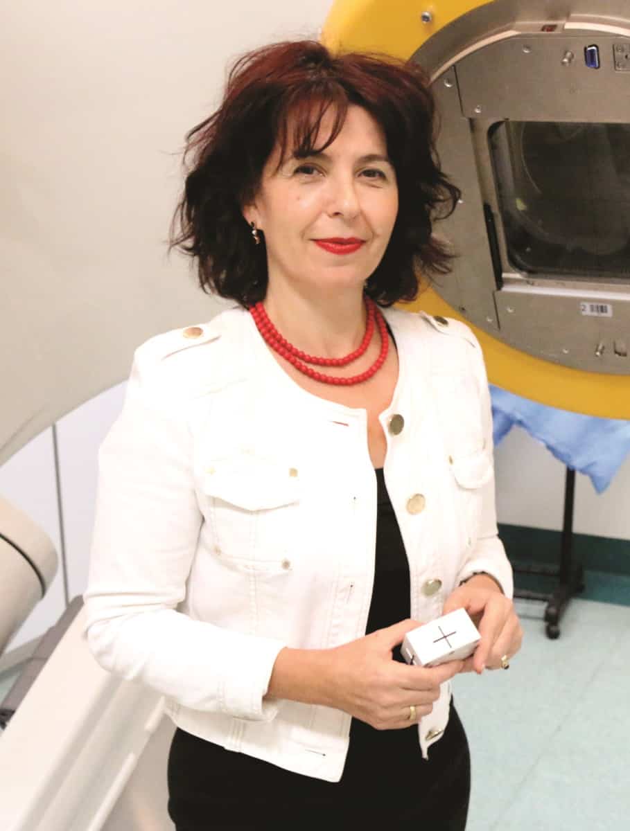
Exceptional contribution: Natalka Suchowerska received the ESTRO honorary membership award for her achievements in innovation. (Courtesy: N Suchowerska)
“I’d like to reflect on the relationship between the successes we’ve had with physics and technology – and the relative neglect of the biology – in radiation therapy.” With these bold words, Natalka Suchowerska opened her presentation at the recent ESTRO 2021 meeting.
Suchowerska, co-director of VectorLAB and associate professor at the University of Sydney, described how the development of new radiotherapy technologies has enabled the introduction of advanced treatment techniques into routine clinical practice. It’s now possible to reduce target margins to zero and escalate prescriptions to more lethal doses. But, she asked, will doubling the accuracy of radiotherapy yield the result we really strive for: a measurable clinical improvement? Moving forward, where are our efforts best directed?
“The real question is whether we can predict patient outcome with the same accuracy as we predict dose,” she explained. “I’ve no doubt that we have the technical ability, but do we have the required understanding of the body’s response to the radiation to move this frontier further?”
One obstacle is that many of the emerging treatment techniques, such as microbeams and FLASH radiotherapy, introduce unprecedented spatial or temporal modulation of the radiation beam. Such approaches have the potential to significantly improve clinical outcomes, but this non-uniform irradiation creates a biological complexity that traditional radiobiology models were not designed to predict. “Even today, we don’t fully understand the biological mechanisms after exposure to highly spatially and temporally modulated beams,” said Suchowerska.
While techniques such as dosimetry, quality management, treatment planning and tumour tracking are tangible technologies that emerge from the clinic and are promoted by vendors, radiobiology is the understanding of the body’s natural response to radiation.
“The commercial sector has no interest in promoting understanding, so whose responsibility is it to lead?” asked Suchowerska. The technical developments needed to create and deliver microbeams and FLASH treatments will keep the physicists busy and the commercial sector happy, she explained. But the true power of this technology lies in understanding the body’s response and how we can harness and drive that response to the best outcome for patients.
More than just the dose
Suchowerska described a study performed by her team, in which prostate cells were irradiated with uniform and spatially modulated clinical radiation beams. The researchers then used mass spectrometry and NMR techniques to examine the metabolites expressed by the irradiated cells. In a normal prostate cell line, they found the expression after uniform or modulated 3 Gy irradiation to overlap. In prostate cancer cells, however, a 1.5 Gy modulated field resulted in a similar metabolite expression as a 2.25 Gy dose from a uniform field.
“So we started to see that the dose is not necessarily a good surrogate for biological response,” Suchowerska explained.
As new technologies emerge, there’s an urgent need to understand the biological effects that they create, in order to make the biology work with us and exploit the opportunities promised by these fledgling techniques. “To improve radiotherapy practice, the next step is to advance our understanding of the biological mechanisms to the same level as we understand the operation of our dosimeters,” said Suchowerska.
To achieve this, medical physicists may have to reach beyond their comfort zones. They will need to work in interdisciplinary teams and collaborate with experts in other fields. “We must value the expertise we have,” Suchowerska told the ESTRO audience. “However, to realise our ultimate goal, our work can’t just end at dose and dose distribution and the optimal operation of the technology. To advance the next frontier, we need to work with other disciplines that bring their expertise to the table. By understanding the biological mechanisms, we have the roadmap of how we need the technology to operate.”
Natalka Suchowerska presented this talk in recognition of being awarded honorary membership of ESTRO for her achievements in innovation. This award is presented as an acknowledgement of the recipient’s exceptional contribution to radiotherapy and oncology, particularly in the field of interdisciplinary or international co-operation.
A multipurpose material that can sense strain and temperature and harvest energy from temperature gradients has been developed by researchers in the UK and the Netherlands. The new material, which could be used to create smart human–machine interfaces and health monitoring devices, was created by Emiliano Bilotti and collaborators at Queen Mary University of London, Imperial College London, Eindhoven University of Technology and Loughborough University.
Current wearable sensors typically have limited mechanical flexibility or require a stiff battery to work. In this research, Bilotti and colleagues have discovered that commercially available Lycra yarns, a flexible material commonly used in textiles, can be modified to show thermoelectricity and strain sensitivity. This is done by adding the conductive copolymer poly(3,4-ethylenedioxythiophene) polystyrene sulfonate (PEDOT:PSS).
Using the new material, the team developed a device that can operate in three different modes to sense strain, measure temperature differences or harvest energy. The strain sensitivity can be useful for creating gloves that track hand movements and the thermoelectric property of the material could be used to power such a glove by harvesting energy from the difference between body temperature and the surrounding ambient temperature.
Low-cost fabrication
The Lycra yarns are given strain sensitivity and thermoelectric properties by immersing them in a solution containing PEDOT:PSS. Upon evaporation of the solvent, the conductive copolymer attaches to the surface of the Lycra yarns, conferring electrical conductivity on the material.
The researchers noticed that, by applying a high strain on the coated fibres, they could induce the formation of cracks across the surface of the PEDOT:PSS coating. These cracks increase the total surface area of the yarns, and provide the strain sensitivity. This is because they create interconnected patches of the conductive copolymer, which separate with the application of a strain.
Increasing the separation between the isolated patches decreases the electrical conductivity of the material, allowing it to be used as a strain sensor. As a result, the material exhibits a large change in resistance even when the sensors are deformed using a low strain of 1%. However, the team found that the thermoelectric properties of the coated yarns are not influenced by strain or the cracks – which do not interfere with the ability of the sensor to measure temperature differences.
Relative temperature
Since the temperature sensitivity of the yarns is based on thermoelectricity, they are not able to sense absolute temperature values. Instead, the material can measure temperature differences as small as 7 °C. This could be used to measure the relative temperature of the human body with respect to the surrounding environment. In addition, the temperature gradients generate a voltage difference due to the thermoelectric effect, which can be used to create electric power.
As a proof of concept, the researchers sewed the yarns onto a glove, and used them to sense the temperature of an object relative to that of the hand. Due to the small temperature differences between a human hand and the environment (around 10 °C), the team calculates that a glove containing 1800 strands of yarn could power the necessary electronics for its practical use. The material could also be used to measure the strain of the glove. This would be useful for creating self-powered wearable devices where, for example, the hand position and its temperature could be measured autonomously.