
Temporary pacemaker regulates heart rhythm then completely disappears

Dissolving pacemaker

Temporary cardiac pacemakers provide essential pacing for patients with short-term heart rhythm disorders following cardiac surgery or while waiting for a permanent pacemaker. Such devices typically require external hardware with leads inserted through the skin, risking infection and limiting patient mobility. In addition, when the implanted device is no longer needed, its removal risks damage to heart tissue.

To overcome these limitations, researchers in the US have devised an implantable cardiac pacemaker that operates without external leads and completely dissolves in the body after completion of therapy. The device is made from materials that fully resorb through natural biological processes in a time-controlled manner, eliminating the need for surgical removal.

Such a bioresorbable, leadless, cardiac pacemaker could provide postoperative control of heart rate and rhythm without the risks associated with traditional temporary pacing systems. The research team – headed up at Northwestern University and The George Washington University – describe the innovative device in Nature Biotechnology.

Device design

To eliminate the need for batteries and thus miniaturize the device, the researchers used wireless energy transfer to deliver power and control commands to the pacemaker. The device’s power-harvesting receiver comprises an inductive coil made from tungsten-coated magnesium (W/Mg), a radiofrequency PIN diode based on a silicon nanomembrane, and a dielectric interlayer of the biodegradable polymer PLGA. The pacemaker also includes flexible W/Mg extension electrodes connected to dissolvable metallic contact pads, which attach to the myocardium to deliver the electrical stimuli.

Device structure

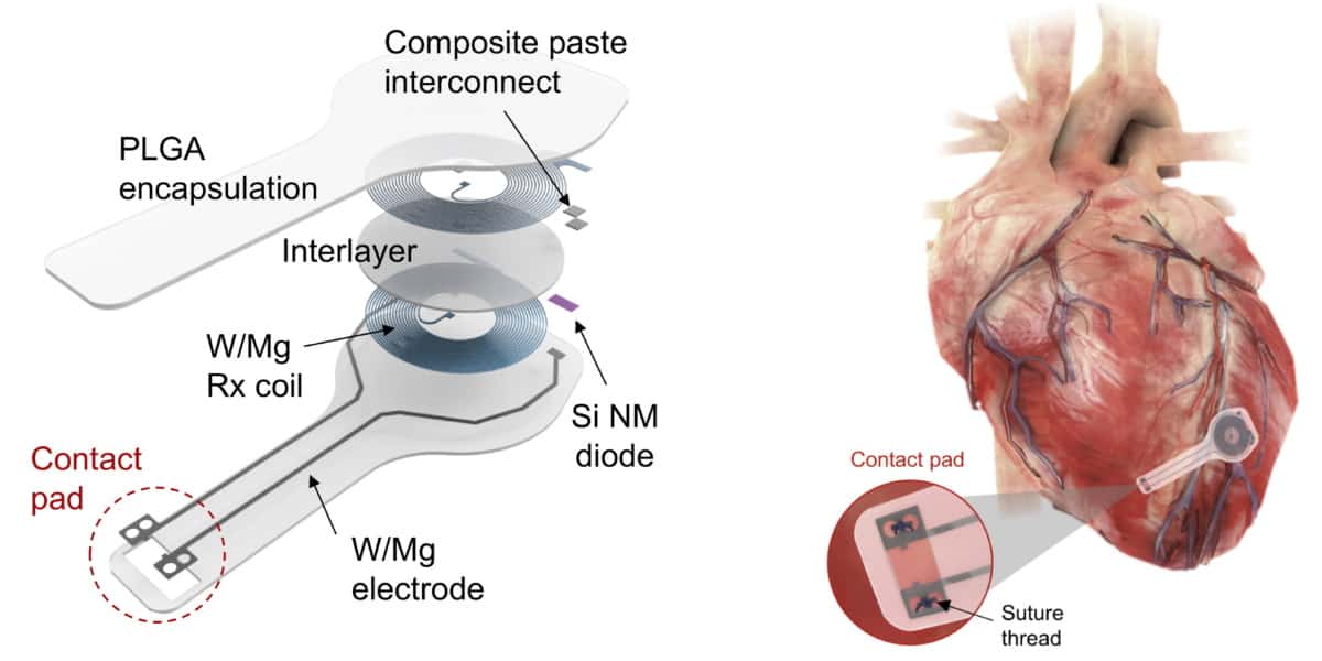
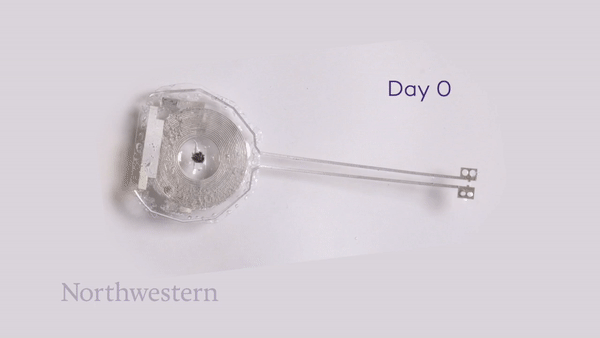
The entire system is just 16 mm wide and 250 μm thick, and made entirely of bioresorbable materials: PLGA dissolves into its monomers, glycolic and lactic acid, while W/Mg and the silicon nanomembrane degrade into nontoxic products. The flexibility of these materials also enables the device to conform to the curved surface of the heart and adapt to its movements.

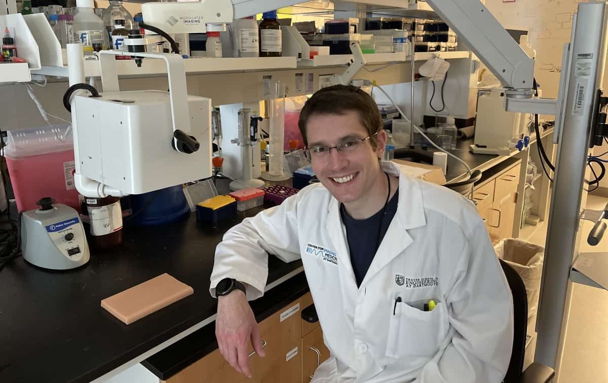
“Significant advances in materials science and organ conformal bioelectronics made this work possible,” explains Igor Efimov, who led the study along with John Rogers and Rishi Arora. “Wireless battery-free pacemakers were developed a while ago. But they suffered from mechanical mismatch between the rigid device and soft tissue of a beating heart, which resulted in high mortality of animals and was not easily translatable to humans.”

“The development of soft organ-conformal electronics by Rogers’ group solved this problem,” says Efimov. “The next step was the development of bioresorbable electronics. This was a game changer for many clinical needs of temporary implantable devices.”

Proof of pacing performance

The team first tested the pacemaker on ex vivo mouse and rabbit hearts, using RF power from a nearby transmitting antenna to initiate pacing. Tests in slice of human cardiac tissue also demonstrated successful pacing.

Next, the group used the pacemaker to treat AV block (impaired signal transmission from the atria to the ventricles) in an ex vivo mouse model. Electrocardiogram (ECG) recordings and measurements of optical action potentials confirmed that the device could deliver effective ventricular pacing, driving the rhythm of the heart to treat the AV block.

The researchers also demonstrated in vivo pacing in an adult dog during open-chest surgery. They recorded a maximum pacing distance (between skin and transmission coil) of 17 cm, validating the pacemaker’s capability for long-range wireless energy transfer. These findings suggest that the device can achieve the necessary power transfer for operation in adult human patients.

Lastly, the team surgically implanted biodegradable pacemakers in rats and performed daily pacing trials on awake or lightly sedated animals. Changes in the ECG signal indicated successful ventricular capture. The device successfully performed pacing for four days after implantation, then its performance started to degrade until after six days the pacing failed. The timescales for stable operation and complete bioresorption can be tailored to meet specific clinical requirements. “Devices can be coated with materials that have a slow rate of resorption, which would ‘program’ the lifetime before the device is resorbed,” explains Efimov.

To monitor the bioresorption process, the researchers recorded CT images of the rats over seven weeks. One week after implantation, the pacemaker maintained its shape and contact with the heart. The device then shrank and collapsed over time, largely dissolving within three weeks, with the remaining residues completely disappeared after 12 weeks. They note that the implantation and resorption of the bioresorbable pacemaker did not impact the rats’ natural physiology.

The researchers are now planning to develop bioresorbable pacemakers for paediatric patients with AV block following septal defect repair, who require a temporary pacemaker for several days after surgery. They will also design devices for adult patients who also frequently experience temporary AV block after valve repair.

“A similar approach can used for many other cardiac, neural and muscle applications,” Efimov tells Physics World. “We also believe that this approach can be used to design a temporary defibrillator to treat post-operative atrial fibrillation, which occurs in 30% of CABG [coronary artery bypass graft] patients and after other cardiac surgeries.”

Exploiting dual-energy CT and DirectSPR software to reduce range uncertainty in proton therapy

From collaboration comes innovation: that’s certainly the mantra of an ambitious, multicentre German R&D initiative which is leveraging the cross-disciplinary expertise of academic researchers, clinicians and industry to deliver game-changing advances in the radiation oncology clinic – enhancing the accuracy, safety and tissue-sparing capability of proton therapy systems in the process. The breakthroughs in question stem from the clinical deployment of dual-energy CT (DECT) for proton treatment planning and the application of so-called DirectSPR software1,2 for the more accurate prediction of proton range in the patient’s body. All part of a unifying vision – think translational research meets clinical application – to reimagine the planning, delivery and management of proton treatments tailored to the unique requirements of individual cancer patients.

The clinical roll-out of DirectSPR is being pioneered by a translational team headed by Christian Richter at the Dresden-based OncoRay. The OncoRay researchers work closely on algorithm development and validation with their colleagues at the German Cancer Research Center (DKFZ) in Heidelberg – the two institutes together form the National Center for Radiation Research in Oncology (NCRO) – while industrial support comes from the Cancer Therapy Business Line at Siemens Healthineers.

OncoRay itself is a publicly funded research centre that focuses on translating new technologies and treatment methods in radiation oncology into clinical application, pooling the strengths of its three founding institutions: Carl Gustav Carus University Hospital Dresden, Technische Universität Dresden and the Helmholtz-Zentrum Dresden-Rossendorf. “While OncoRay’s principal driver is to improve treatment efficacy and patient outcomes,” explains Richter, “we also seek partnerships with leading medical technology manufacturers to ensure that our R&D reaches the wider radiation oncology community – with DECT-based DirectSPR a notable success story in both respects.”

Innovation unpacked

If that’s the back-story, what of the specifics underpinning this new paradigm in proton treatment planning – and in particular, the basis for the more accurate predictions of proton range and stopping behaviour in different tissue types? Key to success is the innovative application of DECT technology and its spectral imaging capability to the prediction of proton range for treatment planning. Unlike conventional CT, this imaging modality involves the acquisition of two separate X-ray energy spectra – an approach that, in turn, allows the characterization of tissues exhibiting different attenuation properties at different energies (though, crucially, without exposing patients to a higher X-ray dose).

DirectSPR, meanwhile, is an algorithm for post-processing of DECT images – and specifically the determination of the individual stopping behaviour of protons in different tissue types. At the same time, DirectSPR maximizes the quantitative quality of the stopping information by using sophisticated noise reduction and by tailoring the application to the scanned body region of interest. In this way, DirectSPR makes it possible for medical physicists to better resolve differences in tissue composition across a given patient cohort – as well as in the same patient – and to take these differences into account during treatment planning. (For completeness, SPR is the stopping-power ratio and expresses the energy loss of the protons versus distance travelled through the patient relative to the energy loss in water.)

See the difference

In Spring 2019, Richter and his colleagues at OncoRay became the first team to deploy DECT-based DirectSPR as part of the routine clinical workflow for proton therapy – a key step in the improvement of treatment planning of static tumour sites at University Proton Therapy Dresden (part of Carl Gustav Carus University Hospital). Since then, for more than 300 patients, DECT-based DirectSPR has enabled the Dresden clinic – on a sustained and repeatable basis – to reduce the volume of irradiated healthy tissue surrounding the target volume (the safety margin) by approximately 35% for prostate cancer and brain tumour treatments.

Clinical impact, of course, is defined along multiple coordinates, not least the accuracy of SPR calculations and related proton-range predictions – both of which dictate the safety margin around the tumour and, by extension, the degree of tissue sparing. Thanks to the clinical introduction of DECT-based DirectSPR, OncoRay has realized significant reductions in the uncertainty of its proton-range calculations (now <2%) versus traditional CT-based planning approaches (where uncertainties have been set at 3.5% of total proton range for the past 30 years). The result: proton-range reductions on average of 3.6 mm for pelvic treatments and 2.6 mm for treatments in the head. “For individual brain tumour treatments,” says Richter, “we are therefore able to reduce dose to critical structures like the brain stem by 16% as well as the optic chiasm and optic nerve by 7%, with 4% reduction overall in mean dose to the brain as shown in a representative patient case. That’s relevant progress in minimizing the patient’s risk of post-treatment side-effects.”

Translation in action

The timeline for translation of DirectSPR into a clinical product can be traced back to early 2015, when scientists from OncoRay and the DKFZ began work on the development and optimization of the DirectSPR approach. Their aim: to minimize the uncertainty associated with proton-range predictions in the patient’s tissues and thereby enhance targeting accuracy and dose distribution accuracy for proton treatments. This four-year, NCRO-funded project – carried out in close collaboration with Siemens Healthineers – began with the introduction of DECT for clinical proton therapy planning at University Proton Therapy Dresden in 2015 – a world-first that made it possible to retrospectively evaluate the DirectSPR approach on a growing database of therapeutic DECT patient scans (rather than just using phantoms or computer simulation).

“Even in the formative stages of DirectSPR algorithm development, OncoRay and DKFZ were focused on translational outcomes,” says Richter. The definition of a sophisticated algorithm early on – and especially the subsequent focus on comprehensive stepwise validation in different scenarios (including an anthropomorphic phantom and biological tissues) – was fundamental to successful translation. “This convinced us, the community and the responsible clinicians of the superiority of the approach,” adds Richter. “Siemens Healthineers came on board in 2016, with the early conversations building trust and understanding around our mutual clinical and research goals.”

It was at this point that the collaboration shifted gears into the preclinical product development phase, with OncoRay, DKFZ and Siemens Healthineers working together on optimization and calibration of the DirectSPR algorithm. With the commercial launch of DirectSPR software in spring 2019, Siemens Healthineers now ensures compatibility of DirectSPR across its full portfolio of DECT scanners with dedicated calibrations for each CT model.

The clinical roadmap

In terms of next steps, Richter and colleagues at OncoRay – as well as their industry counterparts at Siemens Healthineers – are focused on accelerating clinical acceptance of DirectSPR within the proton therapy community (see “Proton perspectives: the DirectSPR opportunity”). OncoRay, for its part, maintains bilateral collaborations with many European proton therapy centres and is also active within the European Particle Therapy Network, a subgroup of the European Society for Radiotherapy and Oncology (ESTRO). “A consensus paper is in preparation to define best-practice guidelines and standardization for the clinical implementation of DirectSPR in the proton therapy clinic,” explains Richter. That document, he hopes, will be brought forward during discussions at the upcoming ESTRO workshop on Clinical Translation of CT Innovations in Radiation Oncology.

Meanwhile, the OncoRay and DKFZ teams are already working with Siemens Healthineers to explore new translational pathways, including the use of DECT-based DirectSPR for proton treatment planning on moving targets – for example, tumour sites in the lung and abdomen. “Our task at OncoRay and DKFZ is to support further improvement of DirectSPR for next-generation CT scanners in a radiotherapy setting,” concludes Richter.

Proton perspectives: the DECT-based DirectSPR opportunity

There’s growing clinical appetite to deploy DirectSPR as a core component of the proton therapy workflow. Here Physics World talks to medical physicists at some of the early-adopting proton facilities to assess progress, next steps and anticipated clinical upsides.

Tianyu Zhao, Washington University School of Medicine in St Louis, Missouri

We are recruiting patients into a National Institutes of Health (NIH)-sponsored study, collecting data using DECT scanners to improve estimates of tissue composition accuracy for tumour sites and surrounding organs. The goal: to reduce the discrepancy between the actual dose delivered versus the planned dose in proton therapy. We plan to quantify the accuracy of DirectSPR for mass density and proton SPR predictions by using phantoms scanned with clinical protocols. This site-specific accuracy information will be used to adjust the uncertainty setting in robustness optimization for proton patients scanned with DECT. Ultimately, the hope is that our work will reduce the range uncertainty for protons and lead to better planning quality and fewer dose prediction errors in proton therapy.

Ming Yang, MD Anderson Cancer Center, Houston, Texas

We are currently upgrading our institutional syngo.via server (from Siemens Healthineers) to VB40 – a prerequisite for implementing DirectSPR. Upon completion, we will start the commissioning process as soon as DirectSPR is available to us. Initial priorities will be to test DirectSPR’s compatibility with our existing treatment planning system and to evaluate the uncertainties associated with the software’s estimation of proton stopping power. Personally, I believe DirectSPR will benefit our patient population in multiple ways. One immediate impact will be a reduction in normal tissues receiving high dose close to prescription dose – so a reduction in normal-tissue toxicity. A smaller range uncertainty parameter will also make it easier to achieve a robust treatment plan meeting the planning objectives. Finally, the reduced range uncertainty might also give us more choices of beam angle to take full advantage of the Bragg peak.

Benjamin Ackermann, Heidelberg Ion Beam Therapy Center (HIT), and Friderike Longarino, Heidelberg University Hospital

HIT completed clinical validation of DirectSPR using a free-of-charge test licence – a convenient and low-risk option for clinics interested in finding out more. The validation process included a complete radiotherapy workflow and dosimetric tests in anthropomorphic phantoms, consisting of a multitude of different tissue-equivalent materials. We are now installing the purchased software licence and looking forward to performing the first patient scans using DirectSPR, starting with body regions like the head and pelvis which exhibit little or no intrafractional movement. With DECT scans and DirectSPR image processing, we expect to achieve better SPR predictions and, in turn, better range prediction in ion beam therapy compared to SECT. Another anticipated benefit is a reduction in safety margins for enhanced sparing of healthy tissue from unwanted irradiation.

Sina Mossahebi, Maryland Proton Treatment Center, US

In our initial DirectSPR study, we aim to quantify dose-calculation discrepancies between conventional SECT and DECT in vivo and on patient plans, with the objective of reducing range uncertainty in the planning of intensity-modulated proton therapy (IMPT). Both SECT and DECT scans are acquired from the same patient and used to evaluate dosimetric differences between treatment plans generated on each image set. The primary goal of the study is to quantify and compare the target coverage and dose to organs at risk (OARs) as well as the plan robustness between SECT and DECT scans obtained for patients undergoing IMPT. In addition, we will validate SECT and DECT dose calculations using patient-specific QA, while exploring practical advantages of DECT with respect to target delineation and implants.

Gabriel Fonseca and Frank Verhaegen, Maastro Clinic, The Netherlands

Our evaluation of DirectSPR is proceeding along multiple fronts. Through extensive phantom testing, we have investigated the SPR information used in the dose calculations by the TPS, comparing SPR predictions obtained via the DECT-based DirectSPR and SECT approaches. The former is more accurate, exhibiting differences of less than half of those obtained by SECT. Another focus is the calibration procedure, which will likely require a range of phantom sizes to match variations in patient size – i.e. extra work in the clinic. Although DirectSPR can provide more accurate SPR values, the treatment plan uncertainties depend on several factors. As such, the Maastro team is evaluating the effect of improved SPR maps on robust optimized plans for a cohort of more than 50 patients. In summary: while we expect DirectSPR to yield improved proton range accuracy from 1 mm up to several mm (for some deep-seated tumours), it’s clear that all sources of uncertainty and risk need to be quantified thoroughly prior to routine clinical deployment. Long term, strict QA procedures will need to underpin the clinical workflow.

Ole Nørrevang, Danish Centre for Particle Therapy, Aarhus University Hospital, Denmark

We’re taking a phased approach to the roll-out of DECT-based DirectSPR here in Aarhus. We started out by implementing virtual monoenergetic images and optimizing the use for both organ and target delineation and stopping-power estimation. With DirectSPR software now available to us, our investigations are proceeding on three fronts: validation of the SPR values produced by DirectSPR; studying the effect of reduced range uncertainty in terms of reduced dose to OARs in the brain; and understanding the workflow, patient safety and transition issues associated with the use of SPR images in the treatment planning of brain tumours. Our focus for now is on the treatment of brain tumours, because we can exclude the effects of motion artefacts in the DECT image. Over time, though, we will develop plans to apply DirectSPR for other treatment sites.

Koen Salvo, PARTICLE Proton Therapy Center Leuven, Belgium

We are currently in the process of defining our DECT simulation protocols, with the adult protocols complete and conversations in progress with Siemens Healthineers on the child protocols. Once those protocols are finalized, we will activate our DirectSPR software and validate it using proton radiography on animal phantoms. With DirectSPR, we hope to calculate the proton range more accurately. If so, we can reduce our target margins and, in turn, reduce the dose to OARs near the target – a significant advance when treating chordomas near the brain stem.

End note: the statements by Siemens Healthineers’ customers described herein are based on results that were achieved in the customer’s unique setting. Because there is no “typical” hospital or laboratory and many variables exist (e.g. hospital size, sample mix, case mix, level of IT and/or automation adoption), there can be no guarantee that other customers will achieve the same results. 1) DirectSPR is an optional feature. 2) DirectSPR calculations require Dual Spiral Dual Energy, Twin Spiral Dual Energy or Dual Source acquisition; TwinBeam Dual Energy data are not supported for DirectSPR calculations. More information on DirectSPR and its requirements can be found here.

Online training attracts new generation of peer reviewers

Peer review is an essential component of scientific learning and progress, and yet researchers who are just starting out on their career are rarely schooled in the skills needed to provide a useful and constructive review. Academic publishers have sought to fill the gap with workshops and training materials that offer guidance to newcomers on how to critique research work that has been submitted to their journals. But IOP Publishing has gone one step further by launching a free online training programme that enables early-career researchers anywhere in the world to gain the skills and confidence they need to achieve excellence in peer review.

“I believe that peer-review training is crucial for experienced and non-experienced reviewers to improve the quality of reports, and eventually, the quality of publications,” says Marijan Beg, a recent graduate from the programme and a teaching fellow in computational data science at Imperial College London, UK. “Most often, we begin peer reviewing in the early stages of our careers without any training. Our understanding of the peer-review process is based solely on the reviews we received for the manuscripts we wrote as PhD students.”

To anyone who may be jaded by the prospect of yet another online training course, the Peer Review eLearning Hub exploits the latest innovations in training software to offer participants an engaging and interactive learning experience. The interface is also fully responsive, allowing trainees to access the training platform on any device and from any part of the world. “It is easy for anyone to sign up and register for the programme,” comments Laura Feetham, reviewer engagement manager at IOP Publishing. “The training portal is free for anyone to use, and registrants gain immediate access to the learning resources.”

The impetus for the project came from an extensive survey of IOP Publishing’s existing pool of peer reviewers, conducted in 2020, as well as in-depth interviews with researchers at all stages of their career. “We know that peer review is not perfect, and we wanted to find out how we could improve it,” comments Feetham.

One of the main themes to emerge from the research was the need for more training in peer review. While that focus was particularly evident in the responses from early-career researchers, Feetham says that even senior academics still remember the stress of writing their first review. “It’s just bizarre that you might typically be invited to write a review towards the end of your PhD, and are just expected to know how to do it,” she says.

To address that need, in September 2020 IOP Publishing launched a series of online workshops under the banner “Peer review excellence: IOP training and certification”. Building on previous experience of delivering live presentations at key conferences and research institutions, Feetham developed an interactive curriculum that engages participants through discussions and practical exercises. “We only include about 50 people in each session, and nearly all of the sessions have been oversubscribed,” she comments. “It has shown us that there is a real appetite for this type of training.”

Based on that success, Feetham sought to recreate the same learning experience in a more scalable and accessible format. To deliver a similar level of interaction and engagement through a purely online portal, she worked closely with Laura Lee Gibbs, a learning consultant at Learn Fox Consultancy, who was able to translate the curriculum into interactive elements that enable participants to learn specific skills. “Laura is an expert in cognition and learning software, and she was brilliant at telling us the most engaging way to get each point across,” explains Feetham.

The comprehensive training programme includes three modules that together take around two hours to complete. The first module focuses on the fundamentals of peer review, including its evolution since it was first introduced in 1665. The second teaches participants how to write a meaningful and constructive review, with clear guidance on how to structure the report and what information to include. And the third one focuses on peer-review ethics, ensuring that new reviewers can spot and report any signs of research misconduct.

Photo of Yuchun Sun

“Before this training I didn’t even know what peer review reports should include, or the most important criteria for evaluating a work,” comments Yuchun Sun of the California Institute of Technology. “I have learned that when I review a manuscript, I need to critically read the article and think about many aspects of the work.”

Each module is taught through a series of elements, which might consist of a few lines of text followed by an interactive exercise such as a flip-card game. “It’s a way of learning that isn’t just about absorbing information and regurgitating it,” explains Feetham. Beg agrees: “I found the experience of using the online training platform concise, enjoyable, and well-structured,” he says.

Trainees must engage with all the elements and pass an assessment at the end of each module to progress to the next one, with a final assessment determining whether they pass the course. Since launching the hub in March, some 900 people have registered for the course and 300 have graduated. Feetham has also been encouraged by the geographic diversity of the participants, with plenty of sign-ups from under-represented communities in Asia, South America and Africa.

But it’s not just about delivering the training. Another important theme to emerge from the initial research is the need for more recognition for peer reviewers. “In physics, at least, researchers see peer review as a way of giving something back to their community,” explains Feetham. “But they do want to be recognized for their work.”

As a result, last year IOP Publishing introduced the concept of a Trusted Reviewer. To kick-start the scheme, this special status was given to anyone who had submitted an outstanding review – as scored by the journal editors – within the past two years. Less than 15% of all reports achieve this highest possible grade, lending an air of prestige to the Trusted Reviewer title, and later this year IOP Publishing plans to provide more public recognition through an external database and badges that Trusted Reviewers can use on their professional profiles.

With the launch of the online training hub, Feetham was keen that graduates from the scheme should also have the opportunity to gain Trusted Reviewer status. Participants who have completed the training have the option of a fast-tracked process, in which the IOP Publishing team helps them to develop a searchable selection of research interests that enables them to be matched to a manuscript in their area of expertise. If the graduate submits a review that is graded as being outstanding or excellent, they are awarded Trusted Reviewer status.

“What’s really impressive is that more than half of those who opt for fast-tracking immediately become an IOP Trusted Reviewer, compared with less than 15% for our general reviewer pool,” comments Feetham. “That suggests we are doing something right. The scientific quality of their reviews is really high, and we are giving them the skills and confidence they need to critique a manuscript in a meaningful way.”

Which brings us to the third element of the ecosystem that Feetham is aiming to create. Graduates who don’t quite make the grade for Trusted Reviewer status are given clear and honest feedback on how to improve, and are then given the opportunity to try again. While that constructive criticism is currently offered on an informal basis, in future there will be an automated system that provides graduates with more structured feedback on the quality of their review and, if necessary, how to improve.

Feetham believes that this powerful combination of training, recognition and feedback will engage a new and more diverse generation of researchers in the peer-review process. “One of the reasons that it’s getting harder to source high-quality reviews in a timely way is that publishers are overburdening a relatively small group of people,” she says. “We want to find early-career researchers who have the time and energy to engage with the process, and give them the skills and expertise they need to produce good reviews.”

And, judging by the response from recent graduates from the programme, that approach appears to be working. “Through the whole training process, I learnt a lot about peer review and how to write a high-quality assessment,” says Nergui Nanding of Sun Yat-sen University in China, who has recently become an IOP Trusted Reviewer. “It will bring long-term benefits to my career, and the Trusted Reviewer status will provide me with more opportunities to review papers in high-quality journals and to build better relationships with the editors and the wider community.”

Plant pioneers, cosmic computer concerns and tips for school presentations: the July 2021 issue of Physics World is now out

Photo of the Cape sundew plant, which is inspiring a robotic limb

With 29 bones, 123 ligaments and 34 muscles pulling the strings, the human hand is a feat of nature’s engineering. It lets us write, touch, hold, feel and interact in exquisite detail with the world around us.

To replicate the wonders of the human hand, researchers in the field of “soft robotics” are trying to design artificial structures made from flexible, compliant materials the can be controlled and programmed by computers.

Trouble is, the hand is such a complex structure that it needs lots of computing power to be properly controlled. That’s a problem when developing prosthetic hands for people who have lost an arm in, say, an accident or surgery.

Designers seeking to make their structures move are, however, finding inspiration from a surprising source: the study of movement in plants. As Daniel Rayneau-Kirkhope explains in the July 2021 issue of Physics World magazine, one strange-looking plant – the Cape sundew – could hold the key to soft, robotic limbs.

If you’re a member of the Institute of Physics, you can read the whole of Physics World magazine every month via our digital apps for iOSAndroid and Web browsers. Let us know what you think about the issue on TwitterFacebook or by e-mailing us at pwld@ioppublishing.org.

For the record, here’s a run-down of what else is in the issue.

• Beware of the “alliars” – While some people claim to want to support under-represented groups, Anya Lawrence says it can be difficult to determine whether they have the right motives

• Bridging the language divide – Pruthvi Mehta says more support should be given to  non-native English-speaking scientists who can feel isolated and disadvantaged in what to them is an unfair system

• Tax breaks – In the second of a series of articles on how to start and fund a business, James Mckenzie looks at government tax incentives, which can be more useful than they first sound

• Vaccination reluctance – Robert P Crease explores the range of factors that turned him from vaccine sceptic to believer

• Replicating how plants move – Once studied by Charles Darwin, the Venus flytrap is perhaps the most famous plant that moves at high speed. But as Daniel Rayneau-Kirkhope explains, researchers are still unearthing new scientific insights into plant motion, which could lead to novel, bio-inspired robotic structures

• Supercomputers face a cosmic challenge – Fast neutrons from cosmic-ray showers can cause significant errors in supercomputers. But by measuring the scale of the problem, physicists hope not only to make such devices less prone to cosmic corruption but also protect everything from self-driving cars to quantum computers, as Rachel Brazil finds out

• How to spark an interest – Rick Marshall shares his advice for giving an inspiring and engaging presentation about physics to pre-university school students

• It’s the little things – Angela Saini and her son Aneurin review Nano: the Spectacular Science of the Very (Very) Small by Jess Wade, illustrated by Melissa Castrillon

• No man is an island – Chanda Prescod-Weinstein reviews Helgoland by Carlo Rovelli

• Stephen Hawking: cosmic commodity – Laura Hiscott reviews Hawking Hawking: the Selling of a Scientific Celebrity by Charles Seife

• Making the switch – Changing career path can be daunting but there are big rewards for those who are brave enough to switch. M S Zobaer describes his unexpected journey from dusty plasmas to neuroscience

• Ask me anything – Mariya Lyubenova is an astronomer researching galaxy evolution at the European Southern Observatory (ESO). She is also head of media relations at ESO

• Sporting chance – With the Olympic Games due to get under way in Tokyo this month, Laura Hiscott brings you 10 physics-related sporting questions about the 10 different events in the decathlon – one of the highlights of the athletics programme.

Non-toxic supercapacitors go fully recyclable

Researchers at the Empa in Dübendorf, Switzerland, have made a high-performance supercapacitor entirely from recyclable, non-toxic materials. The device can withstand thousands of charge and discharge cycles, resists pressure and shock, and works even at freezing temperatures, making it an environmentally-friendly option for powering Internet of Things (IoT) devices.

The development of the IoT has heightened the already-difficult problem of electronic waste. Many electronic components for IoT applications are produced by the million, have a short service life and are powered by lithium-ion or alkaline batteries. While these batteries perform well, they contain toxic materials that need to be collected at the end of their life and then recycled using special processes.

Onto the compost heap

Researchers led by Gustav Nyström have now developed an alternative: an electric double-layer capacitor (EDLC) made from a disposable paper-like material. In the context of electronics, the researchers explain that “disposable” devices are those that can be thrown away in the trash, do not release toxic substances and ultimately fragment into small particles. “At the end of its service our new EDLC can be processed as a noncytotoxic compostable material or simply be left in nature,” says Xavier Aeby of Empa’s Cellulose & Wood Materials lab. “Indeed, the device disintegrates and loses 50% of its mass within nine weeks, leaving only a few visible carbon particles.”

The device, which can also store much more charge than a conventional capacitor, was made using a method called direct-ink writing. In this technique, viscoelastic gel ink is extruded line-by-line and layer-by-layer from a printer nozzle to form three-dimensional objects.

recyclable capacitor

Nyström and colleagues’ ink contains only nontoxic and renewable materials: nanocellulose as a gelling and network-forming agent and as a substrate; carbon powder to make high-surface-area electrodes; glycerol as a plasticizer in the nanocellulose and as the electrolyte; and water as the solvent. The nanocellulose itself contains high-performance bio-nanofibres, which are a finer-scale version of the micron-sized fibres used to make paper.

While the recipe may sound relatively simple, it certainly wasn’t easy to develop. “It took an extended series of tests until all the parameters were right, and each of the components flowed reliably from the printer and the capacitor worked,” Aeby says. “As researchers, we don’t want to just fiddle about, we also want to understand what’s happening inside our materials.”

Ideal for the IoT

To test their device’s suitability for real-world applications, Nyström and colleagues printed an energy-storage circuit composed of six supercapacitors connected in series on a complex 3D surface. They found that this system stores electricity for several hours and is capable of powering a standard digital alarm clock.

In the future, Nyström and Aeby say that such capacitors could power a sensor or microtransmitter for hours after being charged using an electromagnetic field, making them ideal for IoT applications. The number of microdevices is also expected to increase as “point-of-care” diagnostics, such as self-testing devices for diabetics, come into more widespread use. “A disposable cellulose capacitor could be well-suited for these applications too,” Nyström says.

The researchers, who report their work in Advanced Materials, say they now plan to study ways to improve the amount of charge that can be stored in their supercapacitor – “ideally without sacrificing the non-toxicity requirement,” Nyström tells Physics World.

Thermodynamic origins of reaction heterogeneity in lithium battery electrodes

Want to learn more on this subject?

During battery (dis)charging, lithium (de)intercalation in electrodes is usually spatially non-uniform across multiple length scales. Such phenomenon is a major impediment to battery performance and life as it causes energy under- utilization and induces over-(dis)charging, etc. While reaction heterogeneity is often attributed to mass transport limitation, this webinar will highlight the important roles of thermodynamic factors including elastic energy and phase transformations, the understanding of which is important for the development of mitigation strategies.

Through combined modelling and characterization, we elucidate how stress could destabilize the lithium (de)lithiation front in single crystalline and polycrystalline intercalation compounds, and also provide a fundamental driving force for dendrite growth on the lithium metal anode during electrodeposition. Stress relief thus offers a promising approach to improving reaction uniformity at the particle level. At the cell level, we discover that the reaction distribution within the porous electrode is strongly influenced by how the electrode’s equilibrium potential varies with the state of charge.

Two types of prototypical reaction behaviour emerge from common electrode materials with significant impact on the thick electrode performance. This finding leads to an efficient analytical model for optimizing battery configurations in place of common battery cell simulations.

Want to learn more on this subject?

Ming Tang is an associate professor in the Department of Materials Science and NanoEngineering at Rice University, US. After receiving a PhD in materials science and engineering from the Massachusetts Institute of Technology, US, he worked at Lawrence Livermore National Laboratory as a Lawrence postdoctoral fellow and then staff scientist. In 2013, Tang joined Shell Oil as a materials and corrosion engineer, and became an assistant professor at Rice University in 2015. His group is currently interested in applying combined modelling and experimental methods to understand mesoscale phenomena in energy-storage systems and use the acquired knowledge to guide microstructure design. Tang is a recipient of the 2018 Department of Energy Early Career Award.

New gate optimization strategy could boost efficiency in trapped-ion quantum computers

Physicists at the University of Maryland, US and the quantum computing firm IonQ have found a new way to make a central operation in quantum computing more efficient. By slashing the laser power required to perform a so-called two-qubit gate, the collaborators showed that they could speed up the gate’s operation, thereby boosting the performance of their trapped-ion quantum computer.

The building blocks of a quantum computer are qubits – quantum bits that can be in any superposition of two states. In this work, the researchers used ions as their qubits. Rapidly oscillating electric fields trap the ions in a chain, making it possible to perform computational operations by shining laser light on one or more ions.

Two-qubit entangling gates

These computational operations generally divide into two types: single-qubit gates and two-qubit gates. While single-qubit gates are relatively simple to perform and pose no significant challenges, two-qubit gates cost significant time and power. That has consequences for the overall efficiency of the quantum computer, says Norbert Linke, a fellow of Maryland’s Joint Quantum Institute (JQI) and a co-author of the current study. “The performance of two-qubit entangling gates typically limits the overall system since they require the most calibration time and introduce the most error,” Linke explains. “Improving these gates is therefore crucial to boost the performance and eventually scale up these systems.”

Ideally, gate operations would be fast, use minimal laser power, and leave the qubit in the desired state with no errors (maximum fidelity). In the real world, errors in two-qubit entangling gates come from having imperfect control over experimental parameters such as the frequency of the laser and the trapping field. The general technique to achieve the highest fidelity is therefore to take great care in designing the control signal (that is, the laser beam) that interacts with the ions, eliminating all undesirable effects by fine-tuning the parameters of the protocol. This constrains the design space for the control signal.

The IonQ–JQI team’s idea was to sacrifice a small amount of fidelity to save a significant amount of laser power – in some cases an order of magnitude. “We consider the constraints that don’t contribute significantly to the error processes when removed,” explains fellow co-author Yunseong Nam, quantum theory lead at IonQ and adjunct assistant professor at the University of Maryland. “This way, while we sacrifice a minimal amount of fidelity, we can significantly increase the size of the design space, which can then be used to better optimize the power requirement.”

Nam and his colleagues implemented their protocol on the JQI’s programmable trapped-ion quantum hardware with five qubits. When they measured both the power and the fidelity of the gate operations, they found that they could create a maximally entangled state with their method without losing significant fidelity.

Generalizing the technique

Now that the team has carried out a successful proof-of-concept demonstration, its members plan to implement their two-qubit entangling gate in various quantum algorithms. This should allow them to verify whether the newly developed protocol leads to an increase in overall efficiency. Linke adds that they are also exploring ways to generalize their method. “We are working on other schemes for generating entangling gates with different control parameters,” he says. “This will provide the optimal quantum gate mechanism for the particular noise or error characteristics of different devices.”

The paper is published in Physical Review Letters.

Meringue-like material offers lightweight soundproofing for aircraft engines

A new meringue-like material that is a strong absorber of sound over a broad range of frequencies has been developed by Michele Meo and colleagues at the University of Bath. They say that their extremely lightweight aerogel is produced using a low-cost, environmentally friendly process that could soon be replicated on an industrial scale. The material promises to be highly effective in reducing the noise of aircraft engines and could also be used in other advanced engineering applications.

As a sound wave passes through porous materials like cellular foams and fibrous polymers, its energy can be strongly dissipated within microscopic pores. As a result, these materials are highly effective sound absorbers at mid-range frequencies – between roughly 800–2000 Hz. To absorb lower frequencies, however, materials must usually be heavier and bulkier, making them impractical for some soundproofing applications.

To create a light-weight material that dissipates lower-frequency sound, Meo’s team used a graphene oxide-polyvinyl alcohol aerogel (GPA). To manufacture this substance, they first produced a foam by whipping up a blend of graphene oxide sheets and PVA polymer using ultra-high shear mixing. After embedding the foam within a honeycomb scaffold, the researchers then freeze-cast it onto a surface, by exposing one side of the material to liquid nitrogen. This caused ice crystals to grow vertically from the side touching the nitrogen, pushing any larger and lighter air bubbles upwards. Finally, the foam was freeze-dried through sublimation, producing a meringue-like aerogel.

Hierarchical and highly tuneable

The resulting material had a hierarchical and highly tuneable porosity and a greatly enhanced ability to dissipate sound energy compared with other porous materials. In addition, the GPA had a density of just 2.1 kg/m3 – making it one of the lightest acoustic materials ever produced. Having adjusted the composition and thickness of their aerogel to optimize its acoustic properties, and evaluated the influence of different processing times, the team created a material with high sound absorption across the 400–2500 Hz range.

Within this range, average losses in sound transmission reached as high as 15.8 dB – which would reduce the roar of a jet engine to the loudness of a hairdryer. As a result, the researchers say that their GPA would be ideally suited for use as an acoustic insulator within jet engine housings – improving comfort for passengers, while adding very little to the weight of the aircraft.

Alongside aerospace applications, the team is also exploring the potential use of their aerogel in vehicles including cars, helicopters, and submarines, as well as in building construction. Indeed, they say that the aerogel could be available for commercial use within just 18 months.

Beyond acoustics, the researchers predict that similar light-weight materials could be created for other applications including fire resistance and electromagnetic shielding.

The new material is described in Scientific Reports.

Backward-travelling sound wave appears in a metamaterial

An unusual type of sound wave that can travel backwards in space and has previously only been observed in ultracold quantum systems may also exist at ambient temperatures in artificially-engineered materials. Researchers led by Martin Wegener at Germany’s Karlsruhe Institute of Technology (KIT) found evidence for these unusual sound waves, known as rotons, in a so-called “metamaterial” that was designed to shape the flow of acoustic waves. The result might make it easier to manipulate sound in air as well as in solid materials.

In normal sound waves, or phonons, the energy of the sound wave travelling through a medium increases linearly with its momentum. With rotons, however, low energy can be associated with high momentum. Certain frequencies of rotons also generate three different co-existing acoustical modes with the same polarization but different wavelengths. The slowest of these three modes is a backward wave, or “return flow” as the 20th-century physicist Richard Feynman put it.

Until now, rotons – which, like phonons, are particle-like collective excitations or quasiparticles – have only been studied in ultracold quantum systems such as helium-3 and, more recently, Bose-Einstein condensates (BECs). These systems contain electrons that interact strongly with each other in a way that allows them to behave like superfluids – that is, a fluid that flows without any friction. This superfluidic behaviour was first predicted by the Soviet physicist Lev Landau, who suggested that it was due to the presence of phonons and rotons. However, superfluid helium-3 and BECs only exist at temperatures just above absolute zero, which somewhat limits their technical applications.

Roton-like behaviour

Wegener and colleagues designed their model metamaterials such that each unit, or cell, within the material interacts with its third-nearest neighbours. The researchers then used these structures to “mould” the flow of acoustic waves through the material. In their simulations, they observed roton-like behaviour without any quantum effects under normal ambient conditions and at almost any wavelength.

The KIT researchers have now begun making real metamaterials based on their design. To replicate the structures in their simulations, they are using an ultraprecise laser printing technique that can “write” a host of different microstructures with a tightly-focused “pen” of light in three dimensions. “Currently we are working on finding direct experimental proof for the existence of rotons,” Wegener tells Physics World. “We hope to submit our results for publication soon.

The researchers report their present work in Nature Communications.

Optical imaging could reduce recall surgery for breast cancer patients

Samuel Streeter

Patients with early-stage breast cancer often undergo breast-conserving surgery, which involves local excision of cancer with a surrounding margin of healthy tissue. The goal is to remove the entire tumour and minimal healthy tissue, but excision is primarily based on visual inspection and relies on the surgeon’s expertise. Roughly 20% of patients require a second surgery due to incomplete initial removal of cancerous tissue.

Improving the tumour margin assessment during the operation could reduce the number of repeat surgeries and associated health and financial costs. Other options available for intraoperative margin assessment include projection radiography and, more recently, volumetric micro-CT. But these X-ray imaging techniques cannot effectively differentiate between normal, abnormal benign and malignant fibrous tissues.

A team from the Thayer School of Engineering at Dartmouth and Dartmouth-Hitchcock Medical Center is now investigating whether optical imaging could help, reporting their findings in Physics in Medicine & Biology.

“We choose to investigate optical scatter imaging, because it is non-contact, provides rapid scanning and relies only on endogenous contrast in the tissue,” explains first author Samuel Streeter. “The fine patterns of light used by the technique probe only superficial layers of tissue, making the approach particularly well suited for analysing tissue margins.”

Optical scatter imaging

Streeter and colleagues used wide field-of-view optical scatter imaging to assess 57 resected breast tumour slices, containing 13 distinct tissue subtypes, from 57 patients. The optical scatter method employed, known as spatial frequency domain imaging, illuminates the tissue with one-dimensional sinusoidal light patterns and images the reflected light intensity. It rapidly images the top layer of tissue with increased sensitivity to tumour-associated, collagen-rich matrix structures.

For comparison, the researchers also imaged the samples with micro-CT and diffuse white light (DWL) imaging, which is similar to the surgeon’s view in the operating room. To enable quantitative comparison between the three modalities, they converted the colour DWL images to greyscale intensity (luminance) and derived monochromatic optical scatter images from the shortest wavelength (490 nm).

Sample images

In general, the optical scatter images exhibited similar contrast to the micro-CT view of the tissue. The team quantified the similarity between the micro-CT and optical images using two image similarity metrics: mutual information (MI) and the Dice coefficient. For healthy and benign tissue specimens, both optical scatter and DWL images exhibited similar Dice coefficients and similar MI to micro-CT.

However, for the cancerous specimens, optical scatter imaging exhibited greater MI with micro-CT than DWL with micro-CT, as well as greater (for invasive ductal carcinoma) or similar (for invasive lobular carcinoma) Dice coefficients with micro-CT. Analysing all specimens together revealed that optical scatter images exhibited greater similarity with co-registered micro-CT in 89% of specimens using MI and in 81% using the Dice coefficient.

The researchers next analysed the coefficient-of-variation, a measure of the amount of feature content in each image, in all of the wide-field images. In all specimens, optical scatter imaging gave significantly higher values (representing the highest image quality) than either DWL or micro-CT, revealing additional features associated with fibrous tissue structures that may be of diagnostic relevance.

Finally, they analysed micro-CT, optical scatter and DWL images of 2.0 x 2.0 cm regions containing boundaries between malignant tissue and healthy or benign fibrous tissue. In four representative specimens, optical scatter exhibited the highest coefficient-of-variation and the highest contrast ratios across the boundaries, suggesting a greater sensitivity to malignant–fibrous tissue boundaries.

The researchers note that micro-CT is sensitive to microcalcifications, which are related to breast cancer histology and are not visible in the optical images. Along with the fact that micro-CT cannot clearly differentiate fibrous tissues, the optimal approach for margin assessment during breast cancer surgery may be to couple the two modalities.

“This study focused on demonstrating the benefits of optical scatter imaging using tumour slices,” says Streeter. “But to truly combine these modalities in a clinical tool, the optical scatter imaging should be mapped to the three-dimensional surface of the specimen and not just constrained to flat tissue surfaces.”

As such, the team is now using established 3D optical imaging techniques to advance optical scatter imaging by mapping the reflectance to 3D structures. “In so doing, we will be able to overlay optical scatter reflectance from intact specimen margins with the volumetric micro-CT scan,” Streeter tells Physics World. “This multimodal solution could help clinicians identify suspicious margins rapidly – in the operating room – in order to avoid costly re-excision procedures.”

Copyright © 2026 by IOP Publishing Ltd and individual contributors