
Physics in the pandemic: making particles from space tangible for schoolkids

Photo of Andrew Ferguson with a muon detector

Primary-school children, and the rest of us too, are continuously being showered by unseen muons, a heavier relative of the more familiar electron. These muons are created when energetic cosmic rays, including protons and alpha particles, hit our atmosphere and produce a shower of particles as they slow down. At sea level, muons arrive at a rate of about one per square centi-metre per minute.

Muons remain unnoticed unless you have the right equipment to look for them. Several years ago I was therefore excited to read about a successful US-based outreach project called Cosmic Watch. Started by particle physicists, it allows members of the public to make muon detectors for less than $100 and observe these tiny particles for themselves. At the heart of the detector is a silicon photomultiplier chip, which measures the few blue photons emitted by a plastic scintillator whenever a muon passes through.

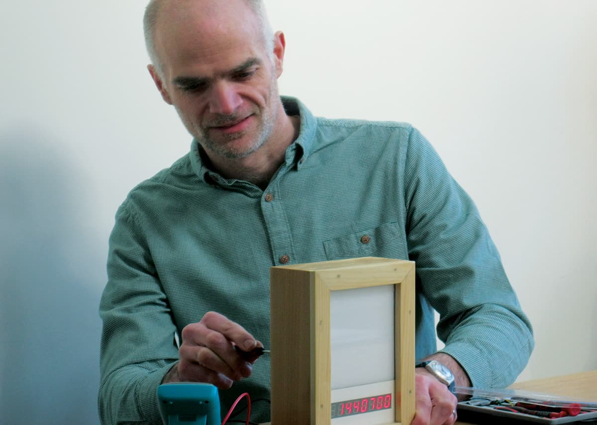
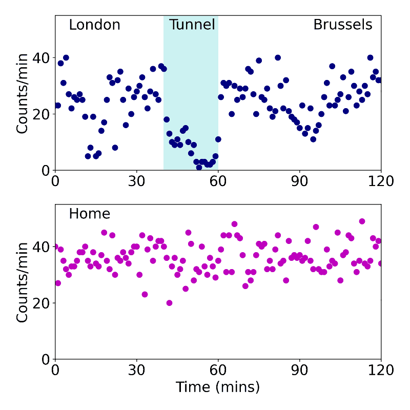
Inspired, I started to build muon detectors based on the Cosmic Watch design. But when I had got one working, I needed something to do with it. A work trip to Belgium on the Eurostar train presented one such opportunity. I took the detector with me (curiously, no questions were asked at security) and, sure enough, as we travelled through the Channel Tunnel between Britain and France, it recorded a lower rate of muons than at sea level. The sea and seabed were shielding the detector.

Muon counter graph in channel tunnel

As fun as that was, muonic measurements are better shared. So, along with another physicist parent, Lisa Ibberson, I got in touch with Kate Cooke, who teaches science at Coton Church of England Primary School in Cambridgeshire, where my son attends. Together, the three of us applied for money from the Institute of Physics School Grants Scheme. Our idea was to work with pupils to teach them about muons, get them to design a muon detector and finally install it in the school.

In July 2019 we were delighted to hear that our grant was successful and the real work began. We held an initial assembly at school that October using a water pistol and a few slides to introduce ourselves and muons. With the pupils in years 3 and 4 (ages 7–9), we drew pictures of the cascading particles resulting from a cosmic-ray air shower. Meanwhile, the children in years 4 and 5 (ages 8–10) were in charge of how the detector looked.

Together we decided that it should have a muon counter and a display that flashed different colours, depending on the muon’s energy. We jointly defined various parameters of the display, including its size, the colours of the flashes and the number of digits on the counter. Finally, we talked with year-6 pupils about the data the detector would produce, with the help of some edible Smartie bar graphs of course. And behind the scenes we were busy ordering printed circuit boards, soldering components and programming microcontrollers.

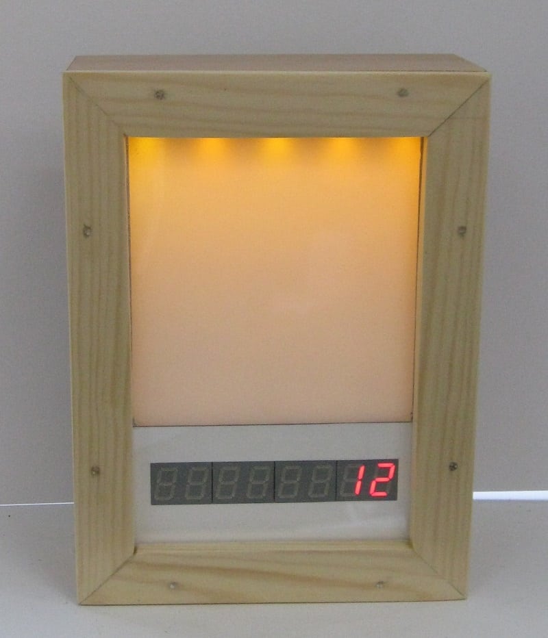
In January 2020 we returned to the year 4/5 class with a red flashing prototype encased in a shoe box. We got some great feedback. The colour red was no good – it was too much like a warning light and too bright. We therefore dimmed the display and democratically chose blue, green and amber for the colours. The pupils also told us we needed a switch to turn off the display when the flashing got too distracting. Finally, we decided to have eight digits in the detector so it could count to 99,999,999 muons – over roughly the time pupils spend in school (at a count every two seconds we were expecting about 15 million counts per year).

Muon detector for primary school outreach project

Unfortunately, when the pandemic struck we couldn’t continue to work directly with the children. Instead, over the summer I worked at home, quietly improving the detector’s electronics. In fact, on walks through the village where I live, people would often ask me what had happened to the muon project. I’d tell them we’d get back to it when we can and fortunately, by September 2020, schools re-opened and we started to think about the final switch-on.

Originally we envisaged a school assembly with a rowdy group countdown to the detector being turned on. We couldn’t do that with social-distancing measures in place, so instead planned a virtual switch-on for December 2020 with pupils from years 4 to 6 in their classrooms, Cooke at school, and me and Ibberson joining from our homes. And so, after revving the pupils up with a quiz to remind them about muons, we turned the device on.

It worked, phew! We then spent a few minutes watching the detector count up to 60, before opening the floor to questions. And, wow, what great questions they asked. How can a muon travel through 10 metres of concrete? Why do they decay into electrons? If the detector had a bigger area, would it count more muons? Has a detector like this ever been made before? Where do the cosmic rays that generate the muons come from?

Overall, the project was a great success. We had wonderful engagement from the pupils, who had contributed to the design of a fun scientific instrument. In fact, the switch-on of the detector, though virtual, was every bit as exciting as if it had happened in school. Looking to the future, we hope that this strange machine from a strange year counts up to 99,999,999 many times over its lifetime – and that it continues to provoke curiosity from primary-school pupils in Coton for years to come.

Flexible detector takes high-resolution X-ray images in 3D

While X-ray imaging is routinely employed in medical diagnosis and in industry for inspecting materials like semiconductors for defects, existing X-ray machines cannot image curved three-dimensional objects with high resolution. A team led by researchers at the National University of Singapore (NUS) and Fuzhou University in China has now developed a new flexible X-ray sensor that can do just this. The device, which relies on a series of nanoparticles that emit light for a long time after being excited with X-rays – a phenomenon known as persistent radioluminescence – might find use in healthcare applications such as portable X-ray detectors for mammography and imaging-guided therapeutics.

The X-ray detectors in today’s X-ray machines are usually flat panels in which each pixel has its own integrated circuit. This set-up makes the pixels bulky and limits the resolution of the detector, explains team member Xiaogang Liu at the NUS’ Department of Chemistry. The panels’ flatness also means the detectors struggle to capture images of curved objects.

Wrap-around device

In their work, Liu and colleagues focused on lanthanide-doped nanomaterials, which have unique luminescent properties that are already widely exploited in X-ray scintillation, optical imaging, biosensing and optoelectronics. They began by doping sodium lutetium fluoride (NaLuF4) nanocrystals with ions of the rare-earth element terbium (Tb3+). They then embedded the doped nanocrystals (which are denoted as NaLuF4:Tb@NaYF4) into silicone rubber to make a highly flexible X-ray detector that can be wrapped around 3D objects.

The next step was to excite the NaLuF4:Tb@NaYF4 with X-rays at an energy of 50 kV. When they did this, the team observed that the material emitted intense light long after the source of X-rays had been removed. The light persisted for more than 30 days, which means it can be used to image objects throughout this time.

Slow “hopping” charge carriers

Liu and colleagues explain that the light is emitted as the lutetium ions in the NaLuF4:Tb@NaYF4 lattice absorb the energy of the X-rays, generating many energetic electrons in the process. When X-ray photons collide with small fluoride ions in the material, flaws known as anion Frenkel defects form in the nanocrystal and trap the energetic charge carriers (electrons and holes) created. The prolonged radioluminescence in the material comes from these electrons slowly “hopping” through the crystal scaffold towards the Tb3+ ions and radiatively recombining with hole-Tb3+ centres, they say.

Liu acknowledges that other persistently luminescent materials already exist. Phosphors are one prominent example, and in 2011 a team at the University of Georgia, US, reported that ZnGa2O4:Cr3+ phosphors have an afterglow lifetime of approximately 15 days. However, Liu notes that these other materials are either not very sensitive to X-rays or are difficult to manufacture at the nanoscale, which makes them unsuitable for making flexible detectors.

Sub 25-micron image resolution

The NUS team’s imaging technique, which they call X-ray luminescence extension imaging (Xr-LEI), can be used to produce images with a resolution of less than 25 micrometres, the researchers say. “Many research groups, including ours, have been taking on challenges in X-ray imaging over the past few years,” Liu notes. “The technology we report on may provide a much-needed solution for imaging highly-curved 3D objects. It could be particularly suitable for applications like point-of-care X-ray radiography and screening mammography without having to compress the breast, which is uncomfortable for the patient.”

As well as healthcare applications, the technique might also be used to detect defects in electronic materials like semiconductors, authenticate works of art and to examine archaeological objects at the micron scale, he adds.

The researchers, who report their work in Nature, say they now plan to optimize the performance of their persistent luminescent nanomaterials to further reduce X-ray dosage and exposure time. “We will also be pursuing the development of dynamic X-ray imaging techniques that would benefit real-time monitoring of biological processes of living organisms,” Liu tells Physics World.

LATTICE radiotherapy plus immunotherapy shows promise for treating advanced bulky tumours

The combination of LATTICE radiation therapy (LRT), a spatially fractionated radiotherapy technique, and immunotherapy dramatically shrank a large metastatic lung cancer mass in one month and resulted in a complete local response within five months, according to a case report in Frontiers in Oncology. The patient, a woman with advanced non-small cell lung cancer and multiple metastases, did not experience any side effects from the high radiation dose she received. The dramatic effectiveness of this combined treatment is spearheading the development of future clinical studies.

Bulky tumours are challenging to treat using radiotherapy due to large tumour sizes and limitations of normal tissue toxicity. Spatially fractionated techniques such as high-dose 2D grid radiotherapy and LRT – a 3D lattice-like reconfiguration of grid therapy – have been employed to safely treat bulky tumours. LRT uses multiple high-dose regions (vertices), distributed within the tumour volume according to its size and shape and the proximity of critical structures, with valleys of much lower dose in between.

The technique’s principal developer, Xiaodong Wu from Executive Medical Physics Associates in Miami, FL, explains that when 3D LRT is delivered using intensity-modulated radiation therapy, volumetric modulated arc therapy or ion beams, highly customized peak-to-valley dose distributions can be generated within the tumour volume, while sparing skin and normal tissue.

Xiaodong Wu and Benhua Xu

LRT is currently used for palliative tumour debulking or boost treatments and to safely deliver high radiation doses to partial volumes of large tumours. It can be administered alone or in combination with conventionally fractionated radiotherapy.

Led by Benhua Xu, chief of the Department of Radiation Oncology at Fujian Medical University Union Hospital in China, this case study represents the first use of LRT in conjunction with immune checkpoint blockade – a treatment that help the body’s immune system recognize and attack cancerous cells.

The 33-year old patient was diagnosed with advanced invasive adenocarcinoma in the lower lobe of her right lung. Within months following surgery and chemotherapy, she had developed multiple metastases in both lungs, the thyroid, the spine and on the posterior chest wall. While the patient was receiving checkpoint inhibitor therapy, the metastatic mass in the posterior chest wall grew from 2.0 to 63.2 cm3, with maximum dimensions of 5.0 x 5.4 x 5.3 cm.

The team administered a single fraction of LRT (20 Gy prescribed to six high-dose vertices) to this fast-growing mass. The patient continued checkpoint inhibitor therapy, receiving six cycles over the next six months. She also underwent stereotactic body radiotherapy to treat multiple metastases and received drug therapies.

Although all of the patient’s metastatic lesions responded to the various palliative treatments, only the posterior chest wall tumour that received high-dose LRT achieved a complete response. While this tumour had not responded to the initial immune checkpoint blockade (anti-PD1), after high-dose LRT, it shrank by 77.84% within a month and continued to regress until achieving complete local response five months later. There were no toxic side effects in the area of the treatment, and the site remained disease free until the patient died several months later.

The researchers note that with only 6.5% of the large tumour’s volume receiving a dose of 20 Gy and higher, the effective uniform dose was calculated as 1.2 Gy. “Based on the traditionally understood mechanism of radiobiology, the probability of achieving complete local control with such a dose for a tumour of 63 cc would be nearly zero,” they write. “The synergetic effect combining high-dose LRT with anti-PD1 becomes a plausible speculation.”

They further hypothesize: “In high-dose LRT, the dose in the vertices are sufficiently high to induce neo-antigen release and initiate the cascade of antigen presenting cell (APC)-based T-cell priming. The dose in between the vertices is low enough to preserve internal tumour circulation/perfusion to potentially facilitate the infiltration of APCs and the primed cytotoxic T-cells. The highly heterogeneous dose configuration could reprogram the immunosuppressive tumour microenvironment to become more immunogenic, and when synergistically treated by checkpoint inhibitors, the primed T cells could attack tumour cells without being exhausted.”

Wu tells Physics World that the researchers are currently preparing systemic clinical studies using this combined treatment for patients with lung cancer, liver cancer, breast cancer and melanoma. He notes that interest in and use of LRT, and spatially fractionated radiotherapy in general, are increasing. Following the early clinical experiences primary made by Innovative Cancer Institute in Miami and Fujian Union Hospital, more institutions, including the Mayo Clinic in Rochester, have begun to offer LRT to their patients.

Strolling in the deep

At the bottom of the North Central Pacific Ocean, some 5000 m beneath the waves, lies a small, fist-sized black rock, with a knobbly surface texture, like a head of broccoli. It holds a secret at its heart – a single tooth, long ago shed by a shark swimming in the waters above. In the manner of a pearl forming around a piece of grit in an oyster, the tooth has become encapsulated by layers of waterborne minerals that settled out of the water around it. It took millions of years to reach its current size – but it shall grow no bigger.

A vast unmanned, electric submersible ploughs across the seabed like a bulldozer, heaving up our rock (among others) with its teeth, before sucking it up a hose to a ship waiting on the surface. These nodules are rich in metals like nickel, copper and cobalt – and industry has come to mine them. But the abyss is not empty, making this activity not without its victims. Our rock and its peers supported an abundance of life, from worms and starfish to crustaceans and ghost-like octopuses. As the mining machine lurches onwards, it leaves a trail of devastation in its wake – not to mention kicking up a lingering muddy cloud that chokes and smothers those survivors such as corals and sponges that are unable to flee and escape it.

Raising the alarm about this ecological vandalism against a realm about which we know precious little is the raison d’être of marine biologist Helen Scales’ beguiling new book The Brilliant Abyss: True Tales of Exploring the Deep Sea, Discovering Hidden Life and Selling the Seabed. With her light and engaging prose, Scales takes the reader on an introductory dive into the mysterious depths to reveal the myriad of life hidden within, from red and green bone-devouring worms that flourish whenever whales fall down to the abyss, to the world’s fishiest-smelling fish. There’s even a hunt for yetis – not of the elusive kind, but tiny, blind, pale crabs that survive living around deep-sea hydrothermal vents and cold seeps by farming bacteria to feast on. Like their abominable namesakes, however, they are very hairy.

As The Brilliant Abyss’ subtitle suggests, the work periodically segues into arresting tales from Scales’ career, from recovering experiments to determine what species of clams, worms and sea cucumbers colonize logs swept out to sea by floods and hurricanes, to weathering out high winds that suspended scientific activity during a research expedition in the Gulf of Mexico.

Fascinating titbits abound in Scales’ writing – including the revelation (to me, at least) that diving mammals such as whales and dolphins have evolved a special, “non-stick” form of the oxygen-carrying, haemoglobin-related protein myoglobin in their muscles. These each have a slightly negative electric charge that repels other myoglobin molecules, allowing the mammals to carry 10 times the protein that we do without the molecules clumping together and causing their bodies to go completely stiff.

Scales’ book also explores such fascinating cases as whether coronal mass ejections from the Sun could have contributed towards stranding numerous young male sperm whales in the North Sea in 2015; as well as explaining why some fish have scales that are blacker than the darkest material man has ever engineered – the multiwalled carbon nanotube, Vantablack, which is up to 99.965% absorbent. One thing that struck me while reading The Brilliant Abyss is that despite being an erstwhile student of geology and having learnt the names given to Earth’s past supercontinents, I don’t recall ever having given thought to the corresponding “superoceans” that surrounded them, such as Mirovia and Panthalassa. A shift of perspective is always fascinating.

It is perhaps in the final third of the book that Scales’ argument for the preservation of the deep from exploitation becomes most clear. She explores the medical potential of deep-sea organisms – such as sponges that harbour anti-cancer compounds – and weighs up the benefits and risks of farming the deep for food and mining it for its mineral resources, before calling for the reader to join her in campaigning for humanity to leave the deep free from excessive interference. It’s a compelling argument – although one that might perhaps have been more strongly seeded in the opening chapters of the work.

One mild disappointment of the book for me is that there are not more illustrations or pictures of the weird and wonderful creatures introduced in the text (at least in my preview copy). Scales’ descriptions may be beautifully written and highly evocative, yet a picture is, as the cliché goes, worth a thousand words. The exception is the gorgeous cover art of various deep-sea species by the artist Aaron Gregory, in a style that seems to evoke the illustrations of the German zoologist and artist Ernst Haeckel, whose work is discussed in the book.

This quibble aside, The Brilliant Abyss is a wonderfully written read that I would highly recommend – it’s the ideal plunge into the depths of Earth’s last great wilderness. 

  • 2021 Bloomsbury Sigma 352pp £16.99hb

Super Earth is astronomer’s dream for atmospheric studies

A newly discovered exoplanet called Gliese 486b could offer the best opportunity yet for studying the atmosphere of a terrestrial planet beyond the solar system. An international team, made up of astronomers at the CARMENES project and NASA’s TESS mission, showed that several aspects of Gliese 486b make it ideal for atmospheric spectroscopy. Indeed, team member Ben Montet of the University of New South Wales says, “This is the kind of planet we’ve been dreaming about for decades”. The discovery could improve our prospects for finding extrasolar atmospheres capable of supporting life.

Exoplanets orbit stars other than the Sun and in the three decades since the first one was found, the discovery of more than 4000 exoplanets have been confirmed by astronomers. Super Earths are among the most sought-after targets of all exoplanet searches. Slightly larger than Earth, these planets have stable, rocky surfaces and could have substantial atmospheres – which astronomers hope could support life in some cases.

Studying atmospheres typically involves observing how light from the exoplanet’s star is affected as it passes through the atmosphere when the exoplanet transits across the star when viewed from Earth – something that Gliese 486b does.

As this light shines through the atmosphere, certain wavelengths are absorbed by its component gases. Astronomers can use these characteristic absorption lines to determine the composition and temperature of the atmosphere. Alternatively, they can study light emitted or reflected by the planet, just before it passes behind the star.

Nearby red dwarf stars

CARMENES and TESS use complementary techniques to discover super Earths and determine whether they are suitable for further analysis. In their study, the researchers searched for exoplanets orbiting nearby red dwarf stars – which are far more likely to host rocky planets than Sun-like stars. Through their search, they detected a particularly interesting super Earth orbiting the star Gliese 486, just 26 light-years away.

The exoplanet Gliese 486b is three times the mass of Earth and 1.3 times the radius, yet it has an orbital period of just 36 hours – placing it extremely close to its star. The astronomers also predict that Gliese 486b has a surface temperature of around 430 °C – making it slightly cooler than Venus.

While these conditions may sound extreme, they are not harsh enough to strip away the exoplanet’s atmosphere. This could mean that Gliese 486b has retained some atmospheric hydrogen and helium from its initial formation. Moreover, the planet’s temperature is sufficiently high to puff out its atmosphere, without any gas escaping, making it ideal for spectroscopic studies. Indeed, the astronomers report that Gliese 486b is the best rocky planet ever discovered for studying with emission spectroscopy; and the second best for transmission spectroscopy.

Although red dwarfs are seen as strong candidates for hosting habitable exoplanets, their high levels of stellar activity also threaten to destroy their exoplanets’ atmospheres. Through further studies of Gliese 486b’s atmosphere, astronomers will be able to better assess whether the search for extrasolar life should be focused on red dwarfs.

The research is described in Science.

The development of new ionic electrolytes for energy storage devices

Want to learn more on this subject?

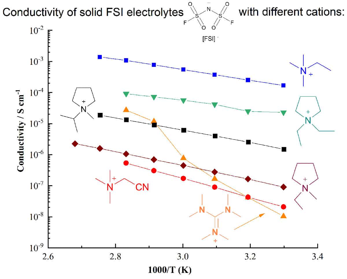
Electrolyte development is a critical component in the quest for higher-performing energy storage devices. Ionic electrolytes such as ionic liquids, plastic crystals and their polymer composites can offer important safety and performance advantages over traditional molecular-solvent based systems, particularly for devices utilizing reactive metals such as lithium or sodium.

An important approach to developing ionic electrolytes that can meet the complex challenges of next-generation electrochemical devices is increasing the range of known and well-characterized electrolyte materials. Understanding how different ion structures affect the physical, thermal, electrochemical properties, and the phase behaviour when combined with Li salts, is vital to optimizing device performance.

This webinar, hosted by Jenny Pringle, will overview our recent work on the design and use of new ionic materials and their application as liquid, quasi-solid state, composite or very high Li salt content electrolytes.

Want to learn more on this subject?

Prof. Jenny Pringle works at the Institute for Frontier Materials at Deakin University, Australia. She is a chief investigator in the ARC Centre of Excellence for Electromaterials Science (ACES) and the ARC Industrial Transformation Training Centre “StorEnergy”. She received her degree and PhD at The University of Edinburgh in Scotland, UK, before moving to Monash University, Australia, in 2002. From 2008–2012 she held an ARC QEII Fellowship, investigating the use of ionic electrolytes for dye-sensitized solar cells. Pringle moved to Deakin University in 2013. There she leads research into the development of new ionic liquids and organic ionic plastic crystals for applications including thermal energy harvesting, gas separation membranes, and lithium and sodium batteries.

Nanoparticle sensors detect arsenic in drinking water

“About 785 million people are living without access to safe and clean drinking water; 140 million people in more than 50 countries have been exposed to arsenic-contaminated water.” These were the stark opening statements from Muhammad Abbas, speaking at the recent APS March meeting.

Arsenic poisoning is one of the most significant public health concerns worldwide. Arsenic is used in semiconductors, pharmaceuticals, wood preservatives, insecticides and chicken feed, and it leaches into the groundwater. Long-term exposure can lead to cancers of the kidney, liver, lungs and skin, as well as causing skin diseases and other health issues such as hypertensive heart disease. As such, the World Health Organization and Environmental Protection Agency recommend a maximum limit of 10 µg/l arsenic in drinking water.

Despite these serious health concerns, testing for arsenic in water currently requires expensive laboratory instruments that cannot be used for on-site detection and are unsuitable for developing nations. Abbas and colleagues at LUMS in Pakistan hope to address this shortfall by designing a low-cost sensor that can detect arsenic in drinking water. “We aim to develop a sensor that’s sensitive and selective, robust and reliable, affordable, portable and easy to use for local technicians,” he said.

The sensor will be based on gold nanoparticles (AuNPs), which are excellent candidates for sensing applications as they absorb in the visible spectrum and change colour according to their size, shape and surface chemistry. To create their sensor, Abbas and colleagues coated AuNPs with dihydrolipoic acid. This coating stabilizes the nanoparticles, which form in a dispersed state in water and are wine-red in colour. Adding an electrolyte such as salt does not affect the AuNPs, which remain dispersed and stay red.

If the water contains arsenic, however, the arsenic will bind to the dihydrolipoic acid, making it unavailable to protect the AuNPs surfaces. In this scenario, adding salt causes the AuNPs to aggregate and change colour. “This aggregation is directly proportional to the amount of arsenic present, which decides the strength in the colour change,” explained Abbas, now a PhD student at the University of Texas at Dallas.

Colour change

To test this approach, the team performed UV-visible spectroscopy on AuNP solutions containing different concentrations of arsenic. “We could see a visual colour change with increased amounts of arsenic, with the nanoparticles changing from red towards blue,” said Abbas. Scanning electron microscopy images of the AuNPs before and after addition of arsenic confirmed the clumping mechanism that caused the colour change.

The sensor’s detection limit was 50 µg/l (50 parts per billion) of arsenic when viewed with the naked eye, or 3 µg/l using UV-visible spectroscopy. This sensitivity is lower than that offered by existing high-tech methods, such as atomic fluorescence spectroscopy, atomic absorption spectroscopy or mass spectrometry, which can detect up to parts per trillion of arsenic. But Abbas emphasizes that these systems are costly, not portable and need trained personnel to operate them.

“The colourimetric method is affordable and portable. The limit of detection is lower right now, but it can be improved,” he said.

The team also investigated potential interference from a range of other metal contaminants and found that, with the exception of mercury, none of the metals interfered strongly, and even mercury only impacted the absorption spectrum slightly.

“This suggests that the sensor would be selective and not disturbed by other elements present in the drinking water,” Abbas explained. “In future, this method may lead to the design of a microfluidic device for detection of arsenic.”

Raman imaging microscopy: The analytical multi-tool

Want to learn more on this subject?

Raman spectroscopic imaging is a powerful, versatile and increasingly common microscopy technique that can quickly identify molecules in a sample and visualize their distribution in three dimensions. This fast, nondestructive and label-free chemical characterization method offers enormous potential to physicists in many fields of research.

Modular Raman microscopes can be integrated with advanced cooling stages for cryogenic measurements, with environmental enclosures for remote operation, and even within vacuum chambers for advanced structural analyses.

This presentation will describe the principles of 3D Raman imaging and the speakers will show in detail how to access chemical imaging at the highest spatial resolution.

Comprehensive analyses of samples often require a combination of different techniques. Structural information on a sample’s surface can be obtained by Atomic Force Microscopy or Scanning Electron Microscopy (SEM). Raman imaging can reveal its chemical composition and by combining the techniques, structural and chemical information can be easily acquired from the same sample position. These approaches will be described and the power of correlative Raman-AFM and Raman-SEM imaging for analysis will be illustrated in the contexts of 2D materials development, cryogenic research, pharmaceutical sciences, geosciences, battery research and life sciences.

Want to learn more on this subject?

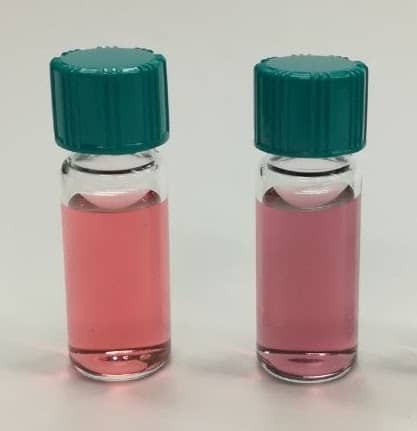
Thomas Dieing is technical product manager for the WITec alpha300 product line and its accessories. He obtained his PhD from La Trobe University, Melbourne, Australia, in 2005 investigating the MBE growth of nitrogen containing III/V semiconductors. In 2006 he joined WITec’s application team and became director of applications and support. In his role as product manager since 2019 he is responsible for all activities related to the product development process.


Ute Schmidt studied physics at the Babes Bolyai University in Cluj-Napoca, Romania, and obtained her PhD from the University of Karlsruhe, Germany. Through her work with in situ scanning tunneling microscopy in an electrochemical environment, she was introduced to scanning probe microscopy. During her postdoctoral scholar position at Karlsruhe and North Carolina State University in Raleigh, USA, she continued to work with metal deposition on different substrates with STM and Atomic Force Microscopy. Schmidt worked for six years as a project manager at Molecular Imaging Corp., Phoenix, USA. She has been an applications manager at WITec since 2003.

Alan Turing £50 note is unveiled, how to get a mortgage on the Moon

The Bank of England has unveiled the final design of its new £50 polymer banknote that features the mathematician and wartime codebreaker Alan Turing. The new £50 – the last of the bank’s notes to go from paper to polymer – will come into circulation on 23 June, which is the 109th anniversary of Turing’s birth. “He was a leading mathematician, developmental biologist and a pioneer in the field of computer science,” says Bank of England governor Andrew Bailey. “He was also gay and was treated appallingly as a result. By placing him on our new polymer £50 banknote, we are celebrating his achievements, and the values he symbolizes.”

As with the other new polymer notes there are a series of security features such as holograms and foil patches to make them more difficult to forge. And to mark the occasion, the UK’s intelligence agency GCHQ has set the “Turing Challenge”, which consists of twelve puzzles that increase in complexity leading to one final answer. The agency says it is their toughest puzzle yet, but surely it can’t be as hard as cracking the Enigma code machine.

Turing was certainly a visionary in several scientific fields, but if he were around today would he invest in a house on the Moon? If you are interested in living in what would surely be an ultralow density development, all you need is a £4.4 million deposit to get on the lunar property ladder. That is the claim of the website Money.co.uk, which reckons that the first house on the Moon would sell for a little over £44 million. You can read more about this hypothetical housing development in “How to get a mortgage on the Moon!”.

Three top atomic clocks are compared with record accuracy

The time kept by three of the world’s best atomic clocks has been compared by connecting them using optical fibres and an over-air link. The comparisons were done by the Boulder Atomic Clock Optical Network Collaboration in the US and are ten times more accurate than previous attempts. The measurements have revealed unexpected variations in the time kept by the clocks, which could provide insights into how the devices could be improved. The research could play an important role in developing a new standard for the second, which would involve distributing and comparing atomic-clock time signals throughout the world.

Atomic clocks use the frequency of a specific atomic transition as an extremely stable time standard. While the second is currently defined by caesium-based clocks that operate at a microwave frequencies, physicists have built much more accurate clocks that are based on light. These optical clocks tick at much higher frequencies than microwave clocks and can keep time that is accurate to about one part in 1018, which is about 100 times better than the best caesium clocks.

The international metrology community aims to replace the microwave time standard with an optical clock, but first must choose from one of several clock designs being developed worldwide. To evaluate and improve these optical clocks – and ultimately create a global network of time standards – researchers must be able to compare their time signals. This can be done using an optical fibre connection or by transmitting optical signals through the air. Indeed, air transmission could play an important role in deploying optical clocks in satellites, where microwave clocks are currently used.

In this latest work, David Hume and colleagues at the US’s National Institute of Standards and Technology (NIST) and the University of Colorado have compared time signals of three optical clocks, which are all located in Boulder Colorado. One clock uses ytterbium atoms, another strontium atoms and the third a combination of aluminium and magnesium ions.

Frequency combs

A 3.6 km optical fibre link was used to compare the ratio of frequencies of the ytterbium and strontium clocks, which were located at NIST and the University of Colorado respectively. The strontium and aluminium–magnesium clocks (the latter located at NIST) were compared using a 1.5 km over-air optical link between two buildings. This involved the use of frequency combs, which allow signals at very different frequencies to be compared.

The over-air technique is also relatively immune to disturbances caused by turbulence in the air. Indeed, the team found that the fibre and free-space links offered similar levels of performance – the exception being when the free-space link was operated during a snowstorm.

The ytterbium and aluminium–magnesium clocks were in different labs at NIST and were compared using a fibre connection.

“State-of-the-art”

The team managed to measure ratios of frequencies of the three pairs of  clocks at an accuracy of one part in 1018, which is an order of magnitude improvement on the previous record of one part in 1017. Hume describes the work as “state-of-the-art for both fibre-based and free-space measurements”. These ratios are natural constants, so the team points out that their results are the three most accurate measurements ever made of natural constants.

The clocks were compared over a period of several months and the researchers found unexpected day-to-day variations in their time keeping. This suggests that the team does not have a complete understanding of what affects the performance of the clocks – which means that further improvements could be possible.

As well as improving the definition of the second, better comparisons of optical clocks could benefit other branches of science. Two clocks at different elevations will run at slightly different rates and this could be used to measure tiny shifts in the Earth’s crust, caused for example by the melting of ice sheets or rising sea level. Differences between clocks could also be used to try to detect dark matter.

The measurements are described in Nature.

Copyright © 2026 by IOP Publishing Ltd and individual contributors