Raman spectroscopic imaging is a powerful, versatile and increasingly common microscopy technique that can quickly identify molecules in a sample and visualize their distribution in three dimensions. This fast, nondestructive and label-free chemical characterization method offers enormous potential to physicists in many fields of research.
Modular Raman microscopes can be integrated with advanced cooling stages for cryogenic measurements, with environmental enclosures for remote operation, and even within vacuum chambers for advanced structural analyses.
This presentation will describe the principles of 3D Raman imaging and the speakers will show in detail how to access chemical imaging at the highest spatial resolution.
Comprehensive analyses of samples often require a combination of different techniques. Structural information on a sample’s surface can be obtained by Atomic Force Microscopy or Scanning Electron Microscopy (SEM). Raman imaging can reveal its chemical composition and by combining the techniques, structural and chemical information can be easily acquired from the same sample position. These approaches will be described and the power of correlative Raman-AFM and Raman-SEM imaging for analysis will be illustrated in the contexts of 2D materials development, cryogenic research, pharmaceutical sciences, geosciences, battery research and life sciences.
Thomas Dieing is technical product manager for the WITec alpha300 product line and its accessories. He obtained his PhD from La Trobe University, Melbourne, Australia, in 2005 investigating the MBE growth of nitrogen containing III/V semiconductors. In 2006 he joined WITec’s application team and became director of applications and support. In his role as product manager since 2019 he is responsible for all activities related to the product development process.
Ute Schmidt studied physics at the Babes Bolyai University in Cluj-Napoca, Romania, and obtained her PhD from the University of Karlsruhe, Germany. Through her work with in situ scanning tunneling microscopy in an electrochemical environment, she was introduced to scanning probe microscopy. During her postdoctoral scholar position at Karlsruhe and North Carolina State University in Raleigh, USA, she continued to work with metal deposition on different substrates with STM and Atomic Force Microscopy. Schmidt worked for six years as a project manager at Molecular Imaging Corp., Phoenix, USA. She has been an applications manager at WITec since 2003.
The Bank of England has unveiled the final design of its new £50 polymer banknote that features the mathematician and wartime codebreaker Alan Turing. The new £50 – the last of the bank’s notes to go from paper to polymer – will come into circulation on 23 June, which is the 109th anniversary of Turing’s birth. “He was a leading mathematician, developmental biologist and a pioneer in the field of computer science,” says Bank of England governor Andrew Bailey. “He was also gay and was treated appallingly as a result. By placing him on our new polymer £50 banknote, we are celebrating his achievements, and the values he symbolizes.”
As with the other new polymer notes there are a series of security features such as holograms and foil patches to make them more difficult to forge. And to mark the occasion, the UK’s intelligence agency GCHQ has set the “Turing Challenge”, which consists of twelve puzzles that increase in complexity leading to one final answer. The agency says it is their toughest puzzle yet, but surely it can’t be as hard as cracking the Enigma code machine.
Turing was certainly a visionary in several scientific fields, but if he were around today would he invest in a house on the Moon? If you are interested in living in what would surely be an ultralow density development, all you need is a £4.4 million deposit to get on the lunar property ladder. That is the claim of the website Money.co.uk, which reckons that the first house on the Moon would sell for a little over £44 million. You can read more about this hypothetical housing development in “How to get a mortgage on the Moon!”.
The time kept by three of the world’s best atomic clocks has been compared by connecting them using optical fibres and an over-air link. The comparisons were done by the Boulder Atomic Clock Optical Network Collaboration in the US and are ten times more accurate than previous attempts. The measurements have revealed unexpected variations in the time kept by the clocks, which could provide insights into how the devices could be improved. The research could play an important role in developing a new standard for the second, which would involve distributing and comparing atomic-clock time signals throughout the world.
Atomic clocks use the frequency of a specific atomic transition as an extremely stable time standard. While the second is currently defined by caesium-based clocks that operate at a microwave frequencies, physicists have built much more accurate clocks that are based on light. These optical clocks tick at much higher frequencies than microwave clocks and can keep time that is accurate to about one part in 1018, which is about 100 times better than the best caesium clocks.
The international metrology community aims to replace the microwave time standard with an optical clock, but first must choose from one of several clock designs being developed worldwide. To evaluate and improve these optical clocks – and ultimately create a global network of time standards – researchers must be able to compare their time signals. This can be done using an optical fibre connection or by transmitting optical signals through the air. Indeed, air transmission could play an important role in deploying optical clocks in satellites, where microwave clocks are currently used.
In this latest work, David Hume and colleagues at the US’s National Institute of Standards and Technology (NIST) and the University of Colorado have compared time signals of three optical clocks, which are all located in Boulder Colorado. One clock uses ytterbium atoms, another strontium atoms and the third a combination of aluminium and magnesium ions.
Frequency combs
A 3.6 km optical fibre link was used to compare the ratio of frequencies of the ytterbium and strontium clocks, which were located at NIST and the University of Colorado respectively. The strontium and aluminium–magnesium clocks (the latter located at NIST) were compared using a 1.5 km over-air optical link between two buildings. This involved the use of frequency combs, which allow signals at very different frequencies to be compared.
The over-air technique is also relatively immune to disturbances caused by turbulence in the air. Indeed, the team found that the fibre and free-space links offered similar levels of performance – the exception being when the free-space link was operated during a snowstorm.
The ytterbium and aluminium–magnesium clocks were in different labs at NIST and were compared using a fibre connection.
“State-of-the-art”
The team managed to measure ratios of frequencies of the three pairs of clocks at an accuracy of one part in 1018, which is an order of magnitude improvement on the previous record of one part in 1017. Hume describes the work as “state-of-the-art for both fibre-based and free-space measurements”. These ratios are natural constants, so the team points out that their results are the three most accurate measurements ever made of natural constants.
The clocks were compared over a period of several months and the researchers found unexpected day-to-day variations in their time keeping. This suggests that the team does not have a complete understanding of what affects the performance of the clocks – which means that further improvements could be possible.
As well as improving the definition of the second, better comparisons of optical clocks could benefit other branches of science. Two clocks at different elevations will run at slightly different rates and this could be used to measure tiny shifts in the Earth’s crust, caused for example by the melting of ice sheets or rising sea level. Differences between clocks could also be used to try to detect dark matter.
Hamish Johnston visited NIST in 2019 and spoke to David Hume and other atomic-clock experts. You can hear those conversations in this episode of the Physics World Weekly podcast.
Reduced radiation dose and shorter exam times for PET/CT scans are big advantages for cancer patients, who may require multiple scans during their treatment and who may find it challenging to remain motionless during a typical 20 min PET scan. Radiologists at the Wright Center of Innovation at Ohio State University Wexner Medical Center have managed to reduce PET scan duration to approximately 2 min, thereby reducing patient discomfort, minimizing motion artefacts and improving patient throughput.
Michelle Knopp.
At the recent European Congress of Radiology (ECR 2021), Michelle Knopp discussed the team’s latest beneficial innovation: lowering the 18F-FDG radiotracer dose from the standard-of-care (480 MBq) to the lowest amount allowable by product label (185 MBq). Results of a phase II prospective study of 105 cancer patients revealed that images acquired by an ultrafast 2 min PET/CT scan, using a 185 MBq radiotracer dose, are of diagnostic quality in patients with a body mass index (BMI) under 35.
The researchers performed 18F-FDG PET imaging 60–75 minutes post-injection using both conventional (90 s per bed position) and investigational (9 s per bed position) acquisitions. For the ultrafast acquisition, the researchers used an organ-, BMI-adaptive regularized reconstruction, which takes into account the bed location and the patient’s BMI. For the 90 s acquisitions, they employed standard EARL reconstruction techniques.
Two blinded readers independently performed visual and quantitative assessment for each data set. Knopp reported that for patients with a BMI under 35, both sets of images were of diagnostic quality. However, for patients with a BMI of 35 or greater, only eight (35%) of the ultrafast, low-dose exams were of diagnostic quality.
“Combining the third-dose with the ultrafast acquisition (9 s versus 90 s per bed position, for a total acquisition time of 2 min versus 20 min) represents an exam time that is one-tenth of the normal time,” explained Knopp. “This is a critical point for many patients, as faster imaging allows for less patient discomfort, leading to less motion artefacts. Additionally, faster acquisition opens the door to dynamic imaging of tracer uptake, which may help reveal additional clinical information about therapy response in cancer patients, and activity of infection in patients with infection.”
“Using these techniques, we are acquiring the source data at about one twentieth of the normal count density,” she added. “Previous reconstruction methodology was generating very noisy and blobby images. The adaptive regularized PET reconstruction approach has opened the door to EARL-equivalent diagnostic image quality.”
Knopp tells Physics World that based on the findings of the phase II study, new research will focus on 15 s per bed position (3 min total acquisition time) whole-body PET scans for patients with higher BMIs. She and her colleagues are currently conducting a phase III clinical trial to investigate this approach.
PET-based lymph node assessment
Surgical sampling of axillary lymph nodes in newly diagnosed breast cancer patients is essential to assess their nodal status and plan suitable treatment. But this procedure can cause discomfort and pain for the remainder of a survivor’s life. Being able to accurately determine nodal status with diagnostic imaging could eliminate unnecessary surgical sampling in node-negative patients.
At ECR 2021, Janna Morawitz, from the Institute of Diagnostic and Interventional Radiology at the University Hospital of Düsseldorf, presented the findings of a study comparing the use of four imaging modalities to assess nodal status. The multi-institute analysis revealed that thoracic 18F-FDG PET/MRI outperformed axillary sonography, breast MR and thoracic MRI in determining the axillary lymph node status of women with newly diagnosed invasive breast cancer.
The study included 44 patients with positive nodes and 68 with negative nodes, based on histopathological diagnosis. All patients underwent all four imaging exams. Thoracic PET/MRI exhibited both the highest accuracy (90.18%) and sensitivity (81.8%) of the four modalities, followed by axillary sonography, with an accuracy of 87.04% and a sensitivity of 69.1%.
Thoracic PET/MRI also had the highest negative predictive value, at 89.0%, followed by axial sonography at 83.3%. However, axillary sonography, the most commonly used imaging exam, had the highest specificity (98.5%).
Morawitz reported that eight patients with positive lymph nodes were missed by PET/MRI, but that these were also missed by the other three modalities. In total, axillary sonography generated 13 false negatives, followed by thoracic MRI (16) and breast MRI (17). Axillary sonography had the lowest number of false positives, with only one case, followed by thoracic MRI with two cases, and then breast MRI and thoracic PET/MRI with three each.
“In a clinical setting, the combination of PET/MRI and axillary sonography might be considered to provide even more safety in making a diagnosis and in reducing the number of unnecessary surgical samplings performed,” said Morawitz. “Radiologists could use PET/MRI as a searching tool because of its high sensitivity, and add axillary sonography afterwards to specific findings if suspicious lymph nodes are identified.”
A high-profile publication that claimed to have discovered the elusive Majorana quasiparticle has been retracted after a re-analysis found no such evidence. In 2018 Leo Kouwenhoven from the Delft University of Technology (TU Delft) and colleagues declared they had found the particle in an extremely thin semiconductor nanowire covered by a superconducting layer (Nature556 74). Yet on 8 March Nature published a retraction after “inconsistencies” with the original analysis came to light. An independent report commissioned by TU Delft, however, found no instances of data fabrication.
The Majorana fermion, a particle that is its own antiparticle, is the brainchild of Italian theoretical physicist Ettore Majorana, who disappeared mysteriously in 1938 after boarding a ferry from Naples to Palermo. It is not a discrete particle but a quasiparticle and consists of two paired electrons. The pair exist in two energy states as well as in a superposition state, just like “qubits” that consist of electrons or photons.
If we do not deliver eye-popping breakthroughs, the entire research direction gets cancelled and we need to do something else
Sergey Frolov
Four years later, Microsoft set up the Microsoft Quantum Lab at TU Delft in 2016, with Kouwenhoven as its director. In 2018 he and his team published the paper in Nature claiming to have detected the Majorana quasiparticle in an extremely thin semiconductor nanowire covered by a superconducting layer. At a temperature of 0.02 K, they showed that two electrons paired up at the end of the wire with one electron in the semiconducting part and the other electron in the superconducting layer. However, the team could only prove the existence of one electron pair and not the existence of the second electron pair that together would have formed a Majorana qubit.
Several physicists, though, were unconvinced. Sergey Frolov at the University of Pittsburgh and Vincent Mourik at the University of New South Wales independently analysed the result, discovering “several inconsistencies” that led them to conclude there was no proof of a Majorana qubit. As a result of these criticisms, Kouwenhoven and co-workers re-analysed the raw data and rebuilt the experimental set-up to recalibrate certain parameters – finding that the results were inconsistent with a quantized Majorana conductance. “We apologize to the community for insufficient scientific rigour in our original manuscript,” the authors wrote in a retraction published on 8 March (Nature 10.1038/s41586-021-03373-x).
On the same day as the retraction, an independent report written by four physicists that was commissioned by TU Delft concluded there were no instances of data fabrication. “There was some degree of data selection in what was published,” says Patrick Lee from Massachusetts Institute of Technology, who was one of the authors of the report. “I don’t think this was done with malice. I think they were caught up at the excitement of the moment.” Lee notes that “some mistakes” were made by the team such as a calibration error, which they discovered after the publication of the paper. “They were aware of it and they came out with it without holding back anything,” says Lee. Indeed, Lee adds that he does not think that Microsoft is in trouble given its links to TU Delft. “It is a setback, but it should not derail the whole enterprise,” adds Lee.
Frolov says that quantum research is vulnerable to mistakes given it is complex science and that many teams are racing to build quantum computers. “We do not have the luxury of being supported to do the same thing for decades – we operate on 3- to 5-year grant-renewal cycles,” says Frolov. “And if we do not deliver eye-popping breakthroughs, the entire research direction gets cancelled and we need to do something else. This is the root cause of hype that we get criticized for.” Frolov adds that researchers will now have to look for different ways of creating Majorana qubits.
Theoretical physicist Michael Wimmer from TU Delft says that some in the field are now using “Majorana mode” for condensed-matter systems to distinguish it from the “fundamental” particle. “From a theoretical perspective, we know that it is possible to make Majorana modes in a suitable system – the theory behind it is well understood,” he says. “The question is whether such conditions were successfully achieved in an experiment so far.” Meanwhile, Das Sarma says that the retraction of the Delft paper “means little to the subject”. He is convinced that his group’s findings stand and says the ones from Delft will too. “The same experiment with better samples should show Majorana [particles],” he says.
A new image showing magnetic fields surrounding the supermassive black hole M87* has been created by scientists working on the Event Horizon Telescope (EHT). The magnetic structure was mapped by measuring the polarization of the light emitted by matter in the hot region around the black hole. Understanding the magnetic properties of that region could provide important insights into how powerful jets of radiation and matter are emitted by some black holes.
In 2019 the EHT made history by capturing the first image of the shadow of a black hole. This is a dark region surrounding a black hole that is expected to be about three times the diameter of the black hole’s event horizon, which is the point beyond which even light cannot escape a black hole. The supermassive black hole is called M87* and is located at the centre of a galaxy about 55 million light-years away. From the image, the team worked out that M87* has a mass of about 6.5 billion times that of the Sun. Back in 2012, astronomers using the EHT were also able to see the base of a powerful jet that blasts out about 5000 light-years from M87*.
Now, scientists working on the EHT have analysed the polarization of light from the bright region surrounding the shadow. There, some matter is being sucked into the black hole while other matter is being blasted out in jets. How these jets are formed is a matter of debate amongst astrophysicists but understanding the magnetic fields near supermassive black holes could provide important clues.
Strongly magnetized gas
The region surrounding the shadow is hot and violent region and therefore large amounts of light are created as matter is accelerated. If strong magnetic fields are present, then the emitted light will be polarized. Using models to analyse the observed polarization, EHT scientists have concluded that only the presence of a strongly magnetized gas can explain their observations.
Team member Monika Mościbrodzka at Radboud University in the Netherlands describes the measurements as “the next crucial piece of evidence to understand how magnetic fields behave around black holes, and how activity in this very compact region of space can drive powerful jets”.
Her colleague Jason Dexter at the University of Colorado Boulder adds, “the observations suggest that the magnetic fields at the black hole’s edge are strong enough to push back on the hot gas and help it resist gravity’s pull. Only the gas that slips through the field can spiral inwards to the event horizon.”
The data used to create the image were acquired in 2017 and Iván Martí-Vidal at the University of Valencia spoke to the challenge of creating the image: “unveiling this new polarized-light image required years of work due to the complex techniques involved in obtaining and analysing the data”.
The research is described in an observational paper and a theory paper, which are both published in The Astrophysical Journal Letters.
This episode of the Physics World Weekly podcast features an interview with the physics PhD student Elham Fadaly, who is a runner-up for 2020 Nanotechnology Young Researcher Award for making an important breakthrough in semiconductor technology. Indeed, that breakthrough was finding the Holy Grail of optoelectronics: a silicon-based material that is an efficient emitter of light. Fadaly chats about her semiconductor research and plans for the future with Physics World‘s Tami Freeman.
Then, we delve into lurid world of publication misconduct with our guide Kim Eggleton, who is tasked with maintaining the trustworthiness of research published by Institute of Physics Publishing. Eggleton explains why some researchers are tempted to inflate their publication records using scams such as buying authorships on papers and peer-review manipulation. She also explains what IOP Publishing is doing to detect and prevent misconduct – including a Peer Review Excellence training and certification programme for reviewers.
“It’s not just about navigating the leap from the lab to market, but also from the market back into the lab,” said Anita Mahadevan-Jansen, at the recent Photonics West BiOS conference.
Presenting in the BiOS Hot Topics session, Mahadevan-Jansen explained that, in addition to the various essential stages required to translate a new medical technique from the laboratory into the clinic, feedback in the other direction is also invaluable to optimize a newly launched technology for patient care. She illustrated this idea with an application developed by her research group at Vanderbilt University – the use of optical spectroscopy to guide endocrine surgery.
One of biggest challenges during surgery of the endocrine glands (usually the thyroid or parathyroid glands) is protecting the parathyroid gland, which is the only organ in body that regulates calcium. The post-operative rate of hypoparathyroidism, which causes blood calcium levels to fall and blood phosphorus levels to rise, can be up to 60%. This problem arose because the only way for a surgeon to find, and avoid, parathyroid glands was by visual inspection.
“In our lab, we discovered that the parathyroid glands have a really strong autofluorescence in the near-infrared,” said Mahadevan-Jansen. The team showed that parathyroid tissue exhibited up to 10 times greater fluorescence signal than other tissues in the neck. In a study of 137 patients having thyroid or parathyroid surgery, detecting this autofluorescence identified both normal and diseased parathyroid glands, in real time, with 97% accuracy.
Mahadevan-Jansen described the device’s progress from the initial discovery in 2008 to a clinical system that was FDA approved in 2018 and CE marked earlier this year. “This was a long journey considering that it is simply based on autofluorescence, no dyes, and the technology is fairly straightforward, just looking at the ratio of signal between two tissue types,” she said.
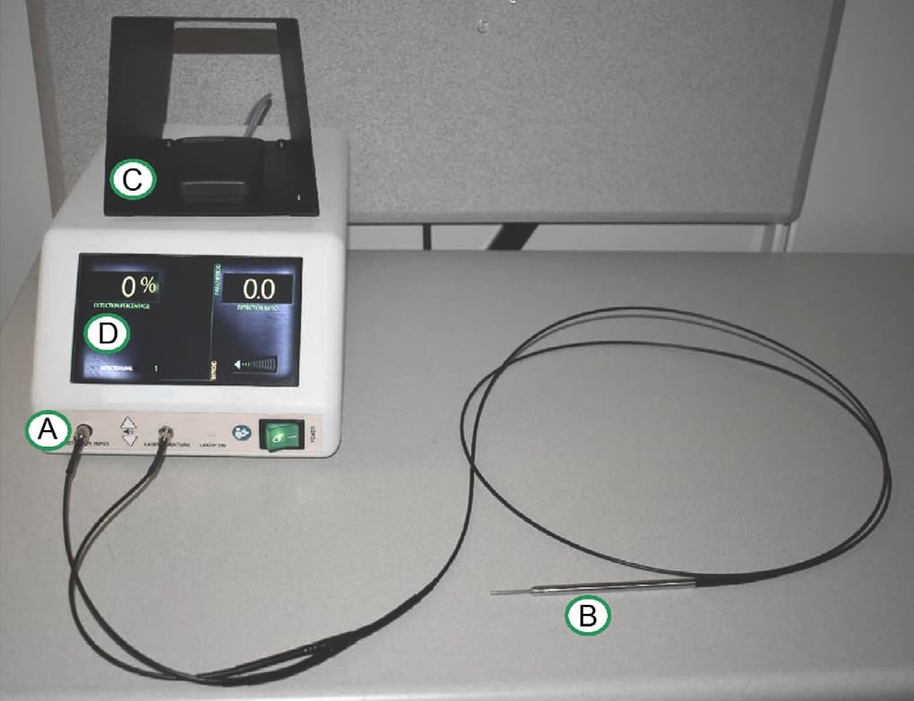
The PTeye prototype: (A) console enclosing the laser and detector; (B) detachable fibre-optic probe, (C) foot-pedal; (D) display. (Courtesy: Surgery 10.1016/j.surg.2018.04.079)
In collaboration with Ai Biomed, the team developed a probe-based clinical system called the PTeye. The device is used intra-operatively, and emits a sound when the fibre-optic probe is near parathyroid tissue. “Yet we came to realise that just because a technology is approved and launched into the surgical community, this does not mean our job is done. The lessons we’re learning since its launch have been tremendous,” Mahadevan-Jansen noted.
The researchers had demonstrated that the autofluorescence technique could find parathyroid glands, but once the device began being used in a clinical setting, they needed to understand how would affect patient care, as well as how surgeons are using the new technology. As such, the PTeye is now being studied in two ongoing clinical trials at four sites, with surgeons using it to guide their surgical procedures in both thyroidectomy and parathyroidectomy cases.
Early results from the trials demonstrate that the technology has an accuracy of 94.3% for identifying parathyroid tissue, with a positive predictive value of 93% and a negative predictive value of 100%. The team saw that the device was definitely helpful, even for an experienced surgeon, particularly in long and complex cases. And for a less experienced surgeon, PTeye had a significant impact, both in building confidence and increasing their efficiency.
“It’s still too early to see the long-term post-operative hypoparathyroidism rate, but I hope to have that over the next year as we continue to accrue patients,” said Mahadevan-Jansen.
And as more surgeons began to use this new device, the team started to see other anecdotal issues arise. For example, the system requires five initial measurements on the thyroid to establish a baseline; but if this baseline is defined improperly, this can result in false positives. Establishing and sharing a procedure to achieve accurate baselining is essential. Another finding is that brown fat, often seen in younger patients, has bright autofluorescence in a similar spectral region.
The team now needs to tackle these issues and understand their impact on the use of the device. Other ongoing challenges include addressing the cost associated with single-use probes, determining whether imaging or spectroscopy is preferable and investigating the identity of the fluorophore – while parathyroid tissue exhibits bright autofluorescence at 822 nm, it’s not clear exactly what this emission is due to.
“We have developed near-infrared autofluorescence as a clinical guidance tool for neck surgery, to confirm the parathyroid,” Mahadevan-Jansen concluded. “This technology is now available commercially, both as a fibre-optic probe-based device and an imaging device. Acceptance by clinicians is still in progress but we are starting to see surgeons really excited about using this.”
A new imaging algorithm devised by researchers in the US could improve our ability to track space junk orbiting the Earth. Through simulated tests, the team has showed how a cross-correlation of the signals reflected by a piece of debris could be used to extract high-resolution, undistorted images of how an object spins as it travels through space – allowing it to be tracked more accurately. Their algorithm could soon prove invaluable in protecting satellite systems from colliding with space junk.
Over the past decades, millions of pieces of fast-moving space debris have accumulated in low-Earth orbit – mostly in the form of discarded satellites and spacecraft. This space junk poses a growing threat to operational satellites, creating a pressing need to precisely monitor the speeds and trajectories of the debris. This is currently done by firing microwave pulses into space and measuring signals reflected from space junk. However, the rapid movement and rotation of the debris can make it difficult to create a high-resolution map of space junk.
The resolution can be improved using higher-frequency microwave signals – but these are more vulnerable to distortion caused by turbulence in Earth’s atmosphere. To an extent, this problem can be solved by cross-correlating the signals picked up by multiple receivers to create a large aperture. However, debris can also rotate over a broad range of timescales – ranging from a just few seconds, to several minutes. To factor out the effects of rotation, parameters associated with rotation are estimated by trial and error – but this is a time-consuming process.
Axes of rotation
To circumvent the issue, Matan Leibovich at New York University, George Papanicolaou at Stanford University and Chrysoula Tsogka at University of California, Merced devised an algorithm that can estimate both the angular speeds and axes of rotation of debris. The calculations they used were like those from two previous algorithms, one of which suffered from low resolution and the other from high levels of atmospheric distortion.
Building on the advantages of these systems, the team constructed and tested their algorithm using a theoretical model of a space imaging system. A real piece of debris such as an old satellite will often appear as a cluster of reflective objects (such as its solar panels) that rotate about a fixed axis. As a result, the team’s simulation depicted debris as clusters of small, highly reflective objects that moved and rotated in fixed arrangements.
The signals reflected by the simulated clusters were picked up by simulated receivers distributed across an aperture area of around 200 km; before being cross-correlated to extract the rotation parameters of the clusters.
Far more accurate
By comparing the results with the outputs of both previous algorithms, Leibovich and colleagues confirmed that their images were far more accurate and well resolved. Through further simulations, they showed that their rotation parameter estimates were less vulnerable to errors than previous approaches. In addition, their images could be fully optimized if receivers were able to view the full rotation of a cluster.
The team now hopes that the algorithm could become a widespread method for imaging rotating space debris. If achieved, this may lead to reliable new protection measures for satellites in low-Earth orbit, ensuring that crucial navigation and communications systems can remain safely up and running.
Over 4500 researchers have signed an open letter warning of significant long-term damage due to cuts made to research funded through international aid money. The reductions, which were announced on 11 March, have been met with outrage from the community, being described as “embarrassing”, “shameful” and “myopic”. Last week six researchers resigned from a UK Research and Innovation advisory group in protest at the move.
We accept that these are difficult times and that challenging decisions have to be made, but this cut will have a disproportionate impact
Matthew Watson, University of Bristol
Official development assistance (ODA) grants support researchers in the UK to work with those in lower- and middle-income countries to develop solutions to complex global challenges including climate change, world peace, natural hazards and human health. The open letter, addressed to UK foreign secretary Dominic Raab and chancellor Rishi Sunak, describes how the £120m cut — approximately half the ODA’s research budget — will “obliterate the hard-won trust with international development partners and governments overseas”. It also highlights that the move will cause a “serious impact on climate-facing research” ahead of the 2021 United Nations Climate Change Conference in Glasgow and undermine “the national security that arises from nurturing sustainable global development”.
Hundreds of research projects and thousands of researchers will be hit, with numerous grants having to be terminated mid-project. “We accept that these are difficult times and that challenging decisions have to be made, but this cut – which is a tiny amount of money to the UK Government – will have a disproportionate impact,” says Matthew Watson, a researcher from the University of Bristol, who is involved in a disaster risk reduction project in Guatemala.
Simon McQueen-Mason, from the University of York, is nearing the end of an ODA-funded project using novel enzymes to reduce industrial waste from sugar mills in India. He fears that all the understanding and knowledge they have gained to date will fail to come to fruition if their funding is pulled at this stage. “It’s devastating because the results so far are really promising,” he says.
McQueen-Mason and his team had high hopes that their work would be able to significantly reduce waste streams from one of India’s major and highly polluting industries, whilst also increasing profitability and creating new jobs. And it won’t just be India that loses out. “The systems we have developed would have been incredibly valuable for the UK with potential for making aviation biofuels, bioplastics, organic acids used in industry and high quality re-cycled textiles,” he says, adding that the industrial partners involved in his project that had invested significant amounts of their own money in the work are livid. “At least one of the companies I work with has already written to Innovate UK to ask for their money back,” says McQueen-Mason.
Building trust
Some of the largest projects to be hit will be research “hubs” such as the Water Security and Sustainable Development Hub, which is led by Richard Dawson from Newcastle University. His team of nearly 130 people address issues including flood, drought and climate change risks, and work to improve water quality, sanitation and hygiene in Columbia, Ethiopia, India, Malaysia and the UK.
I worry for the longer-term perception of UK science, international trade and collaboration, if live contracts are so readily annulled
Richard Dawson, Newcastle University
Recently, Dawson’s team have contributed to tackling antimicrobial resistance and provided guidance to the World Health Organisation on how to manage wastewater safely. They have also run projects to monitor SARS-CoV-2 — the virus that causes COVID-19 — in wastewater to provide information on community infection rates. All invaluable knowledge that cannot be capitalised on if his project is axed.
As well as worrying about the jobs and careers of his team, Dawson is concerned at the impact of the cuts on long-standing and hard-won collaborations with businesses, government and communities. “I worry for the longer-term perception of UK science, international trade and collaboration, if live contracts are so readily annulled,” says Dawson.
That view is backed up by Watson. “This kind of funding is the UK at its best. Working with partners overseas to co-develop solutions through collaboration and sharing of ideas. Strong bonds, built on trust, lead to deep lasting friendships whilst working on a common goal,” he says. “The amount of money saved by the cuts is incomparably small compared to the damage the cuts will have.”


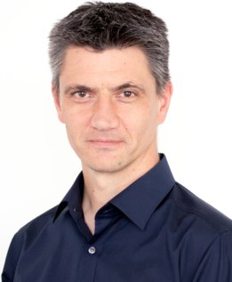 Thomas Dieing is technical product manager for the WITec alpha300 product line and its accessories. He obtained his PhD from La Trobe University, Melbourne, Australia, in 2005 investigating the MBE growth of nitrogen containing III/V semiconductors. In 2006 he joined WITec’s application team and became director of applications and support. In his role as product manager since 2019 he is responsible for all activities related to the product development process.
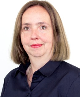 Ute Schmidt studied physics at the Babes Bolyai University in Cluj-Napoca, Romania, and obtained her PhD from the University of Karlsruhe, Germany. Through her work with in situ scanning tunneling microscopy in an electrochemical environment, she was introduced to scanning probe microscopy. During her postdoctoral scholar position at Karlsruhe and North Carolina State University in Raleigh, USA, she continued to work with metal deposition on different substrates with STM and Atomic Force Microscopy. Schmidt worked for six years as a project manager at Molecular Imaging Corp., Phoenix, USA. She has been an applications manager at WITec since 2003.