Contact-free 3D display responds to tiny changes in ambient humidity

Researchers in Korea have developed a new 3D display with a touchless interface that responds to the water vapour in a user’s hovering finger. The display, which relies on structural colour (that is, colour produced by light-scattering nanostructures) rather than reflections from coloured pigments, changes its hue depending on how far the user’s finger is from the screen. The technology might find use in wearable electronics and electronic skins that mimic how human skin can sense pressure, temperature and humidity.

Structural colours are often found in nature – for example, in the wings of some butterfly species. Because they arise from a physical structure (such as photonic crystals or arrays of nanofibers that reflect certain wavelengths of light), they are more durable than chemical pigments, which inevitably fade over time. Structural colours can also be changed by altering the molecular configuration of their surfaces. Both properties are attractive for novel “smart” materials used in interactive displays, and to this end researchers have long searched for ways to mimic the structural colours of the biological world.

Photonic crystal display

Most interactive displays made to date respond to external stimuli by varying the intensity of light they emit, rather than changing colour. The display made by Cheolmin Park and Won-Gun Koh and colleagues of Yonsei University in Seoul is different in that it is based on the multi-order reflection of structural colours in a thin, solid-state block copolymer (BCP) photonic crystal. This material spontaneously develops a layered 1D periodic microstructural film as it forms.

The nanostructured surface of the photonic crystal has a refractive index that varies with a period that is close to the wavelength of visible light. This variation produces a photonic “band gap” that affects how photons propagate through the material, much as a periodic potential in semiconductors affects the flow of electrons by defining allowed and forbidden energy bands. In the case of photonic crystals, light in the wavelength range that corresponds to the photonic band gap gets reflected, while light at other wavelengths is transmitted.

To achieve full-visible-range structural colours, the researchers used a BCP photonic crystal made from alternating layers, including a layer containing a chemically cross-linked interpenetrating hydrogel network. When the domains of this network are filled with a non-volatile ionic liquid (which alters the photonic crystal’s electronic properties), the subtle changes in water vapour levels that occur when a human finger is brought to within 1 to 15 mm from its surface are enough to shift the configuration of the surface structures to produce blue, green and orange colours.

Reflective colour mixing

These colour changes occur thanks to a phenomenon known as reflective colour mixing. When light hits the material’s surface, it couples with surface plasmons – collective excitations of electrons. These plasmons then become trapped in the surface, creating regions in which the dielectric constant of the structure is nearly zero, separated by areas of high refractive index. The presence of the non-volatile ionic liquid changes these dynamics and increases the reflection of different wavelengths of light thanks to two-colour mixing of pairs of reflections.

The researchers also found that they could easily transfer their photonic crystal-based film from a silicon substrate onto another support (in this case a printed one-dollar bill). This means that the technology could be used in printable and rewritable displays, they say.

The touchless sensing display is detailed in Science Advances.
 

Entangled light is unscrambled using entanglement itself

Natalia Herrera-Valencia and colleagues have successfully unscrambled entangled light after it has passed through a  2 m long multimode fibre. Led by Mehul Malik, the team at the Heriot-Watt University in Edinburgh tackled the challenge using entanglement itself. The research was done in collaboration with a colleague at the University of Glasgow and is described in a recent paper in Nature Physics.

Light passing through a disordered (or “complex”) medium like atmospheric fog or a multimode fibre gets scattered, albeit in a known manner. As a result, the information carried by the light gets distorted but is preserved, and extra steps are needed to access it. This gets especially tricky for the transport of entangled states of light because the medium muddles up the quantum correlations. The states get “scrambled” and “unscrambling” becomes necessary to retrieve the original entangled states.

Entanglement rescues entanglement

To understand a complex medium, physicists use a transmission matrix, which is a 2D array of complex numbers that predicts the fate of any input going through the medium. The transmission matrix theory, along with some key developments in technology, has only recently enabled propagation of classical light through complex media. In this work, the Edinburgh team has extended the idea of the transmission matrix to quantum photonics.

A property called the “channel-state duality” allows the researchers to use just a single quantum entangled state – a pair of photons that are correlated in their properties – as a probe to extract the entire transmission matrix of the medium. This is different from the classical way of constructing the matrix, where multiple light probes must be sent in through the medium to get the full matrix.

Once they know how the medium scrambles information, Herrera-Valencia and colleagues could undo its effects using the same matrix. Here again, entanglement offers a neat trick: instead of unscrambling light going through the fibre, the researchers can instead scramble its “entangled twin”, that does not go through the medium, to get the exact same results. They scramble light using a device called the spatial light modulator (SLM) which shapes the light field profile.

Dealing with higher dimensions

Compared to 2D qubits, higher dimensional entangled states have great potential because they can carry more information and are more robust to noise. But such states are also much more susceptible to changes by the environment.

Reporting the preservation of six-dimensional entanglement in space, the research tackles a significant challenge in quantum photonics. “Qubit entanglement already has the technology and deals with degrees of freedom [like polarization] that are not affected by the channel. When it comes to high-dimensional states, there are many issues with spatial mode encoding”, Malik explains. Something as simple as wavefront distortion could scramble the information.

To create and measure high-dimensional entangled states, physicists often use the spatial degree of freedom. In this work, the group uses a spatial “pixel” basis. They divide the continuous position space into discrete regions, or pixels, so if a photon is detected at the first pixel for one arm, its entangled twin will be detected at the same pixel in the other arm. The number of pixels determines the maximum dimension of entanglement that is possible in the system. The pixel basis works great in terms of quality, speed and dimensionality, more so because the SLM enables a precise and lossless control.

Implications for quantum technologies

In addition to increasing dimensionality of states and addressing issues like dispersion in longer fibres, the team is exploring how the idea that a complex channel is equivalent to a quantum state can simplify measurement of quantum states carrying a lot of information.

In their paper, the team also mentions that the technique can be used to transport high-dimensional entanglement even through dynamic media like biological tissues. Entangled light can also be sent through two independent channels, where manipulating any channel would affect the whole state, hence the other channel as well. They write, “Such an ability could be useful in quantum network scenarios or for non-invasive biological imaging, where access to all parts of the complex system may be limited”.

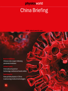
The Physics World China Briefing is now out

Physics World China BreifingThis year has been dominated by one event: the SARS-CoV-2 coronavirus, which was first reported in Wuhan, China, before quickly spreading throughout the world. The severe impact of COVID-19, the disease caused by the coronavirus, has been felt by all and physicists are no exception. Universities and research facilities all shut their doors earlier this year as scientists headed home under lockdown.
In this year’s Physics World China Briefing, we report how universities in China – the first to be affected – are now beginning to cautiously reopen. Despite a few localized outbreaks in the country, which seem to have been contained, things appear to be getting back on track. Without a vaccine on the immediate horizon, however, progress will remain slow and cautious and with international travel limited, collaborations will likely remain online-only for the foreseeable future.

Indeed, the impact of COVID-19 has already hit many scientific conferences, some of which have had to switch at short notice to online platforms. One example is Quantum 2020 – a major international conference in quantum technology organized by the Institute of Physics and IOP Publishing in partnership with the Chinese Physical Society and the University of Science and Technology of China. Due to be held in Shanghai, it will now take place entirely online on 19–22 October. For this year’s briefing, which is free to read, we talk to USTC physicist Chaoyang Lu about managing this virtual shift as well as the future of quantum technologies.

Let us know what you think about the publication on TwitterFacebook or by e-mailing us at pwld@ioppublishing.org.

Multi-user communication network paves the way towards the quantum internet

The concept of quantum communication, with security guaranteed by the laws of physics, took the world by storm when first unveiled in 1984. The traditional protocol, however, allows only two people to communicate securely. Attempts to extend this to “quantum networking” have usually proved either insecure or impracticably complex. Now, however, researchers in the UK and Austria have demonstrated secure information exchange between eight users spaced all around a city.

The canonical quantum communication protocol relies on two parties generating a secure key by exchanging polarized photons. The security of the link is guaranteed by the fact a third party cannot make a measurement of their state without disturbing them and being detected. Though remarkable, this approach is fundamentally limited to pairwise communication: it does not provide a blueprint for the multi-dimensional quantum network, or “quantum internet” that some researchers have dreamed of, in which multiple users connected together can all communicate simultaneously and securely with any other member of the network.

One obvious way to extend the scheme beyond two people is for the second person to simply act as a link in the chain, repeating the procedure and communicating securely with a third person, who in turn passes the message on until the message reaches its ultimate destination. Quantum networking schemes based on such “trusted nodes” have been developed. The security of such schemes is no longer absolute, however, because a trusted node may not be totally secure. Schemes that avoid trusted nodes have generally proved unfeasibly complex and hardware-intensive or have suffered other problems, such as restricting which users can communicate at any one time.

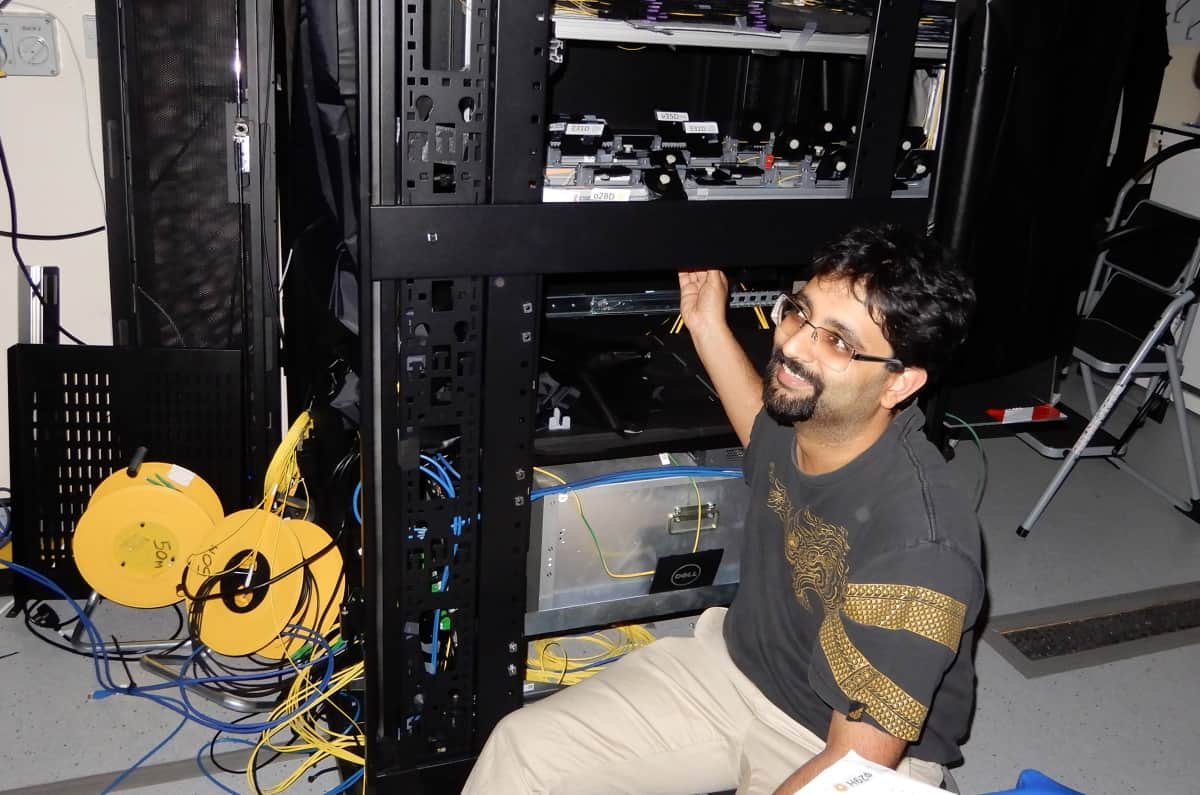
In 2018, researchers at the Institute for Quantum Optics and Quantum Information in Vienna led by Rupert Ursin demonstrated a scheme in which four users received pairs of entangled photons from a single, laser-pumped crystal source. “I generate these two-by-two, but I generate several such two-by-two pairs in a tiny amount of time,” explains project leader Siddarth Koduru Joshi.

Siddarth Joshi

This constant stream of entangled photon pairs from a single, central source allowed each of the four parties to become pairwise entangled with each of the other three parties. “If I’m talking to you, I look at stream one, if I’m talking to somebody else, I look at stream two, and so on,” says Joshi, now at the University of Bristol. The researchers believed that this scheme provided a simpler, more scalable architecture for secure, trusted-node free information exchange between multiple parties.

In the new work, researchers at the University of Bristol, in collaboration with the Austrian scientists, have confirmed that the technique works, demonstrating simultaneous and secure exchange of information between eight users spaced up to 12.6 km away from the central source around the city of Bristol. “This time, we actually demonstrated quantum communication, and we did this through deployed fibres across the city to show compatibility with existing infrastructure,” says Joshi.

Moreover, the researchers added additional multiplexing to simplify the hardware required by each user and make the protocol even more scalable: whereas their original protocol would have required 56 wavelength channels to fully interconnect eight users, their improved version required only 16. The researchers believe their network is the largest trusted-node free quantum network to date.

Quantum and computer engineer Wolfgang Tittel of QuTech at Delft University of Technology in the Netherlands describes the paper as “nice and important work” and is especially impressed by the absence of trusted nodes. An important next step, he says, is the scheme’s integration with quantum repeater technology, which to mitigates photon loss and decoherence and allows entanglement distribution over long distances. This, he says, could “extend the network beyond metropolitan size”.

The research is published in Science Advances.

Using particle physics to foster medical innovation

What are your main areas of research at the Institute of High Energy Physics (IHEP), Chinese Academy of Sciences?

My interest focuses on radiation imaging and its applications in medical physics and industry. This includes the design of X-ray and gamma-ray detectors, imaging algorithms as well as systems development.

How did you get into medical imaging?

For my PhD, I worked on IHEP’s Beijing Electron Positron Collider, which gave me experience in radiation measurements of positron annihilation. When I graduated in 2006, I began research on radiation-imaging technologies such as positron emission tomography (PET), single-photon emission computed tomography (SPECT) and computerized tomography (CT). Since 2008 research into radiation-imaging technology that has been carried out at IHEP’s division of nuclear technology and applications has achieved a number of breakthroughs in PET and SPECT detector development. Such work has especially benefited from the development of electronics and detection technology during the construction of big-science facilities at IHEP.

Do you have an example?

With the rapid development of the Chinese medical-imaging market, our team has co-operated with companies to develop devices such as a dedicated breast-PET (PEMi) scanners. We have now completed more than 500 clinical test runs and in 2015 received certification from the China Food and Drug Administration. Now it is being used for early breast cancer diagnosis in clinical settings – an important addition to improving breast cancer survival rates.

How important is technology transfer at IHEP?

It is very important. In IHEP’s strategic outlook, technology transfer is as critical as the construction of big-science facilities and basic scientific research. Most research centres in IHEP have been involved in technology transfer to some degree, with IHEP also setting up a dedicated technology-transfer office and a spin-out company.

Each of IHEP’s big-science facilities has led to the development of multiple disciplines and technologies.

What are some of the technologies that IHEP has spun out of its research?

The main technologies that have been successfully transferred are accelerator and medical-imaging technologies, such as the PEMi scanner. As IHEP makes large accelerators and their core components, we have also developed high-power electronic irradiation accelerators that are being used for the sterilization of food and medical equipment. These accelerators have already been installed in Yantai, Wuhan, Tianjin and other regions around China.

Many of the technologies at IHEP seem suited for medical applications. Why is this?

 The dominant technologies of IHEP are accelerator, nuclear detection and nuclear electronics technologies, which are exactly the type of technologies that are needed in nuclear medicine, radiotherapy and medical diagnosis. National funding projects give support for these areas of medical physics and there are many companies in this area that we collaborate with.

What kind of innovations do you see in the future?

Given advanced detector and electronics technology, we see developments such as high resolution – i.e. less than 100 picoseconds – time-of-flight PET technology that can increase the resolution of images and reduce the radiation dose to patients. Another aspect in medical applications is “static spectral” CT scanners, which will also allow for high-resolution imaging. In addition, the integration of diagnosis and treatment, especially in precision-particle therapy is another development I see in the future.

Do you collaborate on technology transfer with other countries?

Not yet, but international co-operation is an aspect that we are interested to grow.

With China designing a 100 km collider, do you envisage a similar emphasis on technology transfer when planning that project?

Each of IHEP’s big-science facilities has led to the development of multiple disciplines and technologies. Pixel-detector technology, for example, is involved in almost every scientific project, so we will accumulate more technical knowledge in this field as we advance. Combining the emphasis on detector development with medical-imaging equipment, we plan to focus on pixel-type semiconductor-detector technology by developing an advanced sensing chip and utilizing it in the next generation of CT and SPECT scanners.

Has COVID-19 affected your research?

COVID-19 has not really had any effect on our lab and we have continued to carry on with our research during the pandemic.

Inverted buoyancy makes tiny boat appear to defy gravity

Physicists in France have made small objects float upside-down on the underside of a layer of viscous liquid levitating in air. Although their apparently gravity-defying demonstration breaks no laws of physics, they say it could shed new light on the interaction between air and liquids.

Archimedes’ principle says that an object fully or partially immersed in a liquid experiences the upward force of buoyancy, which is equal to the weight of liquid it displaces. By opposing the force of gravity due to the object’s own weight, buoyancy will cause an object to float if it is less dense (overall) than the liquid – while denser objects will sink.

This is a very familiar phenomenon in our everyday world in which water and other liquids naturally exist at a lower gravitational potential than less dense gases. People on a boat travel through the air, with the sea below them. What the latest research shows is that the same principle would hold for people in an upside-down world – one in which, in effect, the sky lies beneath the sea.

Counter-intuitive effects

The secret to bringing about this topsy-turvy floating is vibration. Since the 1950s, scientists have demonstrated a range of counter-intuitive effects by vibrating fluids at high frequencies. Gas bubbles, for example, can be made to sink, while heavier particles rise. On a larger scale, whole layers of fluid can levitate in air. This is because more dense fluids tend to drip under the action of gravity, eventually displacing the air beneath them. By preventing the formation of drops, vibration keeps the fluid’s lower surface flat and allows it to hover.

In the new experiment, Emmanuel Fort and colleagues at the PSL University in Paris fixed a plexiglass container to a vibrating platform and then filled the container with either silicon oil or glycerol. These viscous liquids have surfaces that remain stable even at high accelerations. After turning the shaker on, the researchers used a syringe with a long needle to inject air towards the bottom of the liquid. With the resulting bubble sinking and growing, it eventually created a layer of air across the bottom of the container – causing the viscous liquid to levitate.

This fluid levitation is nothing new, but what Fort and co-workers then did was to place objects upside-down on the lower interface between the liquid and the air. As they explain in a paper in Nature, this buoyancy is governed by the same basic physics that would be at work on the upper interface. The object’s gravity tending to pull it down while the disturbance its downward motion creates in the liquid tends to pull it up.

Slight displacement from equilibrium

There is an important difference between the normal and inverted flotation, however. In the former, an object is in stable equilibrium because any attempt to move it up or down will be met by a restoring force. When pushed down it displaces a greater amount of liquid and so experiences more buoyancy, while any force pushing it out of the liquid will be opposed by gravity. But in the upside-down case, any slight displacement from equilibrium will see the object accelerate away – thanks to gravity below and the mass of liquid above.

Fort and colleagues found that the vibrations needed to invert the air and liquid also in fact stabilized the exotic buoyancy – at least for objects up to a certain density. They vibrated the container at 100 Hz and showed they could pin a series of small plastic spheres to the lower liquid-air interface, with the spheres remaining in place even when they poked them. To make the demonstration more eye-catching they repeated the feat with a little plastic boat, while floating an identical boat on the upper interface (see figure).

The researchers then found that 2.5 cm-diameter plastic spheres weighing no more than about 6 g only fell to the bottom of the chamber once they had reduced the amplitude of the vibrations to the point where the liquid layer itself succumbed to gravity. Heavier spheres, in contrast, fell first. This behaviour, they say, is consistent with a simple theoretical model that they developed to explain the inverted buoyancy. Only when the spheres’ density approached that of the liquid – meaning a mass of about 8 g  – did significant discrepancies occur. In that case, they add, other more complex effects play a role.

The team based its model on the concept of time-averaged forces, which tend to stabilize the equilibrium states. As they point out, this is the same basic idea underlying what is known as a Stephenson–Kapitza pendulum. In that case, vibrations allow a pendulum fixed from below to oscillate back and forth in a small arc – when otherwise it would fall to the ground.

Writing a commentary piece in Nature, Vladislav Sorokin of the University of Auckland in New Zealand and Iliya Blekhman of the Russian Academy of Science in St Petersburg, Russia, argue that several assumptions – such as a linear relationship between the pressure and height of the air layer – “somewhat limit the accuracy” of the French group’s model. But they reckon that the work might nevertheless lead to practical applications – pointing out that gravity-defying effects in fluids have previously been used to improve chemical reactions and aid mineral processing – and that it suggests other “remarkable phenomena” remain to be discovered in vibrating mechanical systems.

Swarming locusts inspire new collision detector

A new collision detector that mimics the neurological mechanisms that stops swarming locusts from crashing into each other has been developed by Saptarshi Das at Pennsylvania State University and colleagues.  The team’s compact, low-power device could lead to a boost in efficiency for collision detection mechanisms in robots and autonomous vehicles.

Today’s most advanced robots can safely navigate through unfamiliar environments using algorithms that allow them to avoid collisions with surrounding objects. These algorithms can be used for general, non-specific purposes, but this makes them computationally expensive – meaning their hardware requires large energy budgets and spatial footprints. In their study, Das’ team investigated whether navigation could be achieved using more task-specific algorithms.

The researchers turn to the tried-and-tested method of seeking inspiration from nature. In this case, they considered swarms of locusts, which are best known for the widespread devastation they can unleash on crops. The insects fly in dense groups containing millions of individuals, but very rarely collide with each other. This requires individual locusts to carry out complex mathematical calculations in real time, despite their extremely limited brain sizes.

Specialized neuron

Locusts make up for their lack of brain power with a single specialized neuron named the “lobula giant movement detector” (LGMD), which operates using two visual stimuli: the angular sizes and relative angular velocities of approaching insects. The resulting firing frequency of the neuron peaks immediately before collision, driving the locust to change direction.

To mimic this behaviour, Das’ team started from an equation linking the stimuli with the LGMD’s time-varying firing rate. They then incorporated this mathematics into a device containing a molybdenum disulphide monolayer, stacked on top of a programable memory architecture. While the photoconductor increased the device’s current as objects approached, introducing an “excitatory” signal, the architecture underneath decreased its current when no visual stimuli were present – creating an “inhibitory” signal. The signals competed with each other, with the excitatory signal winning out immediately before a collision – creating a signal spike.

Like the LGMD, the team’s device consumes a tiny amount of energy (just a few nanojoules) and occupies a modest spatial footprint of just 1×5 µm. At the same time, it can identify potential collisions from a variety of objects, approaching at a range of different speeds with an efficiency that is lacking in current general-purpose devices.

Das and colleagues now hope to extend the responses of their device beyond head-on collisions, and to incorporate multi-pixel detectors for predicting collisions in 3D. Through these improvements, their technology could be an important step towards safe, affordable autonomous vehicles, and robotics applications including manufacturing and medical surgery.

The detector is described in Nature Electronics.

Clinical experience with the Mercury 4.0 Phantom for CT protocol optimization, including automatic exposure control

Want to learn more on this subject?

Task Group 233 provides new guidance for CT performance assessment and addresses the challenges inherent in established CT imaging quality metrics. The Mercury 4.0 Phantom, designed by Dr Ehsan Samei at Duke University and commercialized by Gammex, now Sun Nuclear Corporation, assists with evaluations outlined in TG-233.

During this webinar, Timothy Szczykutowicz, PhD, DABR, of the University of Wisconsin-Madision, Department of Medical Physics, will present on how his department uses the Mercury 4.0 Phantom for common clinical CT tasks. You will hear about how the Mercury 4.0 Phantom addresses advanced features including automatic exposure control and tube current modulation. This webinar will enable you to understand how this new phantom lets the user check patient size for protocol optimization and proper dose management.

Want to learn more on this subject?

Timothy Szczykutowicz is an associate professor in the University of Wisconsin School of Medicine and Public Health Departments of Radiology, Medical Physics and Biomedical Engineering. His clinical and research activities include: optimizing CT scan protocols, monitoring patient dose, developing new metrics to define image quality in the clinical setting, developing protocol management methodologies, fluence field modulated CT, dual energy CT, and radiology department workflow and quality metrics.

Timothy is also the author of the book The CT Handbook: Optimizing protocols for today’s feature-rich scanners. He is an associate/section editor or on the board for multiple journals including: Medical Physics, Radiographics, Contemporary Diagnostic Radiology, and the Journal of Computer Assisted Tomography.

Nobel laureates endorse Joe Biden for US president

Joe Biden on the campaign trail

Over 80 US Nobel laurates have issued an open letter endorsing Democrat presidential candidate Joe Biden in the US election scheduled for 3 November. Biden – the vice-president under Barack Obama from 2009 to 2017 – will go up against president Donald Trump, who is seeking a second four-year term. The signatories of the open letter include 26 physics laureates, 31 medicine and physiology awardees and 24 chemistry Nobel winners.

The letter – signed by 81 laureates who received their Nobel prizes between 1975 and 2019 – focuses on Biden’s attitude to science. “At no time in our nation’s history has there been a greater need for our leaders to appreciate the value of science in formulating public policy,” the letter states. “During his long record of public service, Joe Biden has consistently demonstrated his willingness to listen to experts, his understanding of the value of international collaboration in research, and his respect for the contribution that immigrants make to the intellectual life of our country.”

They recognize the harm being done by ignoring science in public policy

Bill Foster

One of those to sign the letter is Barry Barish, who shared the 2017 physics prize for his contributions to the LIGO detector. “I support Joe Biden because of his long record of making policy, informed by science, to deal with large, complicated issues like cancer, climate change and nuclear proliferation,” Barish, who is at the California Institute of Technology, told CNN, adding that “we absolutely must elect Joe with his science-based approach to successfully lead us out of the COVID-19 pandemic”.

Facts and science

Nobel laureates’ support of Democratic presidential candidates isn’t new. Hillary Clinton received 70 such endorsements in 2016, while former president Barack Obama received the support of 76 in 2008. Carol Greider, who shared the 2009 prize in physiology or medicine for discovering the enzyme telomerase, asserted that elected leaders “should be making decisions based on facts and science,” adding that she “strongly endorses” Biden, in particular because of his “commitment to putting public health professionals, not politicians, back in charge”.

Democratic Representative Bill Foster of Illinois, the only physicist in Congress, organized the open letter saying it would be an “important” development for the Biden campaign. He says that “a core group” of laureates decided on which issues to raise in the letter. Foster, who received the endorsement of 31 laureates when he ran for Congress in 2007, says that when he started calling the laureates to back the intiative, “it was like pushing at an open door”. He adds that “there was a lot of enthusiasm because of the difference [the laureates] perceive in the scientific understanding” between the two candidates.

Foster believes that the letter reflects the view of much of the US scientific community. “They recognize the harm being done by ignoring science in public policy,” he says. “And it’s not only science; it’s logic and integrity. The scientific community wants to get to a situation in which they trust people’s word.” Foster sees the COVID-19 pandemic as a factor in refocusing voters on the importance of science. “The only reason we’re in a position to develop vaccines rapidly is decades of scientific research,” he says. “This may be an opportunity for the scientific community to remind everyone about long-term investment in science.”

Self-assembled peptides exhibit surprisingly strong diamagnetism

Magnetic forces

Peptides exhibit surprisingly strong diamagnetism when they join together to form microfibres, say researchers in China. Haijun Yang, at the Chinese Academy of Sciences (CAS) in Shanghai, and colleagues measured the force exerted by peptide-containing liquids placed in a static magnetic field. They found that samples in which the peptides had been allowed to self-assemble had a diamagnetic mass susceptibility 11 times higher than samples in which the peptides were dissolved, and 175 times that of pure water. The unexpected result, reported in Chinese Physics Letters, helps to explain the origin of magnetism in biomolecules, and could have applications in magnetically controlled microfabrication, medical imaging and brain–computer interfaces.

A diverse range of biological structures – from entire cells down to DNA and other subcellular polymers – are known to change their orientation in response to magnetic fields. This effect is behind the ability of some animals to navigate using the Earth’s magnetic field. However, many of the biological structures that respond to such fields apparently contain too little iron for the phenomenon to be explained by either ferromagnetism or ferrimagnetism. This suggests that the magnetic sensitivity of these structures is instead due to more subtle magnetic effects.

In an amino-acid chain called a peptide, for example, an external magnetic field induces a weak secondary field with the opposite direction, which pushes the peptide away. This effect, known as diamagnetism, has been attributed to the alignment of electrons in the peptide bonds between amino acids, or to electron currents flowing around cyclic structures called aromatic rings.

To investigate this phenomenon, Yang and colleagues prepared three different samples of a pentapeptide with the sequence AYFFF. A pentapeptide is a polymer consisting specifically of five amino acid units – in this case, one unit of alanine (A), one of tyrosine (Y) and three of phenylalanine (F).

In the first sample, the researchers dissolved the peptide in the organic solvent dimethyl sulfoxide (DMSO). In the second sample, they dispersed the powdered peptide in water. In the third sample, they also dispersed the powdered peptide in water, but then left it to stand for 16 hours. Over this 16-hour period, the peptides self-assembled into fibres more than 10 µm long.

AFM images of peptides

The researchers weighed the samples, and then repeated the measurement in the presence of a magnetic field, which they applied by placing a permanent magnet above the apparatus. They assumed that any difference between measurements with and without the magnet was due to the diamagnetic interaction between the external magnetic field and the magnetic fields induced within the sample.

They found that the peptide dissolved in DMSO exhibited the weakest diamagnetic response, while the sample in which the peptide had been allowed to self-assemble exhibited the strongest. The strength of diamagnetism in the water-dispersed sample was somewhere between the two, which the researchers put down to the occurrence of a limited degree of self-assembly.

When the team performed the same experiment using a different pentapeptide – IIIGK, comprising isoleucine (I), glycine (G) and lysine (K) – they found only a weak diamagnetic response, even for the sample that had been left to self-assemble into fibril structures. Whereas tyrosine and phenylalanine in the AYFFF sequence incorporate aromatic rings in their structures, this is not the case for any of the components of the IIIGK peptide. Yang and colleagues therefore interpret their results as confirmation that the electron dynamics of the aromatic rings, rather than the peptide bonds, are responsible for strong diamagnetism in peptides.

Although the role of aromatic ring currents was not unexpected in itself, the researchers were surprised by how strong the diamagnetism was in the self-assembled peptide samples. They are still investigating the physics underlying the effect, but they do already have an idea of how such anomalously strong diamagnetism might arise.

“Each peptide molecule has an induced magnetic moment in the presence of an applied magnetic field,” says Haiping Fang, of the CAS and East China University of Science and Technology in Shanghai, who led the research. “Thermal forces randomize the orientation of these magnetic moments, weakening the diamagnetism. A group of aromatic peptides in the assembled state – with interactions between them – might reduce the effect of these thermal fluctuations, resulting in the strong diamagnetism.”

Whatever its origin, the researchers foresee several possible applications for the effect. “Peptides with various sequences could be easily assembled into different nanostructures under magnetic fields, widening fabrication strategies,” says Feng Zhang, a collaborator from Guangzhou Medical University. “Assembled peptides modified with biomarkers could also be used as biological tracers, enhancing the contrast between normal and abnormal tissues in magnetic resonance imaging. Or, in novel brain–computer interfaces, the assembled peptides’ strong diamagnetism could be used as a non-invasive sensor to detect weak variations in magnetic signals from brain activity.”

Copyright © 2025 by IOP Publishing Ltd and individual contributors