A network of interconnected atoms could be used to construct a “quantum brain” that mimics how a real brain learns. The new system consists of an array of cobalt atoms on a substrate of black phosphorous, and its developers at Radboud University in the Netherlands say that it could have applications in artificial intelligence.
The human brain contains some 100 billion neurons in connected networks. Whenever we perform a task, these neurons receive electrical signals from other neurons in their network via tiny junction-like structures known as synapses. Once the sum of the signals across the synapses reaches a certain critical value, the neuron “fires” by sending a series of voltage spikes to other neurons. The strength of the connection between different neurons is known as the synaptic weight and can change over time as we learn new things and perform new tasks.
Many of today’s brain-inspired, or neuromorphic, devices use machine learning – the process by which a computer uses software, or algorithms, to train on a given set of examples – to autonomously develop the ability to perform a new task. One such machine-learning model is known as a Boltzmann machine. In physical terms, a Boltzmann machine is an interacting (Ising) system of spins in which randomly fluctuating spins (or magnetic moments) represent neurons.
Magnetic atoms on surfaces are emerging as a platform for realizing such a machine, as it is possible to use them to create tuneable networks of spins that display the necessary random motion. The problem is that magnetic exchange interactions between these atoms usually have a short range, which limits the number of connections to other atoms/neurons that can be formed.
Individually-coupled cobalt atoms
Researchers led by Alexander Khajetoorians and Hilbert Kappen have now created a self-adapting Boltzmann machine by exploiting the orbital dynamics of individually-coupled cobalt atoms placed on black phosphorus. The new work builds on earlier experiments in which they discovered that it is possible to store binary bits of information (0s and 1s) in the electronic state of a single cobalt atom when it is placed on this two-dimensional semiconductor and a voltage is applied to the atom.
Khajetoorians, Kappen and colleagues used the tip of a scanning tunnelling microscope to position the cobalt atoms on the 2D material and create long-range coupling between the atoms. They found that when they applied a voltage to the atom network, it produced an output signal that comes from electrons “hopping” from one cobalt atom to another. This output signal somewhat resembles the firing produced by neurons.
Synaptic weight change
As well as observing spiking behaviour in the output signals, the researchers noticed that the ensembles of cobalt atoms behaved differently depending on what input they received. For example, when the material was stimulated over a longer period with a certain voltage, the synapse-like memory-bearing atoms autonomously reorganized in response – in effect, changing their synaptic weight. “The material learned by itself,” Khajetoorians says.
Wolfram Pernice, a physicist and nanotechnologist at the University of Münster in Germany who was not involved in the study, calls the new work “very nice”. “Particularly exciting is the fact that the learning process is implemented directly in the material,” he tells Physics World. “Using individual atoms to implement artificial neurons and synapses is very elegant.”
Khajetoorians and colleagues say they now plan to scale up their system into a larger network of cobalt atoms. They would also like to study other magnetic atoms in an effort to understand why these atom networks behave the way they do. They report their findings in Nature Nanotechnology.
A compact, integrated light source that simultaneously produces multiple laser beams with different, very high orbital angular momenta has been unveiled by US researchers. The technology may mark a significant step towards orbital angular momentum multiplexing at scale, which could potentially vastly increase Internet speeds.
Interactions between light signals in an optical fibre are negligible, which means that multiple signals can travel down the same fibre simultaneously in a process called multiplexing. As the world’s hunger for faster data transfer grows insatiably, various schemes to use multiplexing to squeeze more data into fibres have been developed.
To ensure that the signals do not get mixed up, they must be sent using independent – or orthogonal – channels. One tantalizing and seemingly simple option is to encode data into the orthogonal angular momentum states of photons: with each state being an independent channel.
The most familiar type of angular momentum photons carry is circular polarization, or spin. This involves the rotation of the axes of the electric and magnetic fields around the wavefront as a photon propagates. Several telecommunications companies are working to incorporate “polarization division multiplexing” into their systems. However, circular polarization has only two orthogonal states so it can at best double a network’s capacity.
Infinite multiplexing
Photons also carry orbital angular momentum (OAM), which involves the wavefronts themselves coiling around the axis of propagation. Symmetry considerations require that this be quantized, but there is no other restriction on how tightly the wavefronts can coil. The OAM – and therefore the potential number of signals multiplexed – is, in principle, infinite.
However, multiplexing signals using OAM in a realistic setup has been tricky. Although light sources can generate beams with high OAM, changing the OAM requires moving parts such as metasurfaces, which is not feasible for a high-speed telecommunications source outside the laboratory. While lasers can achieve ultrafast switching, they produce relatively low OAM.
In 2017, Boubacar Kanté and colleagues at the University of California, San Diego showed how an arbitrarily-shaped laser cavity subjected to a magnetic bias could be produced at the interface between two photonic crystals, allowing light to snake its way around the path but not to escape it. The researchers attributed this to the creation of topologically-protected light paths by the photonic quantum Hall effect.
Edge states
Kanté is now at the University of California, Berkeley where his team is focused on making the light travel along circular paths in the cavity by patterning the photonic crystals to create topologically distinct edge states. Whereas the previous work kept the light confined between the photonic crystals, these edge states are designed to transmit light in one direction.
“At every point in the cavity, these topological rings are actually leaking some of their energy out,” says Kanté.
In their latest experiment, the researchers produced a photonic crystal that simultaneously supported edge states with OAMs of 100, 156 and 276 clockwise, anticlockwise and clockwise respectively. But they could, in principle, have generated any OAM beams with a sufficiently complex resonator. The device is fully integrated with no moving parts – the magnetic field is supplied by etching the quantum wells onto a magnetic substrate.
“This is the first time in history that a laser about the size of a human hair can generate an OAM greater than 200…We can now directly multiplex any number of OAMs of any charge in a simple and compact device,” says Kanté.
Liang Feng of the University of Pennsylvania believes the work marks an important step: “The underlying concept about the generation of the OAM beam is very similar to what we demonstrated in 2016,” he says. “First you need to have unidirectional light propagation in the platform and then you apply the appropriate phase matching condition to guide the light out. In our ring lasers we had confined waveguide modes, so we used gratings to help couple the light out. The beauty of this is that, if you have an angular grating inscribed on the ring cavity, the order number you could generate would be very limited, but in this case the order generated can be huge.”
Andrea Alù of the City University of New York is also impressed by the potential applications of the work: “I believe the final result is interesting, and the response is something people are after,” he says. He notes, however, that the magnetic substrate could complicate fabrication and integration at scale, and may not be strictly necessary: “The same principle may be applied to a reciprocal system which supports these edge states without relying on the quantum Hall effect,” he says: “The open question is what does this magnetic effect buy the authors?”
Does your PhD thesis make you want to dance? An atmospheric scientist from the University of Helsinki has bagged this year’s top prize of $2750 in the annual Dance Your PhD contest. Organized by the American Association for the Advancement of Science and sponsored by the artificial intelligence company Primer, the competition is in its 13th year and asks postgraduate students to explain their research through dance.
With the help of several friends, Jakub Kubecka brought his studies to life with a rap about atmospheric molecular clusters. With trash-talking lyrics like “I’m the first author, you’re just et al”, Kubecka and his mates also carried out some crude dance moves accompanied by computer animations and drone footage.
“To prepare for recording the lyrics, I was running with headphones playing the music at least 30 times per day for the whole month to get it into my blood,” says Kubecka. “We always stayed close to our main goal of showing non-scientific muggles that science can be fun, silly and exciting. And of course, we also didn’t want to miss our opportunity of spitting some scientific roasts.” You can watch the video — filmed while honouring local COVID-19 restrictions – above.
Linking proteins
One thing that has always fascinated me about cooking eggs is why unlike most other liquids, egg whites solidify when heated rather than becoming runnier or evaporating. The reason is that the protein molecules unfold in the heat, enabling them to link up to form a solid. Now Nafisa Begam at the University of Tübingen in Germany and colleagues have used X-ray scattering to gain a better understanding of this process.
One thing they found is that after the white solidified, which takes a few minutes, there is no further solidification. You can read more about their study in “Watching an egg cook with X-rays”, and who knows, maybe it will help you make the perfect poached egg.
Preclinical imaging systems such as positron emission tomography (PET) scanners provide an essential tool for studying disease and assessing therapies, most commonly in mice. But as mouse organs are roughly an order of magnitude smaller than their human counterparts, sub-millimetre resolution is essential for accurate imaging and quantitative measurements within the animal’s organs and tumours.
Many radioisotopes used as PET tracers, however, emit positrons with a large range (several millimetres) and cannot be imaged at sufficiently high resolution. It is also extremely difficult to image more than one PET isotope at a time, as they all create annihilation photons with equal energy.
Now a team headed up at TU Delft aims to solve both of these challenges at once – by utilizing the prompt gamma photons that are co-emitted with positrons by many radioisotopes. Using the VECTor scanner from MILabs, the researchers demonstrate multi-isotope and sub-millimetre imaging of PET isotopes with large positron range, reporting their findings in Physics in Medicine & Biology
Exploiting prompt gammas
PET works by detecting a pair of 511 keV annihilation photons produced when a positron emitted by a radioisotope annihilates with an electron. Coincident detection of these photons enables localization of their source, by forming a line-of-response between the detectors. However, positrons with large ranges will travel in random directions away from the tracer molecule before annihilation, reducing image resolution and quantitative accuracy.
“In fact, a coincidence PET scanner performs tomography of positron annihilations instead of emissions: PAT instead of PET,” explains first author Freek Beekman. “This is not so much of an issue for [the PET isotope] 18F, due to its short positron range. But for many other isotopes important to medical research and diagnosis it results in, sometimes dramatic, blurring effects.”
Fortunately, many PET isotopes with long positron ranges also emit significant amounts of prompt gammas straight from the atom. Detecting these enables more accurate localization of the PET tracer molecules and improves the image resolution. What’s more, different PET isotopes emit prompt gammas of different energies, paving the way towards multi-tracer PET imaging.
Beekman and colleagues tested this approach using a VECTor6CT system equipped with three gamma detectors and a high-energy mouse collimator with 144 pinholes (0.7 mm diameter) organized in clusters of four. The use of a clustered pinhole collimator minimizes several image-degrading effects inherent to electronic collimation.
“VECTor is the only PET technology that can precisely collimate these high-energy prompt photons and detect them,” says Beekman. “A coincidence PET scanner relying on electronic collimation could detect them, but unfortunately without collimation to the so-called line-of-response because it needs two photons in opposite direction for this.”
To demonstrate multi-isotope PET, the researchers injected mice with both 124I-NaI and 18F-NaF before scanning the animals for 60 min. 124I has a mean positron range of 3.4 mm, with a maximum range of 11.7 mm, but also emits large amounts of 603 keV prompt gammas. By using only the 603 keV photons for image reconstruction, sub-millimetre structures in the mouse thyroid were easily resolved.
Using the same scan but a different energy window, the researchers reconstructed high-resolution 18F-NaF images from 511 keV photons. To remove contamination from 124I annihilation photons, they corrected the 18F images by subtracting the estimated 124I signal. They then merged the corrected 18F image with the 124I image to create a clear dual-isotope mouse image showing 124I uptake in tiny thyroid parts and 18F-NaF in bone structures.
Dual-isotope PET can reduce imaging time over two separate scans, limiting the time needed to keep the animal anaesthetized, as well as providing perfectly registered images of different tracer molecules.
Resolution limits
To assess image resolution, the researchers scanned a Derenzo phantom containing 0.45–0.85 mm-diameter rods filled with 124I. Comparing 124I images reconstructed using prompt gammas and annihilation photons (from the same scan) showed that the 0.75 mm rods could be clearly discerned using 603 keV photons, while the 511 keV photons did not resolve any of the rods. Simultaneous dual-isotope PET images of a phantom filled with a mix of 124I and 18F also resolved the 0.75 mm rods.
The team next imaged a quantification phantom with three compartments filled with: (1) 0.98 MBq of 124I; (2) 10.1 MBq of 18F; (3) a mix of 0.98 MBq of 124I and 10.1 MBq of 18F. The measured concentrations of 18F in compartments 2 and 3 were equal after cross-talk correction, as were the amounts of 124I in compartments 1 and 3, demonstrating high quantitative accuracy.
Images of a quantification phantom filled with 18F-NaF, 124I-NaI and a mixture of the two demonstrate excellent separation of isotopes and high quantitative accuracy. (Courtesy: Phys. Med. Biol. 10.1088/1361-6560/abe5fc)
Finally, the researchers imaged a Derenzo phantom containing 89Zr, an important PET isotope with a mean positron range of 1.27 mm and abundant prompt gamma emission at 909 keV. Images based on 909 keV prompt gammas were far clearer than those using 511 keV photons, and could clearly resolve the 0.75 mm rods.
The team has received a grant from the Dutch Research Council (NWO) to develop algorithms that will further improve the images by better combining information from prompt and annihilation photons. “In addition, our partners in academia and pharmaceutical companies that have a VECTor/CT scanner are developing protocols to use this method in a large variety of new applications,” Beekman tells Physics World. “Meanwhile, we are also developing the next versions of the hardware.”
Scientists routinely use laser light to control how an atom’s electrons move from one electronic state to another, but controlling an atom’s nuclear state is far more challenging. Researchers at the Max Planck Institute for Nuclear Physics in Heidelberg, Germany, have now used X-ray light to achieve coherent control over nuclear excitations for the first time. As well as contributing to a better understanding of quantum matter, the work could hasten the development of technologies such as ultraprecise nuclear clocks and batteries that can store huge amounts of energy.
Atomic nuclei are quantum systems in which the component protons and neutrons can quantum-mechanically “jump” from one nuclear quantum state to another when they gain or lose energy. The energy differences in these nuclear jumps are often six orders of magnitude larger than the jumps made by electrons within an atom’s electron shells, says team member Christoph Keitel. “A single quantum jump made by a nuclear component can thus pump up to a million times more energy (into the states) – or get it out again,” he explains. “This has given rise to the idea of nuclear batteries with an unprecedented storage capacity.”
Keitel adds that the quantum states of some atomic nuclei are also much more sharply defined than electronic quantum states. This means the jump frequencies are also more precise – something that could, in principle, be exploited to create nuclear clocks that are far more precise than the atomic clocks used for today’s precision timekeeping and navigation. These ultra-precise clocks could also be useful for fundamental physics studies such as investigations of whether the known physical constants of nature are indeed constant.
Precisely addressing and controlling jumps
Before such applications see the light of day, however, researchers need to find some way of precisely addressing and controlling these jumps. One such technique, which the Heidelberg team has been working on for more than 10 years, involves high-energy X-ray light.
Evers and colleagues sent the first pulse to a “test” target sample made from a stainless-steel foil 1μm thick. The steel in this foil is enriched to contain 95% of the “Mössbauer” isotope iron-57 (57Fe), which has a nuclear (magnetic dipole) transition at an energy of 14.4 keV. The second pulse follows the first after a time delay, and afterwards both pulses encounter the real sample. This sample is also made of stainless steel enriched with 57Fe atoms, but it is 2 μm thick.
Pushing a swing
The researchers explain that their first pulse contains a broad mix of frequencies and is extremely short-lived, lasting just 100 picoseconds (1 ps = 10-12 s). This pulse stimulates a quantum transition in the 57Fe atom nuclei. The second pulse is longer, at 141 nanoseconds, and its energy is precisely tuned to the same quantum transition. The time delay between the two pulses can be adjusted in a way that the researchers liken to pushing a person on a swing. While the first push causes the person to swing, or oscillate, back and forth, the second push either enhances the oscillation or slows it down depending on when it occurs within the oscillation’s phase. The second pulse is thus, respectively, either more constructive or more destructive for the quantum state.
Achieving such a tightly controlled change in the quantum dynamics of an atomic nucleus is a technical feat that took the Heidelberg team years to achieve. Among other factors, it requires the delay of the second pulse to be stable on a time scale of just a few zeptoseconds (1 zs = 10-21 s). Only then can the two pulses work together to control nuclear excitations.
Spurred on by these results, which they report in Nature, the researchers now plan to explore possible applications of their new control scheme. “These include novel spectroscopy approaches and adaptive X-ray optics,” Evers tells Physics World.
In this episode of the Physics World Weekly podcast the science writer Kit Chapman chats about his latest book Superheavy: Making and Breaking the Periodic Table, which is a lively romp through the history of smashing nuclei together to create ever heavier elements. Some of the most talented physicists and chemists of the past 100 years have been involved in the quest for superheavy elements, and Chapman talks about some of his favourite characters. He also discusses a recent feature article he wrote for Physics World about the Facility for Rare Isotope Beams, which will come online next year in Michigan with the incredible aim of doubling the number of known isotopes.
Also in this episode, Physics World’s Margaret Harris explains what to do if you find a meteorite while rambling through the countryside – something that could happen in the English county of Gloucestershire, over which a bright meteor was spotted in the sky on Sunday. We also talk about new evidence that a much larger object from space put an end to the dinosaurs 66 million years ago.
The mechanical properties of Li have recently received considerable attention because of their importance in understanding and enabling Li metal batteries. Although there are numerous models that aim to describe the complex chemical and mechanical processes that occur during Li plating and stripping, virtually all of them assume that the Li is in perfect conformal contact with a separator, whether in liquid or solid cells. This assumption contradicts the widely observed result that the interface resistance (which is inversely proportional to contact area) falls strongly with increasing pressure. This contradiction may call into question how well these models represent realistic systems.
In this talk, Dr Stephen J Harris will take explicit account of the inevitable surface roughness of Li and separators and show how this leads to accurate predictions for the pressure dependence of the critical current density. We also introduce a concept, new to the battery field, that offers to explain how a material as soft as Li metal can penetrate hard ceramic solid electrolytes. This idea comes from an analogous process in a different field of material science, where the penetration problem has already been solved. We suggest that the same solution can eliminate dendrite penetration through solid electrolytes in all-solid-state batteries.
Dr Stephen “Steve” Harris received a BS degree in chemistry from the University of California, Los Angeles, US, and PhD in physical chemistry from Harvard University, US. After a Miller Postdoctoral Fellowship at the University of California, Berkeley (UC Berkeley), US, he began his career at General Motors Research Labs (GM). Apart from a stint at the Ford Scientific Research Labs, Steve worked at GM until 2011, when he was awarded a Miller Visiting Professorship in the UC Berkeley Chemistry Department. Since then, he has worked in the Materials Science and Energy Storage Divisions at Lawrence Berkeley Lab. He is also a Visiting Scholar in the Department of Materials Science and Engineering at Stanford University, US; has consulted for battery and venture capital companies; and is on the advisory board for several battery start-ups.
Dr Harris’ work has ranged widely, including studies of combustion chemistry, the kinetics, and thermodynamics for growth of CVD films, aerosol dynamics modelling, fatigue failure in gears, and fracture mechanics in cast aluminium. When he returned to GM from Ford, he started working on Li-ion batteries, focusing on how the presence of heterogeneities and flaws affect ion transport, durability and energy density. Presently, he is looking at the mechanics and electrochemistry at Li metal interfaces, and has proposed a general solution to the dendrite penetration problem.
Think back to your childhood. What did you want to be when you grew up? In primary school I wanted to be a special-effects designer. I even wrote a letter to the BBC asking how to be one, to which they replied I should study physics and design technology as well as draw and make models in my spare time. Fast-forward many years and – although I am not a special-effects designer – I am a physicist working at NUSTEM, which aims to increase the diversity and number of young people choosing STEM (science, technology, engineering and maths) careers.
If you ask young children today what they want to do for a job, they will give a relatively narrow range of possible careers. Last year, we asked more than 600 children between the ages of seven and 11 that question, finding that the 20 most mentioned jobs accounted for 75% of all aspirations. They included doctor, vet, sports coach and police officer. Of course, this makes sense – children will only know about a few jobs, often those they hear about or see around them at home or at school.
Children will only know about a few jobs, often those they hear about or see around them
More worryingly, however, was that although some jobs were mentioned by boys and girls, career aspirations were strongly gendered. Footballer and YouTuber, for example, were the most popular careers given by boys, while vet and teacher were most popular with girls. In fact, our earlier research on career aspirations in primary school showed that children are making choices about what they don’t want to do before they are eight years old (International Journal of Science Education 42764). We therefore must talk to children about future jobs and careers much earlier than has traditionally been the case.
It’s good to talk
Parents and families are strong influences on a child’s attitudes and interests. Those who did not study science beyond the age of 16 can feel that science is about “knowing stuff”. This might mean that when their child wants to talk to them about an aspect of science or asks a good science question, families are uncomfortable and close the conversation down. Indeed, many pre-school children are fascinated by science topics such as space, and might want to “become an astronaut” to follow up their interest, but parents might not feel confident talking to their child about space and its mysteries, or about how there are lots of jobs linked to space exploration that go beyond astronaut. Without encouragement, the child may subconsciously think this means that science is not for them or their family. Over time, these interactions steer children away from a career related to science.
At NUSTEM, we have been developing story time activities for nursery-aged children and their families. Although we do not directly talk about careers in the session, we try to make conversations about science, technology, engineering and maths “normal” in the family. By working and playing together, the children and adults enjoy the story and activity and then ask each other questions. The aim is to help families to discover that science – and of course physics – is about asking good questions and deciding on good ways of finding out answers to those questions.
With funding from the UK Space Agency, I recently wrote a storybook called Are We Nearly There Yet? It features the ExoMars Rosalind Franklin rover that is set to launch in 2022, arriving at Mars a year later. In the story, Rosie the Rover is heading to Mars but wakes up due to a solar storm. To help her get back to sleep, Mission Control tells her about all the other robots on other missions. This simple story is easy enough for children to understand and links to a love of space that many of them already have.
During a family session, a facilitator first reads the story before encouraging parents and children to re-read it. They then use building blocks to create their own space robot and make a pretend surface of Mars for it to travel on, with the facilitator modelling the sorts of questions that families could ask to help them explore the story and the science a bit more.
When we evaluated the project, we found that after the parents and carers had taken part, they felt more confident talking about space with their children. Of course, this sort of activity does not have to be an organized event. Families can encourage children to ask questions about their storybooks, build models or draw pictures about whatever they are reading. It can be any story book, and we have gathered together a list of more picture books with a science theme for young children.
Starting young
Although career choices seem a long way off, I believe parents and families can help their children to be aware of the broadest range of possible careers as they grow up. We know that in more normal times, many primary schools welcome visitors to talk to children about their jobs and what they do, often under the title of “people who help us”. However, these visitors tend to be people such as local police officers, healthcare workers and fire-fighters. If you are a STEM professional, I would encourage you to talk to children in primary schools about yourself, and your job. In that way, we can help children see that science is often about asking questions and finding solutions to problems and that it can be done by “people like them”.
As the leader of a materials science and engineering laboratory, my office – situated in the middle of a sea of experimental spaces, white boards, and group member desks – was rarely quiet before the pandemic. The murmurs of derivations from the white boards and the swinging of the doors to our optics and synthetic chemistry labs provided a constant background hum. And although I am trained as a physicist, the members of my group come from numerous scientific and engineering backgrounds, creating a rich, dynamic research environment.
During the past year, these noises and interactions have vaporized. As I work from the isolation of home while my group members perform research (at significantly reduced capacity) in the lab, our entire research environment and lab culture have been transformed. New synthetic pathways no longer decorate the wall outside my office door, and students no longer rush into my office to show me their latest laser threshold data. But the daily coffee breaks and walks also stopped. More recently, the more significant celebratory milestones, like graduation and holiday parties, have been postponed. And while this stark, sudden shift had a huge and immediate impact on productivity, it also led to a slow, continued erosion of mental health.
Seeking practical solutions
Universities are trying a wide range of strategies to address this erosion, some of which are more successful than others. Loneliness is already a challenge for science and engineering students for many reasons, including struggles with imposter syndrome, issues with advisor(s) and being unable to travel home due to visa restrictions. COVID-19 is amplifying these issues, making it more important than ever to try to find practical solutions.
In my case, I took inspiration from the healthcare sector and decided to bring in a mental health therapist to lead a weekly discussion group for my lab. There were several practical challenges associated with this decision, including finding someone who is skilled in and willing to work with the STEM/education sector (and PhD students specifically), paying that person and getting student buy-in.
To overcome the first hurdle, I reached out to my personal network as well as a range of potential contacts on social media (yes, I said social media). Through them, I found a person who had previously run similar groups. As far as payment, I paid “out of pocket”, meaning I paid them directly. As a principal investigator, I value my students’ health and well-being, and I feel it is my responsibility to ensure their success. To get student buy-in, I dedicated workday time to the discussion group to emphasize that I believed it was important. Although attending the group was completely optional, I explained why I felt group sessions and the group format were a good approach and why I valued therapy, as part of an attempt to normalize it.
Specialized support
We are now finishing our second month of these sessions, and when I asked my group if they wanted to continue, the answer was a resounding “yes” for several reasons. Although the university does offer similar groups, they are typically attended by students from many disciplines. As a result, the therapists are not trained in engineering-specific challenges, and the discussants are not all engineers. The result is that the students end up feeling even more isolated than they already were.
While I do not attend our group’s sessions, I know that my group members discuss a wide range of topics, both pandemic-related and otherwise. Given the pressure that many of them are feeling, it is very important to provide them a place to express their concerns related to screening/qualifying exams, research and academic progress, housing and finances, and family. And this environment needs to be with their colleagues who can empathize with and support them, as well as with a therapist who can lead a constructive discussion. I also encouraged my students to use the therapist to alert me to any concerns they have with the university or with me that they do not feel comfortable bringing to my attention directly. In other words, the therapist can act as their advocate. This advocacy role (as well as my lack of experience in psychology) is one reason why I decided not to lead these groups myself.
Creating this resource for my group has presented many challenges, but I am hopeful that the school of engineering will continue the initiative post-pandemic. To ensure our students’ academic and research success, it is critical to care for both their mental and physical health. This requires engaging the students to provide them with the resources they need, not the resources we think they need.
In February 2020, some 20,000 scientists, engineers and business insiders filled the cavernous halls of the Moscone Center in San Francisco for Photonics West, one of the last major scientific conferences to be convened before the pandemic took hold. This year the halls will be empty, but the organizers have had the best part of a year to reimagine the event as a digital meeting place for discussing the latest advances and innovations in lasers, photonics and biomedical optics.
Using several different technology platforms, including the ubiquitous Zoom, the Slack messaging service, and the event’s dedicated app, the Photonics West Digital Forum is designed to provide attendees with the same content and networking opportunities as the live event. The popular plenary and Hot Topic sessions will be live-streamed and available to watch on demand, while attendees will be able to access thousands of technical presentations in four major conference tracks. New for 2021 is Quantum West, a four-day event that will explore the role that photonics plays as quantum technology moves from R&D to engineering products for the commercial marketplace.
Alongside the technical programme, delegates will be able to take part in live networking sessions and social meet-ups. Industry events including presentations and panel discussions will be hosted online, as will the annual Start-Up Challenge and SPIE’s Prism Awards. Meanwhile, a digital marketplace will enable attendees to explore the most innovative technology solutions from suppliers from all over the world, watch product demonstrations, and engage directly with company representatives. Some of the products being featured in the marketplace are highlighted below; to find out more visit the companies’ virtual booths.
Upgrade boosts SuperK performance and reliability
The SuperK FIANIUM series of supercontinuum lasers from NKT Photonics offers the highest efficiency of any on the market, as well as the highest power in the visible range. Newly upgraded electronics and improved fibre technology have boosted performance and reliability still further, while also making the lasers even easier to use.
The industry-leading SuperK FIANIUM series of supercontinuum lasers now feature upgraded electronics and improved fibre technology. (Courtesy: NKT Photonics)
SuperK lasers deliver high-brightness diffraction-limited light over the entire 390–2400 nm range, providing single-mode, broadband collimation at high power. The modular design achieves power levels of up to 2 W at visible wavelengths and a total output power of up to 6.5 W, while the high efficiency improves reliability and reduces any unwanted residual pump power at the output. Adding a filter also converts the SuperK into an ultra-tunable laser.
A pulse picker option makes it possible to change the repetition rate of the laser during operation. A range of 0.15–78 MHz is available as standard, with custom options available on request, providing maximum flexibility for lifetime applications such as fluorescence lifetime imaging.
The lasers are based on a monolithic fibre architecture, which ensures excellent reliability and a lifetime extending to thousands of hours. The design eliminates the need for regular maintenance, and also enables alignment-free operation. No laser expertise is needed to generate a high-quality beam, and functions can be changed on-the-fly from a computer interface with no need to power down the system. In standby mode, the laser remembers the latest power or current setting and returns to the same level when the emission is re-activated.
Sources deliver polarization-entangled photons for quantum research
OZ Optics, a leading supplier of fibre-optic products for telecommunications and industrial and medical applications, has expanded its product line for quantum photonics. The company now offers two sources of polarization-entangled photons for applications in quantum sensing, quantum communication, and quantum computing, and with both source types, OZ can now produce high-quality polarization entanglement throughout the near-infrared and short-wave infrared frequency bands.
Polarization-entangled photon sources from Oz Optics are designed for applications in quantum sensing, quantum communication, and quantum computing. (Courtesy: OZ Optics)
The first source, the EPS-1000, is an all-fibre generator of broadband polarization-entangled photon pairs, produced at telecom wavelengths with more than 80 nm of bandwidth. Based on periodically-poled silica fibre (PPSF) technology, it features turn-key, room-temperature operation, while the all-fibre design makes it environmentally stable for challenging applications such as space-based instruments.
The latest addition is the EPG-1000 series of crystal-based polarization entangled photon sources, which have been designed to meet the diverse phase-matching needs of the quantum R&D community. This source generates polarization-entangled photon pairs through the well-established process of spontaneous parametric down conversion (SPDC), and it features a compact interferometer that supports several phase-matching techniques. With additional supporting equipment, the EPG can either be used to produce photon pairs or to create polarization entangled pairs with a fidelity of more than 95%.
“These two types of sources diversify our quantum photonic capabilities and positions OZ Optics to become a global market leader in the quantum light source space,” commented OZ Optics CEO Ömür Sezerman.
For detailed specifications and additional information about these and other products, visit www.ozoptics.com.
Virtual reality enables photonics learning
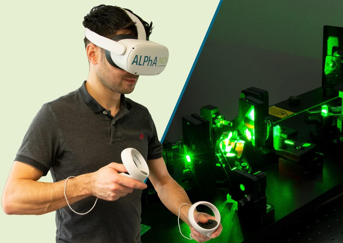
The Immersive Photonics Lab from ALPhANOV is an innovative training tool that engages trainees in a virtual reality photonics lab. The immersive learning environment helps participants to master the professional and technical know-how they need for their role, whether they are new to the industry or learning to use a new piece of equipment. By helping to disseminate training programmes, it enables companies to tackle the shortage of skilled labour in the photonics industry.
The Immersive Photonics Lab from ALPhANOV exploits virtual reality to teach photonics skills and procedures. (Courtesy: ALPhANOV)
The virtual reality application, which won an SPIE Prism Award, faithfully reproduces physical phenomena and enables fast and effective development of procedural skills, whether in industrial or educational settings. The tool emulates all the equipment needed to train professionals and students, without risk of injury or damaging valuable optical components.
The Immersive Photonics Lab training application can be used anytime, anywhere, which makes it ideal for remote learning and customer training. The tool gives participants easy access to the latest generation of photonics technologies, while also reducing the need for equipment downtime to support technical training.
ALPhANOV works with each customer to developing a tailor-made training programme as well as a dedicated technical environment within the Immersive Photonics Lab.
TOPTICA rises to new laser challenges
TOPTICA Photonics will be showcasing its wide range of cutting-edge laser systems for demanding scientific and industrial applications in biophotonics, industrial metrology and quantum technology. The company prides itself on providing lasers covering the widest wavelength range on the market – going from 190 nm in the ultraviolet to 3 mm at 0.1 THz – with high power outputs even at exotic wavelengths.
The CTL laser is a widely tunable laser that offers mode-hop free tuning up to 120 nm. (Courtesy: TOPTICA Photonics)
The company will be hosting exclusive educational sessions throughout Photonics West via Zoom Webinar. These will cover leading product innovations, such as the CTL continuously tunable laser with mode-hop-free tuning up to 120 nm, terahertz systems for both time-domain and frequency-domain techniques, and the TOPO laser system for mid-infrared spectroscopy and applications. Also featured in the webinar line-up is the iChrome FLE, designed to be a “flexible laser engine” for diverse applications in biophotonics, and highly sensitive linewidth analysers for controlling both ultra-narrow and broadband lasers.
Representatives from TOPTICA Photonics will also be taking part in the inaugural Quantum West conference. CTO Wilhelm Kaenders will give a presentation on the use of lasers in quantum applications on Wednesday 10 March, while the next day a panel discussion on photonics technologies for the emerging quantum market will feature TOPTICA’s Mark Tolbert.
To learn more about TOPTICA and its product range, visit the company’s virtual booth in the Digital Marketplace.
Phase-only SLM targets small-scale solutions
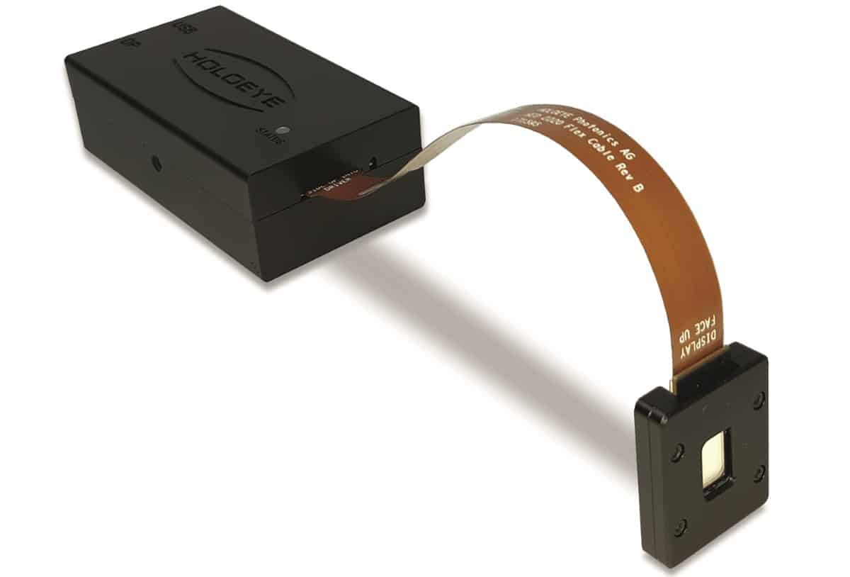
HOLOEYE has released a new series of compact phase-only spatial light modulators (SLMs) that are designed to be integrated into small-sized or even portable solutions. The LUNA SLM features a liquid-crystal-on-silicon (LCOS) microdisplay with an active area diagonal of 0.39 inches, and which offers full high-definition resolution of 1920 x 1080 pixels and 4.5 µm pixel pitch.
The LUNA phase-only spatial light modulator from HOLOEYE is compact enough to be integrated into portable solutions. (Courtesy: HOLOEYE)
The SLM provides linear 8-bit phase levels and offers fast digital addressing via the DisplayPort video interface, which provides an input frame rate of 60 Hz. The display can even accept video data via the high-speed MIPI digital serial interface, a novel approach that offers new possibilities for industrial implementations of phase-only SLMs.
The driver ASIC is embedded in the LCOS microdisplay itself, saving board space and allowing easier integration. The standard driver box measures just 85 x 47 x 28 mm, and also features a USB connector for power and advanced configurations.
HOLOEYE currently offers two versions of the LUNA phase-only SLM: one operating at visible wavelengths, which offers a phase shift of 2π over the 420–650 nm range, and one for the telecommunication waveband operating at 1400–1700 nm.
During this year’s virtual Photonics West exhibition, PicoQuant will unveil its latest multichannel event timer. The MultiHarp 160 is a plug-and-play time tagger and time-correlated single-photon counting (TCSPC) unit that is optimized for applications requiring up to 64 timing channels. It offers an outstanding time resolution of 5 ps and an ultrashort dead time of less than 650 ps.
The MultiHarp 160 from PicoQuant offers time tagging and correlated single-photon counting for up to 64 synchronized channels. (Courtesy: PicoQuant)
“We developed the MultiHarp 160 to meet the challenges of future TCSPC applications using many channels,” commented Rainer Erdmann, managing director of PicoQuant. “Our device provides internal synchronization of all inputs without need for additional hardware or software tools, and extraordinary throughput can be achieved thanks to a special FPGA link.”
The MultiHarp 160’s number of synchronized input channels can be scaled from 16 up to 64. A common synchronization channel, supporting sync rates of up to 1.2 GHz for periodic signals, is available as timing reference for all channels. Time tags from all input channels are combined into a single data stream that is accessible via a USB 3.0 interface. The data stream is also accessible to external FPGA boards via a dedicated interface, enabling great flexibility in tailoring the way that data is preprocessed for the specific needs of an application.
For an opportunity to preview the capabilities of the MultiHarp 160, a free webinar will be hosted by Dr Torsten Langer, sales and application specialist at PicoQuant, on 1 March 2021. Registration is possible on PicoQuant’s webinar website.
You can also find out more about the MultiHarp 160, as well as PicoQuant’s full range of pulsed diode lasers and instrumentation for time-resolved data acquisition, single-photon counting, and fluorescence imaging at the BiOS and Photonics West digital marketplaces.
Real-time laser simulation offers new capabilities
BeamXpert, which introduced the simulation software BeamXpertDESIGNER at last year’s Photonics West, has now upgraded the application to allow the export of optical setups to mechanical CAD software, improve the useability, and provide an optimized and enlarged component database.
BeamXpertDESIGNER is now being used by TRUMPF to develop laser systems for EUV lithography. (Source: TRUMPF Group)
The software enables the rapid development of optical systems for the propagation of laser radiation. By using two consecutive approaches, it is also possible to rapidly evaluate aberrations and their influence on M². All output results are based on the relevant ISO standards.
Customers of the software use it, among other things, for developing laser systems for ophthalmology and satellite communications, and for the design of high-power lasers and optics for solar-cell research and manufacturing.
One notable example is TRUMPF, which uses BeamXpertDESIGNER for developing high-power CO2 lasers that are used to generate extreme-ultraviolet radiation for lithography equipment. This application requires the simulation of optical systems with hundreds of components and a propagation length of more than a kilometre.


 Dr Stephen “Steve” Harris received a BS degree in chemistry from the University of California, Los Angeles, US, and PhD in physical chemistry from Harvard University, US. After a Miller Postdoctoral Fellowship at the University of California, Berkeley (UC Berkeley), US, he began his career at General Motors Research Labs (GM). Apart from a stint at the Ford Scientific Research Labs, Steve worked at GM until 2011, when he was awarded a Miller Visiting Professorship in the UC Berkeley Chemistry Department. Since then, he has worked in the Materials Science and Energy Storage Divisions at Lawrence Berkeley Lab. He is also a Visiting Scholar in the Department of Materials Science and Engineering at Stanford University, US; has consulted for battery and venture capital companies; and is on the advisory board for several battery start-ups.