
Biodegradable sensor opens door to real-time monitoring of key disease biomarker

An international team of researchers has developed a novel biodegradable electronic sensor capable of measuring trace amounts of nitric oxide (NO) species. In a paper published in NPG Asia Materials, the team – from Korea University, the University of Cambridge, The Pennsylvania State University and Korea Institute of Science and Technology – outline how the implantable technology could be used to record NO concentration from the surface of organs before safely degrading into materials cleared by the body.

Nitric oxide and nitric dioxide, referred to collectively as NOx, play vital roles in vascular, nervous and respiratory health. Currently, commercial sensors monitor NOx concentration externally via exhalation. Such devices, however, may not be sensitive enough to sufficiently quantify the presence of these gases within the patient.

“It might be much more beneficial to monitor the gas levels from internal organs,” explains study author Huanyu “Larry” Cheng. Measuring NOx levels from inside the patient offers increased accuracy and sensitivity over traditional sensors.

Nevertheless, making such a device is not an easy task. The sensor must be pliable, operating under mechanical loads without compromising electrical performance. It must selectively measure NOx, even at very low concentrations. Finally, it must be removable after use. In this work, the twist – and the focus of the team headed by Chong-Yun Kang and Suk-Won Hwang – is to exploit non-toxic, biodegradable materials that are completely absorbed by the body. This means that detection is a one-and-done operation, and following implantation, no further surgery is required.

The highly flexible device uses silicon-based technology to detect NOx concentrations as low as 0.0005%. What’s more, the team determined the sensor to be 100 times more sensitive to NOx than to other well-known physiological substances – providing an edge over other promising sensors made of graphene and metal oxides.

Nitric oxide: putting the ‘NOx’ in noxious

NOx gas is a notorious air pollutant. Exposure to environmental NOx can trigger respiratory conditions such as asthma and chronic obstructive pulmonary disease (COPD). Despite this, physiological quantities of NO are critical to cardiovascular health – a discovery so important that it won the 1998 Nobel Prize for Physiology and Medicine.

Nitric oxide

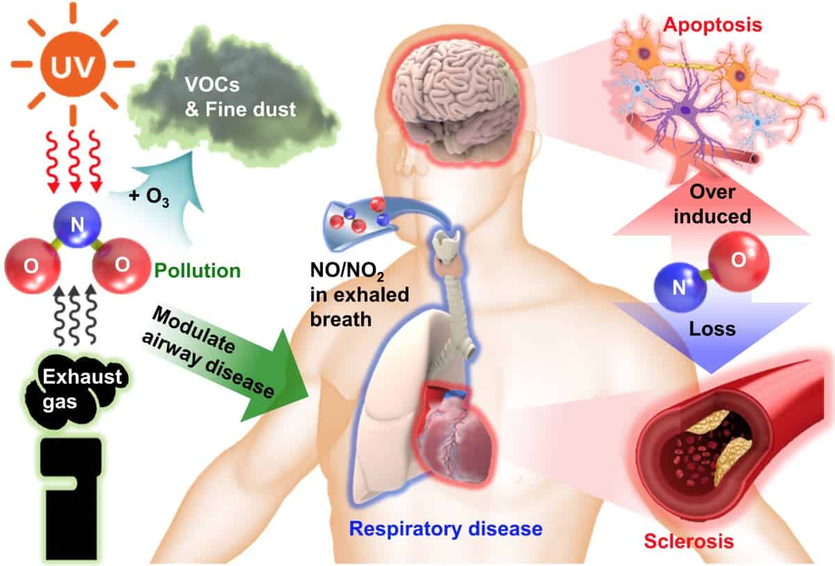
Our bodies use NO as a chemical messenger, regulating blood flow to control oxygen and nutrient transport. An NO deficiency can lead to high blood pressure and the onset of atherosclerosis, or the narrowing of arteries due to fatty plaque build-up.

But NO is also toxic to nerve cells in high quantities. Excess NO production is linked to neurodegenerative conditions like Alzheimer’s disease and Parkinson’s disease.

How does the sensor work?

To create their sensor, the researchers patterned the electronic components onto a silicon nanomembrane (approximately 100 nm thick) and transfer printed them onto the biodegradable polymer poly(lactic-co-glycolic) acid (PLGA).

Silicon, as Cheng puts it, “is the building block for modern electronics”. The element is also highly sensitive to NOx. When gaseous NOx molecules adsorb onto the surface of the sensor, they deplete the top layer of electrons in the silicon. This increases the electrical resistance of the gas with respect to the baseline response (dry N2/air).

The study recognized that gas absorption is not the only parameter that affects electrical resistance. Mechanical forces can also impact how current flows through the system. If the sensor is to conform to the surfaces of different organs, it must remain functional under deformation.

A large-scale sensor array showed little change in the relative response rate when subjected to 1000 cycles of bending or stretching. Furthermore, the researchers used computer simulations to probe the stress and strain response of each layer within the device. They concluded that the array remains functional when stretched up to 40% of its length.

The team assessed the selectivity of the sensor to NOx by comparing the intensity of the resistance signal to its responses to other common gaseous biomarkers, including carbon oxides and ammonia. Not only does the technology show exceptional selectivity, but it does so incredibly quickly. When introduced to the sensor, the gas was detected in just 30 s.

Crucially, the device biodegrades over a timescale suitable for tracking a patient’s NOx levels. When submerged in a body-fluid mimic (phosphate buffered saline, pH 7.4, 37°C), all electronic components degraded to non-toxic end products in just 8 hr.

The results demonstrate that the sensor operates effectively under the harsh conditions of the body. Next, the researchers plan to look at tweaking the design to monitor other bodily functions for various disease detection applications.

Cosmic Bell test probes the reality of quantum mechanics

In this interview, Johannes Handsteiner describes an experiment to test the foundations of quantum mechanics. Handsteiner was part of a team at the Institute for Quantum Optics and Quantum Information (IQOQI) in Vienna, Austria, that designed a Bell test experiment using light from now-ancient quasars. Handsteiner explains why his team went to such lengths to close an important loophole in this famous test designed to rule out the possibility of a classical explanation.

The interview was recorded in 2019 before the COVID-19 pandemic. Handsteiner now works at Quantum Technology Laboratories, a company involved in contract research in the field of Quantum Key Distribution (QKD).

Single molecules keep to the straight and narrow

A change in the position of a single molecule can determine the outcome of a chemical reaction, but studying such movements is difficult because molecular motion is random at the atomic scale. Researchers at the University of Graz in Austria, together with colleagues in Germany and the US, have now succeeded in controlling the motion of single organic molecules by using the sharp tip of a scanning tunnelling microscope (STM) to shift them 150 nm along a flat surface.

In the work, researchers led by Leonhard Grill placed dibromoterfluorene (DBTF) molecules on an extremely flat silver surface under ultrahigh vacuum at cryogenic temperatures (7 K). Since the orientation of a molecule on a surface can affect how it diffuses along that surface, Grill and colleagues decided to try rotating individual molecules of DBTF using the sharp tip of the STM. To their surprise, they found that the molecules became extremely mobile when their orientation matched that of a specific atomically close-packed configuration of the silver surface. This increased mobility appears as a bright and smooth “stripe” in the STM images that runs across a single row of atoms over the entire region scanned.

“Sender-receiver” experiment

The team then used the STM tip to apply a local electric field to single molecules. They found that the electrostatic forces induced by this field caused the molecules to move along a perfectly straight track, in a direction that depended on whether the electrostatic forces were attractive or repulsive. In this fashion, molecules could be sent or received over distances of up to 150 nm, with a final position that could be controlled within 0.01 nm. The researchers also measured the time required for the molecules to travel this distance, and calculated the speed of a single molecule to be roughly 0.1 mm/s.

In a further experiment, Grill and colleagues created a “sender-receiver” set-up in which they successfully transferred a single molecule between two independent probes – like two people throwing and catching a ball. To do this, the “sender” tip applies a repulsive force to the molecule, which then moves to the exact position of the “receiver” tip. The information encoded within the molecule (such as its elemental composition and atomic arrangement) is thus transmitted, too.

Members of the team, who include researchers from Oak Ridge National Laboratory in the US, the Humboldt-Universität zu Berlin, the Leibniz Institute for Interactive Materials and Aachen University, say they now plan to study how molecular speed correlates with chemical and structural properties. “Such experiments should allow us to determine the kinetic energy and momentum of molecules and help us measure energy dissipation as the molecules diffuse or after they collide with other molecules,” Grill tells Physics World.

Full details of the present research are reported in Science.

Arecibo Observatory destroyed as metal platform collapses onto the iconic telescope

The iconic Arecibo Observatory in Puerto Rico has been destroyed after a 900 tonne metal platform suspended above the telescope collapsed around 8 a.m. local time today. The National Science Foundation (NSF) says that no injuries have been reported and that it is now “working with stakeholders to assess the situation”.

Since it opened in 1963, the Arecibo Observatory has been crucial for radio astronomy and at 305 m wide was the world’s second-largest single-dish telescope. Although the telescope’s main uses were focused on radio astronomy, space weather and atmospheric science, it was also renowned for its planetary radar facility, which NASA used for near-Earth asteroid tracking and the characterisation of planetary surfaces.

On 10 August, however, one of the six 8 cm-wide auxiliary steel cables that support the telescope’s platform failed, tearing a 30 m gash through the main reflector dish as it flailed. The auxiliary cables were added in the 1990s to help balance the increased weight when the reflecting system was upgraded. Part of that upgrade involved installing the Gregorian Dome, which was also damaged as the cable crashed down.

Then on 6 November one of four main cables supporting the platform snapped with investigations showing that another two had wire breaks, increasing the likelihood of the tower platform falling and destroying the telescope. On 19 November, the NSF – one of the organizations that manages the observatory together with the University of Central Florida, Universidad Ana G Méndez and Yang Enterprises – decided to decommission the telescope on safety grounds.

Following the news, a “save the Arecibo Observatory” campaign began on social media as astronomers and members of the public also shared their thoughts on the closure using the Twitter hashtag #WhatAreciboMeansToMe. There was also a petition to the US government calling for emergency action to stabilize the telescope, which had been signed by almost 60,000 people.

“[Arecibo’s] demolition or unplanned collapse presents the potential of an environmental emergency as it lies on top of an aquifer and would affect the nearby population,” the petition states. “We urge emergency action to have the Army Corps of Engineers or another agency evaluate the telescope structure and search for a safe way to stabilize it, to provide time for other actions to be considered and carried out.”

However, that now appears impossible after reports that the structure had collapsed (see below). “The Arecibo radio telescope platform has collapsed onto the dish, presumably the scenes are very messy on the ground,” noted James O’Donoghue, a planetary astronomer at the Japanese Aerospace Exploration Agency. “It’s a sad day for astronomy.” 

The NSF says it is “saddened” by the development. “As we move forward, we will be looking for ways to assist the scientific community and maintain our strong relationship with the people of Puerto Rico,” it adds.

RSNA 2020: AI highlights from an all-virtual annual meeting

RSNA 2020, the annual meeting of the Radiological Society of North America, showcases the latest research advances and product developments in all areas of radiology. Here’s a selection of studies presented at this year’s all-virtual event, all of which demonstrate the increasingly prevalent role played by artificial intelligence (AI) techniques in diagnostic imaging applications

Deep-learning model helps detect TB

Early diagnosis of tuberculosis (TB) is crucial to enable effective treatments, but this can prove challenging for resource-poor countries with a shortage of radiologists. To address this obstacle, Po-Chih Kuo, from Massachusetts Institute of Technology, and colleagues have developed a deep-learning-based TB detection model. The model, called TBShoNet, analyses photographs of chest X-rays taken by a phone camera.

Deep-learning diagnosis

The researchers used three public datasets for model pre-training, transferring and evaluation. They pretrained the neural network on a database containing 250,044 chest X-rays with 14 pulmonary labels, which did not include TB. The model was then recalibrated for chest X-ray photographs by using simulation methods to augment the dataset. Finally, the team built TBShoNet by connecting the pretrained model to an additional 2-layer neural network trained on augmented chest X-ray images (50 TB; 80 normal).

To test the model’s performance, the researcher used 662 chest X-ray photographs (336 TB; 326 normal) taken by five different phones. TBShoNet demonstrated an AUC of 0.89 for TB detection. With optimal cut-off, its sensitivity and specificity for TB classification were 81% and 84%, respectively.

The team conclude that TBShoNet provides a method to develop an algorithm that can be deployed on phones to assist healthcare providers in areas where radiologists and high-resolution digital images are unavailable. “We need to extend the opportunities around medical artificial intelligence to resource limited settings,” says Kuo.

Algorithm predicts breast cancer risk

Researchers at Massachusetts General Hospital (MGH) have developed a deep-learning algorithm that predicts a patient’s risk of developing breast cancer using mammographic image biomarkers alone. The new model can predict risk with greater accuracy than traditional risk-assessment tools.

Analysing mammograms

Existing risk-assessment models analyse patient data (such as family history, prior breast biopsies, and hormonal and reproductive history) plus a single feature from the screening mammogram: breast density. But every mammogram contains unique imaging biomarkers that are highly predictive of future cancer risk. The new algorithm is able to use all of these subtle imaging biomarkers to predict a woman’s future risk for breast cancer.

“Traditional risk-assessment models do not leverage the level of detail that is contained within a mammogram,” says Leslie Lamb, breast radiologist at MGH. “Even the best existing traditional risk models may separate sub-groups of patients but are not as precise on the individual level.”

The team developed the algorithm using breast cancer screening data from a population including women with a history of breast cancer, implants or prior biopsies. The dataset included 245,753 consecutive 2D digital bilateral screening mammograms performed in 80,818 patients. From these, 210,819 exams were used for training, 25,644 for testing and 9290 for validation.

The researchers compared the accuracy of their deep-learning image-only model to that of a commercial risk-assessment model (based on clinical history and breast density) in predicting future breast cancer within five years of the mammogram. The deep-learning model achieved a predictive rate of 0.71, significantly outperforming the traditional risk model’s a rate of 0.61.

Eye exam could provide early diagnosis of Parkinson’s disease

A simple non-invasive eye exam combined with machine-learning networks could provide early diagnosis of Parkinson’s disease, according to research from a team at the University of Florida.

Parkinson’s disease, a progressive disorder of the central nervous system, is difficult to diagnose at an early stage. Patients usually only develop symptoms – such as tremors, muscle stiffness and impaired balance – after the disease has progressed and significant injury to dopamine brain neurons has occurred.

Fundus eye image

The degradation of these nerve cells leads to thinning of the retina walls and retinal microvasculature. With this in mind, the researchers are using machine learning to analyse images of the fundus (the back surface of the eye opposite the lens) to detect early indicators of Parkinson’s disease. They note that these fundus images can be taken using basic equipment commonly available in eye clinics, or even captured by a smartphone with a special lens.

Using datasets of fundus images recorded from patients with Parkinson’s disease and age- and gender-matched controls, the researchers trained support vector machine (SVM) classifying networks to detect signs of disease on the images. They employed a machine-learning network called U-Net to select blood vessels from the fundus image, and used the resulting vessel maps as inputs to the SVM classifier. The team showed that these machine-learning networks could classify Parkinson’s disease based on retina vasculature, with the key features being smaller blood vessels.

“The single most important finding of this study was that a brain disease was diagnosed with a basic picture of the eye. You can have it done in less than a minute, and the cost of the equipment is much less than a CT or MRI machine,” says study lead author Maximillian Diaz. “If we can make this a yearly screening, then the hope is that we can catch more cases sooner, which can help us better understand the disease and find a cure or a way to slow the progression.”

Science must listen to opposing views

This year’s Nobel Prize for Physics celebrates a huge achievement in astronomy. Andrea Ghez from the University of California, Los Angeles, and Reinhard Genzel from the Max Planck Institute for Extraterrestrial Physics in Garching, Germany, shared half the prize for providing the first conclusive evidence of a supermassive black hole at the centre of the Milky Way. The other half went to the University of Oxford mathematical physicist Roger Penrose for his theoretical work on the origin of black holes.

Though Ghez and Genzel’s achievements are obviously warranted, one cannot – and should not – ignore the controversies surrounding the observatories that were so critical to the discovery. Every article I read about this year’s Nobel prize failed to even hint at the debate surrounding the observatories that were key to the work. Genzel used telescopes in Chile, while Ghez worked on the W M Keck observatory on Maunakea – the most sacred place in the Hawaiian Islands.

Maunakea is short for Mauna a Wakea or “Mountain of Wakea” – Wakea being one of the progenitors of the Hawaiian people. It is the home of divine deities of the Hawaiian people and is the burial ground and embodiment of ancestors, including high-ranking chiefs and priests. To many Hawaiians and Hawaiian cultural organizations, the observatories on Maunakea are sacrilegious, destroying Hawaiian family shrines.

Currently the island is home to 13 observatories, the existence of which is due to, and benefits from, the colonization of Hawaii. In 1898 the US annexed the Hawaiian Islands and made them into its territory. The Republic of Hawaii was created by Western settlers who led a rebellion against the queen regent of the Hawaiian Kingdom. The annexation of these lands was widely opposed in Hawaii and many US scholars still debate whether these lands were taken in proper accordance with the US constitution.

Due to these legal complexities, the lands are referred to as “ceded lands”, unlawfully and violently taken, and it is this ceded land that Maunakea is part of. “Hawaii was never acquired lawfully,” said Kealoha Pisciotta, a former employee of the Mauna Kea Observatories, in an article in the Atlantic in 2015. As leader of the Mauna Kea Anaina Hou – a group dedicated to protecting Maunakea from further development – Pisciotta added “No money can buy sacredness.”

Looking back at the history of the Maunakea observatories, however, apparently money can and has bought sacredness. In the 1970s John Jeffries, a physicist at the University of Hawaii, discussed at town hall meetings the economic advantage of further development on Maunakea to convince those who were opposed. Such developments were eventually allowed, even though the land is supposedly protected by the US Historical Preservation Act due to its significance to Hawaiian culture.

Taking a moral position

We are in a similar position today regarding the proposed Thirty Meter Telescope (TMT), which, if built, would be the largest visible-light telescope on Maunakea. For years, it has faced continuing opposition by activists, including Pisciotta. Yet the TMT’s website provides little information about the valid concerns and outrage Native Hawaiian activists feel about building the TMT on Maunakea and fails to mention the mountain’s sacredness. Instead, the legitimate peaceful protests are called “unforeseen challenges” and it includes vague statements about “wanting to better understand the island’s issues as well as the cultural and natural significance of Maunakea”.

There is also no mention of the videos that show astronomers failing to stop state violence at peaceful protests, or show Hawaiian elders being arrested. There is no denouncement or admonishment of Western astronomers who have made racist characterizations of Hawaiians who oppose the development. Retired astronomer Sandra Faber at the University of California, Santa Cruz, for example, wrote in an e-mail to colleagues in 2015 that the TMT has been “attacked by a horde of native Hawaiians who are lying about the impact of the project on the mountain”.

Astrophysicists – and indeed the wider physics community – must ask themselves if Western science should continue to trample over the ideas and beliefs of native people at the insistence that it is for the “greater good”. Why, after centuries of struggle, can Indigenous people still not have a say over what happens to their own home? Why is the complete refusal to respect the millennia-old sacred beliefs of a nation in favour of an experiment somehow an accepted moral position to take?

Telescopes have already been built on the graves of Hawaii’s beloved ancestors. These people have been hurt enough. As Steve Lekson, curator of archaeology at the University of Colorado Museum of Natural History, commented to the New York Times in 2014: “Given that the US was founded on two great sins – genocide of Native Americans and slavery of Africans – I think science can afford this act of contrition and reparation.” I agree. If the TMT isn’t built on Maunakea, the physics community will be just fine.

I watched Ghez’s public lecture following the announcement of the Nobel prize unhappy, but not surprised, that the controversy over the TMT was not addressed. Instead, its virtues were extolled: the insistence that Maunakea is the perfect location due to its elevation, that it suffers little light pollution and lacks turbulent air around the peak that can spoil observations.

Enough has been done to harm the beliefs of the people to whom the land belongs. There is already a deep, poisonous vein of systemic racism that runs through many scientific fields – let’s not try to cement it.

MR-Linac commissioning – improving accuracy and efficiency with BEAMSCAN MR

Want to learn more on this subject?

The commissioning and quality assurance of an MR-Linac presents medical physicists with many technical and dosimetric challenges. Having a 3D water phantom system is beneficial for saving time and for enhanced data acquisition.

In this webinar, Joshua Kim of Henry Ford Hospital, will share his experience using the BEAMSCAN MR 3D water phantom for commissioning and quality assurance measurements of the ViewRay MRIdian MR-Linac. He will provide an overview of the ViewRay MRIdian system, address the technical challenges in beam data acquisition during commissioning, and explain how these can be overcome using the BEAMSCAN MR 3D water phantom.

Key topics covered in the webinar include:

  • Technical overview of the ViewRay MRIdian system.
  • Dosimetric challenges of MR-Linacs and how to overcome them using BEAMSCAN MR.
  • Current workflow for commissioning and acceptance testing, including areas for increasing efficiencies.
  • Quality assurance workflow using BEAMSCAN MR:
    – Phantom set-up for measurements.
    – Beam data acquisition – profile and PDD measurements with different detectors.
    – Small field measurements using PTW’s microDiamond detector.
    – Transmission reference detector.
    – Comparison and validation of data.
  • Routine machine QA measurements.

Don’t miss this opportunity to gain valuable tips and advice on commissioning and QA measurements on the ViewRay MRIdian MR-Linac.

Want to learn more on this subject?

Joshua Kim holds a PhD in biomedical sciences/medical physics from Oakland University. He is board-certified in therapeutic medical physics, and currently serves as a medical physicist at Henry Ford Health System, Department of Radiation Oncology, Detroit (MI), USA, where he is also responsible for commissioning and quality assurance of the ViewRay MRIdian MR-Linac. His clinical work and research interests include new modalities for simulation imaging and image-guided radiotherapy, with a focus on online adaptive radiotherapy.

Neutron-rich tantalum offers a view of how heavy elements are forged

A beam of neutron-rich tantalum ions has been created for the first time by an international team of physicists working at RIKEN’s KEK Isotope Separation Facility (KISS) in Japan. The feat was achieved by Philip Walker at the University of Surrey and colleagues, who used state-of-the-art isotope separation techniques to isolate and study the ions. Their research could soon shed new light on how nuclear processes in dying stars create the heavy elements we observe in the universe today.

Rapid neutron capture, also known as the “r-process”, is a series of nuclear reactions that astrophysicists believe is responsible for about half of the elements heavier than iron in the universe. The process is thought to occur in core-collapse supernovae and neutron-star mergers. It involves the successive capture of neutrons by nuclei to create neutron-rich isotopes that eventually become stable heavy nuclei.

To gain a better understanding of the r-process, physicists study short-lived, neutron-rich nuclei that are made in accelerators. Neutron-rich tantalum nuclei are of particular interest because they could offer a way of studying what happens when a nucleus acquires 126 neutrons, where a neutron shell should close and the r-process should come to a temporary halt.

Array of detectors

In this latest study, researchers were able to isolate and study tantalum-187 nuclei, which have 114 neutrons and 73 protons. The isotope was created by firing xenon ions into a tungsten target. The collision products are then stopped in high-pressure argon gas and a tuneable laser is used to ionize the tantalum. The tantalum ions are then extracted and isolated in a beam that transports the ions to an array of detectors to study the radiation emitted when the nuclei undergo radioactive decay.

The collision process can create tantalum nuclei in high angular momentum states (called isomers) and Walker and colleagues focussed on one such isomer. By looking at the radiation given off when the isomer decayed to the ground state of tantalum-187 they found that the rapidly rotating nucleus adopted both prolate (American football shaped) and oblate (squashed sphere) structures.

The team say that its results clearly show that the KISS instrument can measure the properties of heavy, neutron-rich nuclei. In future studies, they will now aim to investigate how the addition of more neutrons could tip the shapes of tantalum nuclei into the fully oblate.

They also hope to study tantalum-199, which is expected to have a closed neutron shell that would temporarily halt the r-process. If this could be achieved, the reward would be new insights into how heavy elements are forged in supernovae and other violent astrophysical events. “It now seems to be a real possibility to go further and reach uncharted tantalum-199, with 126 neutrons, to test the exploding-star mechanism,” says Walker.

The research is described in Physical Review Letters.

New family of quasiparticles appears in graphene

Researchers at the University of Manchester in the UK have identified a new family of quasiparticles in superlattices made from graphene sandwiched between two slabs of boron nitride. The work is important for fundamental studies of condensed-matter physics and could also lead to the development of improved transistors capable of operating at higher frequencies.

In recent years, physicists and materials scientists have been studying ways to use the weak (van der Waals) coupling between atomically thin layers of different crystals to create new materials in which electronic properties can be manipulated without chemical doping. The most famous example is graphene (a sheet of carbon just one atom thick) encapsulated between another 2D material, hexagonal boron nitride (hBN), which has a similar lattice constant. Since both materials also have similar hexagonal structures, regular moiré patterns (or “superlattices”) form when the two lattices are overlaid.

If the stacked layers of graphene-hBN are then twisted, and the angle between the two materials’ lattices decreases, the size of the superlattice increases. This causes electronic band gaps to develop through the formation of additional Bloch bands in the superlattice’s Brillouin zone (a mathematical construct that describes the fundamental ideas of electronic energy bands). In these Bloch bands, electrons move in a periodic electric potential that matches the lattice and do not interact with one another.

Hofstadter’s butterfly

In 2013, the Manchester team led by Andrei Geim and Alexey Berdyugin, along with two independent groups at the Massachusetts Institute of Technology and Columbia University in the US, observed a stunning fractal pattern in plots of electron density versus magnetic field strength in these graphene-hBN superlattices. This pattern, known as “Hofstadter’s butterfly”, emerged when the teams determined the energy spectrum of the superlattices by measuring their electrical conductivity in strong magnetic fields of up to 17 Tesla.

The Manchester researchers now report another surprising behaviour of electrons in such structures, again under strong magnetic fields. “It is well known that in a zero magnetic field, electrons move in straight trajectories and if you apply a magnetic field they start to bend and move in circles, which decreases the conductivity,” explain team members Julien Barrier and Piranavan Kumaravadivel, who carried out the experimental work. “In a graphene layer aligned with hBN, electrons also start to bend, but if you set the magnetic field at specific values, the conductivity increases sharply. It is as if the electrons moved in straight line trajectories again, like in a metal with no magnetic field anymore.”

Novel Brown-Zak quasiparticles

Such behaviour is “radically different from textbook physics”, Kumaravadivel says, and he and his colleagues attribute it to the formation of novel quasiparticles that represent a new class of metallic state. These quasiparticles are known as Brown-Zak fermions, and according to Berdyugin, they move at exceptionally fast ballistic speeds throughout the graphene-hBN structure despite the extremely high magnetic field. This is because, unlike electrons, which rotate with quantized orbits in the presence of a magnetic field, the Brown-Zak fermions follow a straight trajectory tens of microns long in magnetic fields of up to 16 T.

“Under specific conditions (that is, whenever the ‘cyclotron radius’ of the fermions is a multiple of the moiré lattice constant), we found that the fast-moving quasiparticles feel no effective magnetic field,” Barrier tells Physics World.

Implications for device engineering

The graphene used to prepare the Manchester team’s device is very pure, which makes it possible for the charge carriers within it to achieve mobilities of several million cm2/Vs. Such high mobilities imply that the charge carriers could travel straight across the entire device without scattering, and they are much sought after when fabricating 2D materials because they could make it possible to develop ultrahigh frequency transistors. Computer processors containing devices of this type would be able to perform a greater number of calculations in the same amount of time, resulting in a faster machine.

The researchers say that the Brown-Zak fermions they observe are new metallic states that should be generic to any superlattice system, not just graphene. This makes their findings important for fundamental electron transport studies, as well as for characterizing and understanding novel superlattice devices based on 2D materials other than graphene.

Spurred on by this result, Barrier and his colleagues say they now plan to explore anomalous features of Brown-Zak fermions that do not match the Hofstadter theoretical framework. Full details of the research are reported in Nature Communications.

Could 4D MRI be a major leap forward for foetal imaging?

© AuntMinnieEurope.com

Clinicians could have better information about foetal heart health thanks to a new imaging method developed by researchers from King’s College London. The team described how they used 4D MRI to measure volumetric blood flow in a recent paper.

To achieve 4D imaging, the researchers reconstructed multiple 3D images into cine loops that simulate foetal heartbeats. The loops allowed two cardiologists to visualize foetal blood flow, a component of heart health that can’t be easily observed with current prenatal imaging technologies.

While the MRI technique is similar to ones used for adult cardiac imaging, the team had to overcome numerous technical limitations for prenatal imaging, including correcting for foetal motion and a much faster heart rate.

The result is a “massive leap forward” for foetal cardiac MRI, according to Kuberan Pushparajah, the study’s second author and a senior lecturer in paediatric cardiology at King’s College London. “We will now be able to simultaneously study the heart structures and track blood flow through it as it beats using MRI for the first time,” Pushparajah said in a press release.

4D MRI

The research team tested the new 4D imaging method with seven foetal cases, including three healthy subjects, two subjects with right-sided aortic arches and two with other cardiac abnormalities. The subjects ranged from 24 to 32 weeks in gestational age.

When two foetal cardiologists assessed the 4D MR images, they observed pulsatile blood flow through the entire cardiac cycle. The blood flow patterns appeared as expected on both 2D and 3D visualizations, the authors noted in a paper published in Nature Communications.

The readers used the 4D images to successfully delineate 96% of 140 possible vessel segmentations. They also used the regions-of-interest to create flow curves, which had a 97% success rate and showed pulsatile, phasic blood flow in major arteries for most subjects.

One drawback of the imaging method was that it performed relatively poorly for visualizing flow through the ductus arteriosus (DA). When the readers graded their confidence in their delineation of all structures, they gave DA the lowest score by far, indicating the vessel boundary was poorly visualized. All other structures received scores showing that at least part of the vessel boundary was clearly defined.

The trouble with DA could be attributed to the MRI method’s low spatial resolution and long temporal resolution, which can result in blurring for the smallest boundaries. Other problems with the MRI technique included some inter-repeatability bias, including that one reader scored blood flows slightly faster than the other.

Despite these drawbacks, the readers still observed flow fastest in the outflow tracts and slower in the inferior vena cava and superior vena cava. This demonstrates the method’s efficacy for identifying flow abnormalities, according to co-lead study author Tom Roberts.

“The results in this paper are exciting because no one has been able to look at the foetal heart using MRI in four dimensions like this,” stated Roberts, a perinatal imaging research associate at King’s College London. “Doctors can start to measure how much blood is pumped out in each heartbeat, which can be used to tell how effectively the heart is performing.”

The method still needs to be optimized and tested at different field strengths, the authors noted. They also called for studies that compare their 4D imaging method to Doppler ultrasound.

The team hopes to one day use the method to perform better diagnosis of congenital heart disease (CHD). This is particularly important because some forms of CHD are difficult to diagnose on ultrasound.

“If CHD is detected prior to birth, then doctors can prepare appropriate care immediately after birth, which can sometimes be life-saving,” stated Roberts, “It also gives parents advance time to prepare, when otherwise the CHD might have been discovered at birth, which can be very stressful.”

  • This article was originally published on AuntMinnieEurope.com ©2020 by AuntMinnieEurope.com. Any copying, republication or redistribution of AuntMinnieEurope.com content is expressly prohibited without the prior written consent of AuntMinnieEurope.com.
Copyright © 2026 by IOP Publishing Ltd and individual contributors