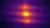
Reconstructed vortex rings inside a magnetic micropillar. Credit: Claire Donnelly
Researchers have observed three-dimensional magnetic vortex rings in a real-world magnetic material for the first time. Contrary to theoretical predictions, these rings – which are spin configurations within the material’s bulk – are remarkably stable and could move through the material like smoke rings move through air. If such movement can be controlled, they might have applications in energy-efficient 3D data storage and processing.
In a ferromagnetic material, the spatial distribution of the local magnetization is responsible for the material’s magnetic properties. These spatial distributions can be very complex, and intricate magnetic “textures” are behind many modern technologies, including hard disk drives. A vortex is one such distribution, and it forms when the material’s magnetization circulates around a central core.
Vortex rings are more sophisticated still, and occur naturally in physical systems such as fluids, plasmas and turbulent gases in the Earth’s atmosphere. However, while they have long been predicted to exist in ferromagnets, they have never been observed there until now.
X-ray magnetic tomography
Scientists led by Sebastian Gliga of the Paul Scherrer Institute in Switzerland discovered these doughnut-shaped ring patterns in nanoscale structures of gadolinium cobalt. The result comes thanks to a technique the group developed in 2017 called X-ray magnetic tomography, which enables them to observe nanoscale magnetic configurations in 3D, deep within a micron-sized sample. Before this advance, researchers were only able to visualize magnetization structures a few layers below a material’s surface.
Claire Donnelly, the study’s co-lead author and a physicist at the University of Cambridge in the UK, says that new data analysis techniques were also crucial. These new techniques allowed the researchers to pick out topological structures – such as rings – within their dataset via calculations of the magnetic vorticity vector. “Calculating this quantity and observing that it circulates around loops (just as a smoke ring’s vorticity vector would) allowed us to identify the magnetic vortex rings,” Donnelly says.
The stability of these rings was unexpected, notes Konstantin Metlov, the study’s other co-lead author and a researcher at the Donetsk Institute for Physics and Engineering and the Institute for Numerical Mathematics RAS in Moscow, Russia. This is because they were predicted to be dynamic, moving objects. Their stability appears to come from the long-range interaction between electron spins in the material – a phenomenon that had not been considered before. “This is very exciting, since it implies that such complex 3D magnetic structures – and possibly other more topologically non-trivial ones – may be easier to stabilise than originally thought,” the researchers tell Physics World.
According to Gliga, Donnelly, Metlov and colleagues, the rings’ stability could be important for practical applications because it means they could move through magnetic materials. Learning how to control these structures within the volume of a magnet might thus aid the development of 3D magnetic data storage and processing.
The researchers, who report their work in NaturePhysics, say they plan to extend their investigations using, for example, time-resolved techniques they developed earlier this year. This would give them a glimpse of how vortex rings actually move. “We also want to find out how they are created in the first place – and how they collapse,” Donnelly says. “Now that we can observe these systems in experiments, we’ll also be looking out for more complex structures, like knotted vortex rings.”
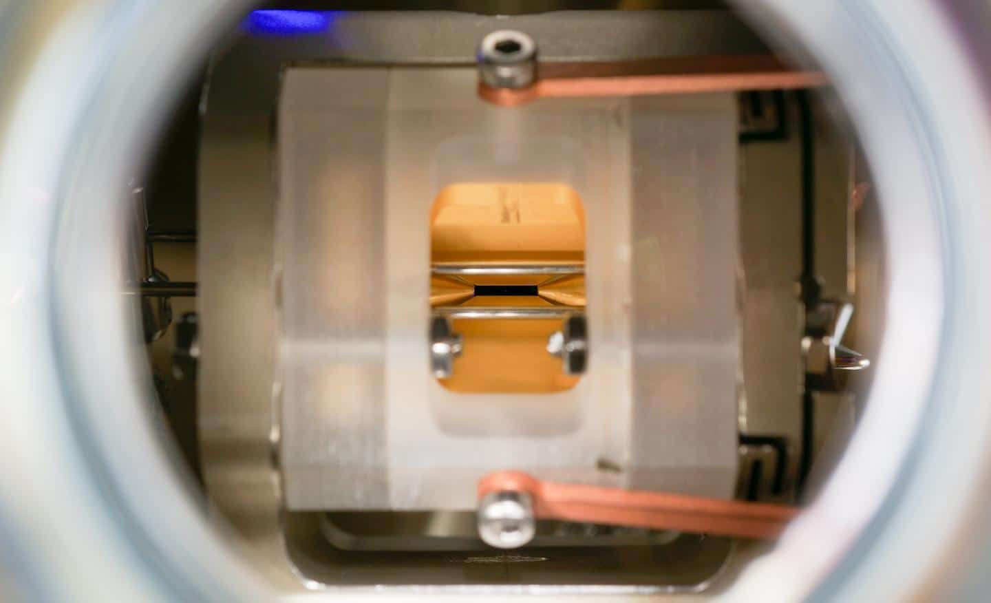
A new technique to cool reactive molecules to temperatures low enough to achieve quantum degeneracy – something not generally possible before – has been created by researchers in the US. In this temperature regime, the dominance of quantum effects over thermal fluctuations should allow researchers to study new quantum properties of molecules. As a first example, the researchers demonstrated how a slight change in applied electric field can alter the reaction rate between molecules by three orders of magnitude. The researchers hope their platform will enable further exploration of molecular quantum degeneracy, with potential applications ranging from quantum many body physics to quantum information processing.
When atoms are cooled close to absolute zero, the blur created by thermal effects that govern their behaviour in the classical world around us is removed, making their quantum nature clear. This has led to some fascinating discoveries. In ultracold quantum bosonic or fermion-pair quantum gases, for example, all the atoms in a trap can simultaneously occupy the quantum ground state, resulting in a wavefunction that is macroscopic.
Cooling and trapping molecules is much trickier because they are inherently more complex than atoms. Whereas atoms can only contain quanta of energy in electronic excitations, the chemical bonds in molecules can stretch, rotate and bend – and cooling molecules involves removing energy from all of these degrees of freedom. Moreover, the complexity of molecules increases the complexity of their collisions. Although elastic collisions are necessary to knock the fastest-moving molecules out of a trap and cool it, inelastic collisions dissipate heat in the trap.
Huge rewards
The rewards for success, however, are huge according to Jun Ye at JILA in Boulder, Colorado. “As is always the case in life, something negative has a positive side. Once you have molecules under control, you suddenly have so much more flexibility to control their quantum state.”
This week, Ye and colleagues published two new papers – one in Nature and one in Science. In the Nature paper, they applied an electric field to compress potassium and rubidium atoms in a 3D optical trap, inducing the atoms to pair up and thereby forming a 2D cloud of polarized potassium-rubidium molecules. Side-to-side collisions were elastic, whereas the head-to-tail ones were inelastic. As the molecules were polarized and confined to two dimensions, they were much more likely to collide side-to-side than head-to-tail. This allowed the researchers to achieve about 200 elastic collisions for every inelastic one, driving out the hotter molecules and cooling their sample to quantum degeneracy.
Scientific milestone
“When my former colleague Deborah Jin [who died in 2016] was still alive, we had a collaboration showing that, when you turned on the electric field, the molecules just got lost because you were enhancing the inelastic collisions,” explains Ye, “This year, we were able to reverse that process, and that’s the milestone that the scientific community has been looking forward to.”
In the Science paper, the researchers studied the effect of electric fields on the reaction rates of the cooled molecules with each other. They allowed the molecules to collide at multiple orientations in 2D, and monitored the number of potassium-rubidium molecules in the trap. As the researchers had suppressed simple inelastic collisions effectively to zero, the only remaining way for a particle to leave the trap was to react with another potassium-rubidium particle to form K2 and Rb2 molecules.
As expected from theoretical predictions, the researchers found that, as they tuned the electric field by a few per cent across a specific resonant frequency, the rate of reaction increased by three orders of magnitude as two energy levels became degenerate, reducing the activation energy barrier.
The researchers now intend to study other, more exotic phenomena using the new tool they have developed. “We are very interested in studying collective behaviour of molecules and quantum correlations in these fantastic low entropy systems,” says Ye. “That’s something we’re working on right now.” Further into the future, he says, “quantum information might be a really interesting direction to look. If you can control quantum coherence, if you can synthesize a system with low enough entropy that you have quantum control, and now you have this extra ingredient that we can tune the interaction – which is always really important to program a computer – all these ingredients are falling into place. We are certainly not there yet, but one can dream big.”
John Doyle of Harvard University in Massachusetts sees two major advances in the works: “One is this sample of Fermi-degenerate molecules that are kept away from each other due to long-range interactions…This opens up the door to polyatomic quantum gases,” he explains. “The other is the more fundamental idea that you can control chemistry exquisitely by tuning these long-range interactions. Their results showing that if you change the field by just a teeny amount you can drive a system from unstable to stable will, I think, be viewed as an archetypal achievement.”
In this episode of the Physics World Weekly podcast, Jun Ye is in conversation with Physics World‘s Hamish Johnston about another of his research interests, atomic clocks.
Protein folding is a process that is crucial to life and understanding its intricacies is an important challenge of computational biology. In many fields of science, converting data into sounds has helped researchers deal with complex patterns. Now, an international team of researchers has created a method to represent folded protein nanostructures as musical compositions.
“We explore different avenues of artistic creation, interpolating between human design, natural or evolutionary design, and designs from a deep recurrent network model that was trained against musical scores of known three-dimensional protein structures,” they write in a paper that has been accepted for publication in Nano Futures.
“Artistically, our work offers a new perspective on the limits of scientific understanding, and allows human players to interact with nanoscale phenomena, providing a tool for STEM outreach, and use of nanoscopic phenomena for artistic expression.”
Personally, I love Star Trek – well, the original series – so I have no problem with scholarly papers on the subject. But perhaps not in a journal described as being “concerned with the continuity of foetal and postnatal life”.
Indeed, I would like to see a paper on the most pressing philosophical question facing Star Trek fans – who is better, Captain Kirk or Captain Picard?
Recruitment and retention of specialist physics teachers remains a long-standing problem in England, with supply consistently falling short of national demand. Official figures from the Department for Education (DfE) in England, for example, show that there were 29,580 new entrants to postgraduate initial teacher-training (ITT) courses in the academic year 2019/20 – a slight increase on the 29,215 postgraduate trainees in 2018/19. Yet while subjects like biology, history and geography exceeded government recruitment targets, it’s notable that take-up was well below par in other subjects such as computing (79% to target), mathematics (64%) and physics (43%). What’s more, the shortage of physics teachers is even more acute for schools serving low-income communities with a history of academic underachievement.
As part of its strategy to address the shortage of candidates for physics ITT programmes, the Institute of Physics (IOP), which publishes Physics World, is aiming to encourage talented graduates and postgraduates in physics and engineering disciplines to enter the teaching profession via its Teacher Training Scholarship scheme. Funded by the DfE, the scholarships represent a compelling proposition, headlined by a tax-free financial package that helps would-be teachers transition through their one-year ITT course in England.
Support is substantial, wide-ranging and sustained. The IOP’s 2021/22 scholarship scheme, for example, is now open for applications and has 200 scholarships on offer to the next ITT cohort. Successful candidates will each benefit from tax-free funding of £26,000, with payment being phased throughout the training year and reinforced by a structured programme of continuing professional development (CPD) to complement trainees’ core ITT learning.
From industry to teaching
With the emphasis fixed squarely on recruiting outstanding physics teachers, it’s clear that IOP is casting the net wide, aiming to attract not just recent physics and engineering graduates into teaching but also established professionals with experience across diverse physics-based industries. A case study in this regard is Alastair Miatt, an IOP Teacher Training Scholar who completed his ITT course over the summer ahead of taking up a new physics teaching post in September.
After graduating with a mechanical engineering degree from the University of Cambridge in 1991, Miatt spent just short of three decades working in the automotive industry – a career that spanned a range of engineering management roles at Jaguar Land Rover (JLR) in the Midlands and the north of England. It was when Miatt turned 50, however, that he arrived at what he describes as one of those stereotypical “what am I going to do with the rest of my life?” junctures. Turns out the answer was teaching, a decision informed by his experience of voluntary work in the classroom – part of a JLR collaboration with All Saints Catholic High School in Knowsley that saw him mentoring sixth-form students with their engineering, design and technology projects.
Alastair Miatt: “The IOP combines this deep understanding of physics education with great ideas about how to engage young people.” (Courtesy: Alastair Miatt)
“I’m a naturally conservative character, but my motivation was to do something fresh and take a leap with the next stage of my career,” he explains. “Although I’m an engineer by training, physics teaching was the natural choice. The fascination of physics is in helping young people to understand how the world works at a more fundamental level – that’s a powerful thing.”
For other mid-career scientists and engineers considering a similar transition, Miatt says the key is to recognize how all that accumulated professional experience can underpin success in the classroom – and, longer term, in making science relatable to young people. “There’s all sorts of expertise that you build up throughout your career and it’s easy to take that for granted or underestimate it,” he explains.
In Miatt’s own case, the industry perspectives from JLR offer so many different ways of relating fundamental physics concepts to real-world scenarios – the use of ultrasound sensors in self-driving cars, for example, as an applied case study illustrating the principles of wave theory. “The move into teaching is a big challenge for sure,” he adds, “but all the domain knowledge and skills from my time at JLR – whether that’s technical know-how, project planning or public speaking – has helped me rise to the challenge and embrace the change.”
A framework of support
For Miatt, and other career-changers like him, it’s evident that the journey from manufacturing plant back to the classroom would be that much harder – if not impossible – without access to the IOP Teacher Training Scholarship scheme. On a purely practical level, there’s the financial buffer to support industry professionals through ITT and into their formative years as newly qualified teachers. Equally important, notes Miatt, is the validation and recognition from one of the world’s foremost learned societies: “IOP is very supportive during the application process. Securing the scholarship was the real clincher – a massive vote of confidence in my potential as a physics teacher.”
That IOP support continues throughout the ITT year, with a series of physics-based online CPD events running alongside the day-to-day inputs that scholars get from their university ITT provider (the University of Chester in Miatt’s case) and in-school teaching placements. Prior to the coronavirus lockdown in March, for example, Miatt attended a masterclass on space science and gravity at the National Space Centre in Leicester. As well as providing an opportunity to compare notes and discuss common challenges with fellow IOP teaching scholars from around the country, he says the masterclass showcased the benefits of “venue-driven learning”, yielding all manner of simple, creative teaching ideas to incorporate into his lesson plans. “The IOP combines this deep understanding of physics education with great ideas about how to engage young people and communicate physics more effectively and creatively,” he adds.
With the pandemic forcing school closures nationwide throughout the Spring, Miatt concedes that the disruption made for “a teacher training year like no other”. Nonetheless, with schools back open again and readjusting to the “new normal” since September, Miatt remains excited – and optimistic – after completing his first term as a newly qualified physics teacher – a post at Neston High School in Cheshire. “The notion that physics teaching can unlock a new world for young people is not too idealistic – it’s possible,” he concludes.
MRI is the standard modality for assessing neurological disorders, due to its ability to image intracranial anatomy with unparalleled soft-tissue contrast. Conventional high-field MRI scanners, however, are costly, immobile and require dedicated power and cooling infrastructure. As such, MRI is unavailable to critically ill patients who cannot be safely transported to the scanner or patients in low-resource settings.
A low-cost, portable brain MRI scanner could expand access to MR neuroimaging, as well as enabling point-of-care diagnostics for neurological emergencies. With this aim, researchers at Massachusetts General Hospital/Harvard Medical School are developing a portable scanner based on a compact, lightweight permanent magnet. Writing in Nature Biomedical Engineering, the researchers describe the design and testing of their prototype system.
“There are cases where MR brain imaging would be diagnostically useful, but it is not feasible because of the logistical burden and cost,” says first author Clarissa Cooley. “To address this, we wanted to develop a truly portable MRI brain scanner that could be used in new locations, like a patient’s bedside or rural clinic. Our design is meant to be a very accessible MRI option for detecting brain abnormalities that are visible at a lower field and lower resolution.”
Optimized design
The team’s portable MRI scanner is based around four key design points. First, by creating a dedicated brain scanner with a small-diameter bore that fits around the head, rather than a full-body system, the scanner size and cost can be reduced.
At the heart of the scanner is a permanent magnet made from an array of neodymium (NdFeB) rare-earth magnets that generate an 80 mT static field. Unlike the bulky superconducting magnets used in conventional MRI systems, or previously used electromagnets, the permanent magnet does not require external power or cryogenic cooling.
Arranging the magnet segments in an optimized Halbach cylinder configuration creates a transverse field inside the magnet and zero field outside the magnet. This intrinsic self-shielding is ideal for portable applications where stray fields could pose safety hazards. The constructed magnet assembly is 49 cm long, with an outer diameter of 57 cm and 27 cm bore opening.
The third design factor is that, rather than designing a homogeneous magnet, the team shaped its magnetic field variation into a built-in field gradient (of 7.6 mT/m) for readout encoding. This reduces magnet size and cost, and eliminates the need for a traditional readout gradient coil, lowering the acoustic noise, power and cooling requirements. With the built-in field variation used for image encoding in the x dimension, switchable gradient coils provide phase encoding in the y and z directions. The RF transmit/receive coil is incorporated into a compact helmet.
Finally, the researchers used advanced reconstruction techniques to correct for image distortions that arise from the non-linear field gradients used to encode the image. “Our image reconstruction method utilizes measured magnetic field maps to correct for these distortions,” Cooley explains.
Proof-of-principle
The prototype scanner – including the 122-kg magnet, coils, amplifiers, console and cart – weighs approximately 230 kg. Replacing the general-purpose console, amplifiers and cart with custom lightweight designs could reduce this to roughly 160 kg, the team notes. With no refrigeration systems for a superconducting magnet, power requirements are low, enabling the scanner to be operated from a standard power outlet.
Cooley and colleagues used their prototype scanner to record MR images from three healthy volunteers. The scanner successfully generated T1-weighted, T2-weighted and proton density-weighted brain images – standard brain scans routinely used for detection, diagnosis and monitoring of clinically important brain pathology. Each image was acquired in roughly 10 min and had a spatial resolution of 2.2 × 1.3 × 6.8 mm.
Although the scanner’s spatial resolution and sensitivity are both lower than that of a high-field MRI, the researchers emphasize that its performance is sufficient to detect and characterize serious intracranial processes, such as haemorrhage, hydrocephalus, infarction and mass lesions. Preliminary work also suggests that diffusion-weighted imaging, which is critical to applications such as acute stroke detection, should also be possible.
These initial images were acquired in an RF shielded room to eliminate external electromagnetic interference (EMI). “For true portable imaging, we are integrating EMI detectors into our scanner for EMI mitigation,” says Cooley. “This will greatly increase the image quality when our scanner is operated at the point-of-care.”
“We are also excited to begin work on a point-of-care MRI scanner specially designed for neonatal patients in the [neonatal intensive care unit] NICU,” she tells Physics World. “The transport and scanning of sick neonates is logistically very difficult and can be dangerous. The availability of a bedside MRI scanner in the NICU could have tremendous benefits for diagnostics and monitoring of neonatal brain injury.”
Balloon-borne telescopes can observe a wealth of astrophysical phenomena that ground-based instruments cannot, but onerous cooling requirements limit how much equipment can be taken aloft. Researchers at NASA’s Goddard Space Flight Center found a way to minimize this problem by drastically reducing the weight of a telescope’s cooling system. The researchers have tested their approach on a mission called the Balloon-Borne Cryogenic Testbed (BOBCAT) and have a follow-up mission planned to study it further.
Distant galaxies and star- and planet-forming clouds of gas and dust emit photons in the infrared region of the spectrum. Because the Earth’s atmosphere blocks most of this infrared radiation, these objects are hard to study from the ground. While space missions are the ideal option, they are extremely expensive. Balloons that carry telescopes way up into the stratosphere are a good alternative because they cost much less.
Near absolute zero temperatures required
The mirrors of balloon-borne telescopes can be huge, measuring up to 3 to 5 m across – “the size of a living room”, says team leader Alan Kogut. This presents a challenge because the mirrors, like the rest of the telescope, need to be cooled to near absolute zero during the mission. If they aren’t, their heat can wipe out the infrared light from deep space “like overexposing a camera”, Kogut says.
“Liquid helium can easily cool the telescope, but keeping it cold means putting the entire telescope into a giant thermos bottle called a dewar,” he says. “A thermos bottle the size of a living room would weigh several tonnes – more than even the largest balloons can carry.”
Standard dewars need to be this heavy because their walls must sustain a vacuum against sea-level air pressures, Kogut explains. However, he and his colleagues reasoned that a balloon-borne dewar could be much lighter since the pressure at the balloon’s operating altitude of 40 km is only 0.3% of that at sea level.
Extremely thin stainless-steel walls
The dewars developed for the BOBCAT mission comprise an inner cup, which contains the liquid coolant, surrounded by an outer shell. The gap between the two layers is under vacuum, preventing air from carrying heat from the outside into the cold interior. This “bucket” design is conventional, but the walls of the cup and shell are not, being made of stainless steel which, at 0.5 mm thick, is “not much thicker than a soda can’s”, says Kogut.
The new dewar can be launched at room temperature, and it has an integrated valve that allows the vacuum gap between the inner cup and outer wall to vent continuously during ascent. This permits air to escape, thereby eliminating any pressure gradient across the walls.
Once the balloon reaches an altitude of around 40 km, the valves closes to seal the dewar’s vacuum, explains Kogut. The telescope is cooled by pumping liquid nitrogen or liquid helium into the ultralight dewar from separate storage tanks, which themselves are of standard construction, are small and don’t weigh much.
Successful first test
The team tested the new design on an 827-kg-payload flight launched in August 2019. The goal of this initial test was two-fold. First, it was meant to prove that cryogenic liquids (14 litres of liquid nitrogen and 268 litres of liquid helium in the test) could indeed by transferred at float altitudes. Second, it was designed to measure the total amount of heat leaking to the receiving dewar. The researchers calculated this to be around 2.7 W, which is larger than the 1 to 2 W measured for the same dewar in ideal laboratory conditions. This value will be compared in a follow-up flight using a lighter dewar of identical size, they say.
Each December, Physics World selects its Top 10 Breakthroughs of the year. Watch this video to find out which research has made it onto this year’s shortlist. On Thursday 17 December, one of the ten will be crowned Physics World’s Breakthrough of the Year 2020.
In addition to having been reported in Physics World in 2020, our selections must meet the following criteria:
Significant advance in knowledge or understanding
Importance of work for scientific progress and/or development of real-world applications
Of general interest to Physics World readers
Come back next week to discover the winning breakthrough – and in the meantime you can read about the Top Ten, or listen to Physics World editors discuss their choices in the Physics World Weekly podcast.
This episode of the Physics World Weekly podcast features a lively chat about some of the best physics done this year as we unveil our Top 10 Breakthroughs of 2020. Our choices run the gamut from medical physics to particle astrophysics – and we even have two “Holy Grails” in the fields of superconductivity and semiconductor physics.
The Top 10 serves as the shortlist for the Physics World Breakthrough of the Year award, which will be announced on 17 December. Links to all the nominees, more about their research and the criteria for the awards can be found here.
One of the highlights in the Physics World calendar is the announcement of our Breakthrough of the Year, which will be made this year on Thursday 17 December.
Today, we are revealing the 10 finalists for 2020, which serves as a shortlist from which we will pick the Breakthrough of the Year.
This year’s Top 10 Breakthroughs were selected by a crack team of five Physics World editors, who have sifted through hundreds of research updates published on the website this year. In addition to having been reported in Physics World in 2020, our selections must meet the following criteria:
Significant advance in knowledge or understanding
Importance of work for scientific progress and/or development of real-world applications
Of general interest to Physics World readers
Here are the Physics World Top 10 Breakthroughs for 2020, in no particular order. Come back next week to find out which one has bagged the Breakthrough of the Year award – and in the meantime you can listen to four of the judges talk about the Top 10 in the Physics World Weekly podcast.
Ideal system: A strontium ion trapped in an electric field. The measurement on the ion lasts only a millionth of a second. (Courtesy: F Pokorny et al., Stockholm University)
To Markus Hennrich and colleagues at Stockholm University, Sweden, together with researchers at the universities of Siegen in Germany and the Basque Country and Seville in Spain, for using a series of “weak” measurements (the subject of Physics World’s 2011 Breakthrough of the Year) to probe the nature of superposition collapse in quantum mechanics. While the act of measurement usually forces quantum systems into definite classical states, the work of Hennrich and colleagues showed that some measurements do not destroy all quantum information. By taking a series of “snapshots” during experiments on a single ion of strontium, the team revealed that measurements are not instantaneous, but instead gradually convert superposition states into classical ones. Because weak measurements could in principle allow errors to be detected in quantum states without destroying those states in the process, the work might be used to improve error correction in quantum computers.
To Haocun Yu and Lee McCuller of the Massachusetts Institute of Technology and their colleagues on the LIGO Scientific Collaboration for showing that quantum-scale correlations can leave their mark on macroscopic objects weighing tens of kilograms. The researchers explored the exquisite interplay between the laser beam of a LIGO interferometer and its mirrors – each of which weighs 40 kg. They observed that radiation noise contributes to the motion of the mirrors, which is a result of Heisenberg’s uncertainty principle. When using squeezed vacuum states of laser light they showed that the quantum noise drops below the standard quantum limit, which demonstrates quantum correlations between the laser beam and the mirrors. The research could lead to the improved detection of gravitational waves by LIGO, Virgo and future observatories.
To the Borexino collaboration for observing neutrinos from the carbon–nitrogen–oxygen (CNO) cycle in the Sun. To do so the team had to first painstakingly minimize the effects of background radiation in the Borexino detector, which comprises 278 tonne of ultrapure liquid scintillator located deep inside a mountain at Italy’s Gran Sasso Infn Laboratories. The observation confirms a theory of stellar nucleosynthesis first proposed over 80 years ago. It should also encourage physicists to use the next generation of neutrino detectors to try to resolve the “metallicity puzzle” of the Sun – a mystery regarding the abundance of carbon, nitrogen and oxygen in the star.
To Noel Clark and colleagues at the University of Colorado Boulder and the University of Utah in the US, for observing a ferroelectric nematic phase of matter in liquid crystals more than 100 years after it was predicted to exist. In this phase, all the molecules within specific patches, or domains, of the liquid crystal point in roughly the same direction – a phenomenon known as polar ordering that was first hypothesized by Peter Debye and Max Born back in the 1910s. Clark and colleagues found that when they applied a weak electric field to an organic molecule known as RM734, a striking palette of colours developed towards the edges of the cell containing the liquid crystal. In this phase, RM734 proved far more responsive to electric fields than traditional nematic liquid crystals. Although further work is required to identify materials that display the phenomenon at room temperatures, ferroelectric nematics could find applications in areas from new types of display screens to reimagined computer memory.
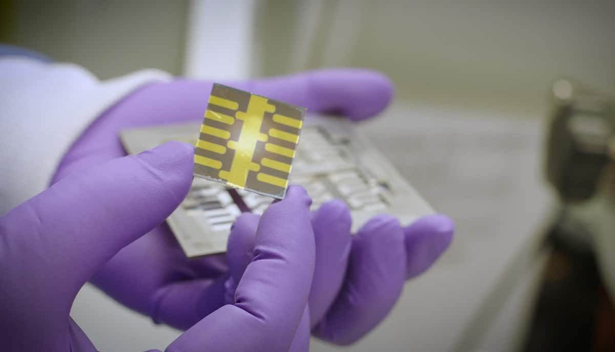
The perovskite thin-film X-ray detector is 100 times more sensitive than conventional detectors and requires no outside power source. (Courtesy: Los Alamos National Laboratory)
To Wanyi Nie and colleagues at Los Alamos National Laboratory for using thin-film perovskites to create an extremely sensitive X-ray detector. Using a synchrotron beamline to characterize their thin-film perovskite detectors, the researchers found that the X-ray absorption coefficients of the perovskite materials were on average 10 to 40 times higher than that of silicon for higher-energy X-rays. They also demonstrated that the new X-ray detectors are 100 times more sensitive than conventional silicon-based devices. This new type of solid-state X-ray detector could enable medical and dental imaging at extremely low radiation dose, enabling the same quality image to be generated using a much-reduced X-ray dose, making scans safer for patients. Nie also notes that it should be possible to fabricate large-scale detector arrays at far lower cost than for semiconductor detectors.
To Kostya Trachenko of Queen Mary University of London, Bartomeu Monserrat and Chris Pickard of the University of Cambridge and Vadim Brazhkin of the Russian Academy of Sciences for calculations showing that the upper limit on the speed of sound in solids and liquids depends on just two dimensionless quantities – the fine structure constant and the proton-to-electron mass ratio. The team’s theoretical prediction is backed up by experimental data of the speed of sound in a range of solid materials and a calculation of the speed of sound in metallic hydrogen – a material that is yet to be created in the lab but should have the fastest speed of sound. The research provides insight into how fundamental constants impose bounds on physical properties.
To Andrea Alù, Qiaoliang Bao, Cheng-Wei Qiu and an international team of collaborators at the City University of New York, National University of Singapore, Monash University, China University of Geosciences and the University of Texas at Austin, for showing that dispersion- and diffraction-free propagation of light is possible, with a resolution that beats the diffraction limit by more than an order of magnitude, in twisted layers of 2D molybdenum trioxide. Their work builds on the discovery of “magic-angle” graphene – Physics World’s Breakthrough of the Year in 2018 – by using twisted layers of 2D materials to change the behaviour of propagating photons, rather than electrons. Just as the electron version of twistronics has led to a flurry of research on superconductivity and electron states, the new photonics variant has important implications for nano-imaging, quantum optics, computing and low-energy optical signal processing.
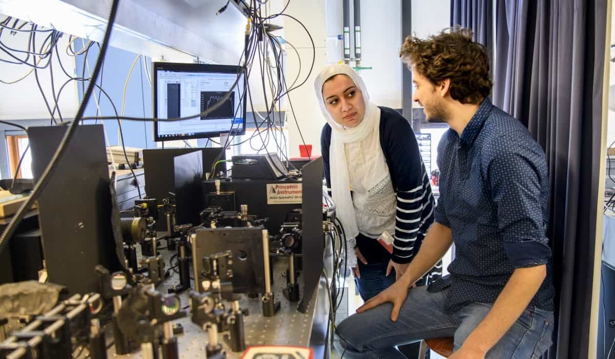
Silicon sees the light: Elham Fadaly (left) and Alain Dijkstra in their Eindhoven lab. (Courtesy: Sicco van Grieken/SURF)
To Elham Fadaly, Alain Dijkstra and Erik Bakkers at Eindhoven University of Technology in the Netherlands, Jens Renè Suckert at Friedrich-Schiller-Universität Jena in Germany and an international team for creating a silicon-based material with a direct band gap that emits light at wavelengths used for optical telecommunications. Normally, silicon has an indirect electronic band gap, which means that it is a poor emitter of light and must be integrated with other semiconductor materials to create optoelectronic devices. To create a direct band gap, the researchers had to grow crystals of silicon-germanium alloy with a hexagonal crystal structure, rather than the usual diamond-like structure. They did this by creating nanowires of the alloy, which emitted infrared light. As well as having applications in optical telecoms and optical computing, the new silicon-based material could be used to create chemical sensors.
To a team headed up by Joao Seco at the German Cancer Research Centre in Heidelberg and Simon Jolly at University College London, for demonstrating how a mixed particle beam could enable simultaneous cancer therapy and treatment monitoring. The idea is to use a beam containing both carbon ions, which provide therapeutic irradiation of the target tumour, and helium ions, which travel straight through the patient and can therefore be used for imaging. In experiments at the Heidelberg Ion Beam Therapy Center using pelvis phantoms, the researchers showed that even small inflations of an air balloon inside the phantom caused an observable change in helium range. They also demonstrated that small phantom rotations changed the measured signal. The experiments reveal the potential of using a mixed beam to monitor intra-fractional anatomy changes, enabling more accurate delivery of particle therapy and, ultimately, providing better outcomes for cancer patients.
This research by Ranga Dias and colleagues is described in a paper in Nature. The paper has since been retracted by the journal.
To Ranga Dias and colleagues at the University of Rochester and the University of Nevada Las Vegas in the US for observing superconductivity at temperatures up to 15 °C in a hydrogen-rich material under immense pressure. Superconductors carry electrical current with no electrical resistance and have a range of applications from the high-field magnets used in MRI scanners to particle accelerators. Practical devices based on superconductors must be chilled to very cold temperatures, which is costly and can involve the use of helium, so a long-standing goal of condensed-matter physicists has been to develop a material that is a superconductor at room temperature. The carbonaceous sulphur hydride material made by Dias and colleagues shattered the previous high-temperature record by about 35 degrees and is the first to claim room-temperature superconductivity. While a pressure of 2.6 million atmospheres was required to achieve room-temperature superconductivity, the researchers think it may be possible to reduce the pressure by changing the chemistry of the material.
China has successfully launched a space telescope to study some of the most energetic events in the universe. Lifting off today from the Xichang Satellite Launch Center at 4.14 a.m. local time, the Gravitational wave high-energy Electromagnetic Counterpart All-sky Monitor (GECAM) represents one of the first new “all-sky” devices that will monitor fast-radio bursts, high-energy neutrinos and magnetars. The craft is set to begin operations as soon as it enters orbit, doing so for three years.
We’re really excited to see GECAM fly on time, as we overcame numerous technical difficulties and made it through the pandemic
Shaolin Xiong
To study these events, GECAM consists of two satellites – each weighing 160 kg – that will orbit on opposite sides of the Earth at an altitude of about 600 km. Each GECAM satellite features a dome-shaped array of 25 gamma-ray detectors and eight charged particle detectors. They will search for cosmic events happening in the energy range of 6 keV – 5 MeV and, within a couple of minutes of detection, GECAM will send out alerts to telescopes around the world for follow-up observations.
The idea of GECAM emerged following the announcement in February 2016 of the first detection of gravitational waves by the US-based Laser Interferometer Gravitational-Wave Observatory (LIGO). A year later and LIGO, working together with the VIRGO gravitational-wave detector in Italy, spotted the first gravitational wave produced by the merger of two neutron stars – an observation that was followed up by the Fermi Gamma-ray Space Telescope and other observatories around the world, kick-starting the era of multimessenger astronomy. Likewise, GECAM spot the gamma-rays bursts that are related to the production of gravitational waves by such comic mergers.
“We’re really excited to see GECAM fly on time, as we overcame numerous technical difficulties and made it through the pandemic,” says GECAM’s principal investigator Shaolin Xiong from the Institute of High Energy Physics, Chinese Academy of Sciences (CAS) in Beijing.
GECAM is the first of a line-up of space science missions to be launched by China in the coming five years. Other missions include the Advanced Space-based Solar Observatory, the Einstein Probe as well as the Solar wind Magnetosphere Ionosphere Link Explorer – a joint mission between CAS and the European Space Agency.