The US physicist Arthur Ashkin, who shared the 2018 Nobel Prize for Physics for his contributions to laser physics, died on 21 September aged 98. His most famous work concerned the use of laser light to manipulate microparticles, which led him in the 1980s to create “optical tweezers” that could be used to trap and control atoms, viruses and bacteria. For this development he shared the 2018 Nobel prize with Gérard Mourou and Donna Strickland for their “groundbreaking inventions in the field of laser physics”.
Ashkin was born on 2 September 1922 in New York City, US. He completed a degree in physics from Columbia University in 1947 followed by a PhD from Cornell University in 1952. After his PhD, he moved to Bell Labs in New Jersey where he remained for the rest of his career. It was at Bell Labs where he pioneered the use of laser to manipulate objects. In 1970 Ashkin showed that forces generated by laser beams can trap tiny dielectric particles in air or water. The scattering of light pushes the particles in the direction of beam propagation so two counter-propagating beams will stop a particle from moving along the axis of propagation.
A major improvement came 16 years later, when in 1986 Ashkin showed that it is possible to trap particles using just one laser beam – rather than a counter-propagating pair. This optical trap, known as optical tweezers, could hold particles ranging in size from tens of nanometres to tens of microns – a range that allowed the study of viruses, bacteria and other biological cells. To manipulate such living organisms in a way that would not damage them, Ashkin switched the laser light from green to infrared light. Optical tweezers have since proved invaluable to biophysicists, who have used them to measure the forces involved in biological processes such as the transport of organelles within living cells, how bacteria are propelled by rotating flagella and how forces affect large biological molecules such as DNA.
Ashkin retired in 1992 and held 47 patents through his career. In 2013 he was inducted into the US National Inventors Hall of Fame and in 2018, at 96, became the oldest person ever to receive the physics Nobel prize. Given Ashkin’s health, he was unable to attend the Nobel prize ceremony and his Nobel lecture was instead delivered by his colleague René-Jean Essiambre following a short video greeting by Ashkin.
On the nanoscale, objects warm up faster than they cool down. That is the surprising conclusion of Alessio Lapolla and Aljaž Godec at the Max Planck Institute of Biophysical Chemistry in Germany, who have predicted this asymmetry using mathematical models of confined nanoparticles.
One basic assumption in thermodynamics is that an object that is either hotter or colder than its surrounding environment will cool down or heat up, respectively, at the same rate. So, an object that is slightly warmer than room temperature will reach room temperature at the same time as an identical object that started slightly below room temperature.
Lapolla and Godec tested this principle in their study using a mathematical model of a tiny nanoparticle trapped inside a one-dimensional box. As the particle undergoes Brownian motion, its position is mapped using a probability distribution – which peaks at the centre of the box, where the particle is most likely to be found. Since Brownian motion increases with temperature, the duo predicted that heating should cause the probability distribution to spread out during heating, and become narrower during cooling. However, this was not expected to result in a difference in how hot and cold particles reached equilibrium with their surroundings.
Off the walls
Contrary to expectations, however, Lapolla and Godec observed that warmer particles took longer to cool down than cooler objects did to warm up. To explain this asymmetry, the duo suggests that the more dynamic motion of a warmer particle means that it bounces off the walls of the box more often. As a result of this bouncing, the warm particle tends to drift towards the centre of the box more readily. This counteracts the spreading out due to Brownian motion – thereby narrowing the particle’s probability distributions to the centre of the box. In contrast, this bouncing became less pronounced at cooler temperatures. With less opposition to their Brownian motions, colder particles could relax to their equilibrium states more readily than their warmer counterparts.
Lapolla and Godec believe their results will improve our understanding of temperature changes in nanoscale systems. It could also provide fresh insights into phenomena like the Mpemba effect – whereby water appears to freeze more quickly when its starting temperature is warmer. The duo now hope to verify their results through practical experiments, which could be carried out relatively easily by confining particles within optical traps. The insights gained through these studies could also help to improve efficiency in devices including micromotors and heat pumps.
The announcement of the 2013 Nobel Prize for Physics is memorable for the hour-long delay in announcing the winners – François Englert and Peter Higgs – and for the long citation.
The pair won “for the theoretical discovery of a mechanism that contributes to our understanding of the origin of mass of subatomic particles, and which recently was confirmed through the discovery of the predicted fundamental particle, by the ATLAS and CMS experiments at CERN’s Large Hadron Collider”.
Theory, experiment or both?
I can vividly remember the morning of 8 October 2013 while I sat at my desk at Physics World headquarters waiting for the prize announcement. The Higgs boson had been discovered at CERN in July 2012, and it was pretty obvious that the 2013 prize would be related to the discovery. The big question was would it go to the theorists who predicted the Higgs back in 1964 or to the experimentalists who discovered it – or both?
Now when it comes to announcing the winners of each year’s Nobel prize, the Nobel Committee usually does so precisely at the scheduled time. But as the minutes ticked by, and no news was forthcoming, I started to wonder if 2013 was going to be the year when the committee finally dispensed with the rule that no more than three people can share the prize.
I imagined that, in an opulent, portrait-lined room at the Swedish Royal Academy of Sciences, a huge row had broken out about whether or not to give at least a portion of the award to the thousands of physicists working on ATLAS, CMS and the LHC. After all, the Higgs was not found by accident at CERN – there had been a decades-long sustained and focussed effort to detect the particle. Furthermore, modern science is highly collaborative, so recognizing the CERN physicists, I felt, would be the perfect way to update a prize that is more than a century old.
Out-and-about in Edinburgh
But alas, it wasn’t to be. The hour-long delay in announcing the prize merely occurred because the committee could not contact Peter Higgs. He was out-and-about in Edinburgh and in the end only heard about his win after lunch, several hours later, when he was congratulated in the street by a former neighbour.
What is more, I don’t think any scientific collaboration will be winning the Nobel Prize for Physics any time soon. Last year I interviewed Lars Brink – a Swedish particle theorist who served on the Nobel Committee for Physics on eight separate occasions and who served as chair for the 2013 award. Brink says that the Academy is hesitant to open the physics prize up to organizations or collaborations such as CERN. “We don’t want 5000 people calling themselves Nobel laureates,” he told me.
What the Nobel Committee did do, however, was to beef up that year’s citation. By mentioning ATLAS, CMS and the LHC, the committee did go at least some way to recognise the legion of experimentalists who confirmed calculations done by Higgs and independently by Englert and Robert Brout – the latter having died in 2011 and who therefore also missed out on the prize.
But given that the Higgs prediction and subsequent discovery is a classic example of how theory and experiment work hand in hand, I still think it is a shame that all those hard-working experimentalists were unable to share the prize. My fantasy citation for the 2013 prize would therefore have been “to François Englert, Peter Higgs and to all the physicists working on ATLAS, CMS and the LHC for the theoretical discovery of a mechanism that contributes to our understanding of the origin of mass of subatomic particles and the experimental discovery of the predicted fundamental particle”.
Still, who knows, perhaps the experimental discovery of the Higgs will be honoured this year after all – or in years to come.
In recent years, the pharmaceutical industry has been interested in biocatalysis (enzymes) for the production of drugs and drug intermediates due to the high selectivity of enzymes. However, many of these enzymes require expensive cofactors. Electrochemistry provides an opportunity to do cofactor regeneration to decrease the cost and the sustainability of biocatalysis for synthesis.
This webinar will introduce bioelectrocatalysis and discuss its current and future applications in electrosynthesis.
Shelley Minteer is a USTAR Professor in the departments of chemistry, and materials science and engineering at the University of Utah. She received her PhD in analytical chemistry at the University of Iowa in 2000 under the direction of Professor Johna Leddy. After receiving her PhD, she spent 11 years as a faculty member in the Department of Chemistry at Saint Louis University before moving to the University of Utah in 2011. She is also an Associate Editor for the Journal of the American Chemical Society. She has published more than 350 publications and more than 450 presentations at national and international conferences and universities. She has won several awards, including the Luigi Galvani Prize of the Bioelectrochemical Society, the Missouri Inventor of the Year, International Society of Electrochemistry Tajima Prize, Fellow of The Electrochemical Society, and the Society of Electroanalytical Chemists’ Young Investigator Award. Her research interests focus on electrocatalysis and bioanalytical electrochemistry. She has expertise in biosensors, biofuel cells, and bioelectronics.
Sometimes, the answers to difficult questions are within plain sight. That is what researchers at the University of Southern California realised as they looked for new ways to find and see cancer cells inside the human body and discovered that the dyes they could use are all around us every day.
Early detection of cancerous cells gives patients the best chance of successful treatment and survival. However, finding those cells can be challenging. In their latest research, reported in Biomaterials Science, Cristina Zavaleta and her team found that they could use common dyes and pigments, like food colouring and tattoo ink, combined with nanoparticles to essentially paint cancer cells – making them easy to spot when compared with normal cells.
Paint by cell type
Finding cancer cells without imaging agents is a difficult job. “For instance, if the problem is colon cancer, this is detected via endoscopy,” Zavaleta explains. “But an endoscope is literally just a flashlight on the end of a stick, so it will only give information about the structure of the colon – you can see a polyp and know you need to take a biopsy.”
But with imaging agents that can specifically paint the cancerous cells, medical professionals have a much easier job. “If we could provide imaging tools to help doctors see whether that particular polyp is cancerous or just benign, maybe they don’t even need to take it,” says Zavaleta.
Inspiration for this work came from an unusual source: Zavaleta’s imagination was sparked in an animation class with Pixar artists. “I was thinking about how these really high-pigment paints were bright in a way I hadn’t seen before,” she says. This led to a trip to a tattoo artist and the realization that these everyday inks could have exciting properties for medical imaging.
Putting the pieces together
The researchers created optical imaging agents using liposomal nanoparticles. These nanoparticles are tiny spheres made of a membrane of lipid, or fat, molecules – the same kind of structures that form the boundaries of human cells – and the colourful dyes can be housed inside. Using nanoparticles made of fats and dyes that are already FDA-approved and used in everyday life is a clever approach for potentially accelerating their clinical translation.
The researchers found that not only do nanoparticles containing these common dyes exhibit improved fluorescence signals compared with the clinical dyes, because more of the dye can be loaded into each nanoparticle, but that they are also significantly more visible inside tumours than in adjacent normal cells. The researchers suggest that this is because the tumours have leakier blood vessels feeding them, letting in the nanoparticles, which cannot enter into normal cells.
The new dyes also have unique spectral fingerprints, which could aid in their specific detection. It could even be possible to use multiple different dyes together to paint different types of cells in unique ways.
Whilst the initial findings in mice models are encouraging, there is still some way to go until this work might help patients in the clinic. However, Zavaleta and her colleagues are hopeful that this study could provide a foundation towards better cancer detection in the future.
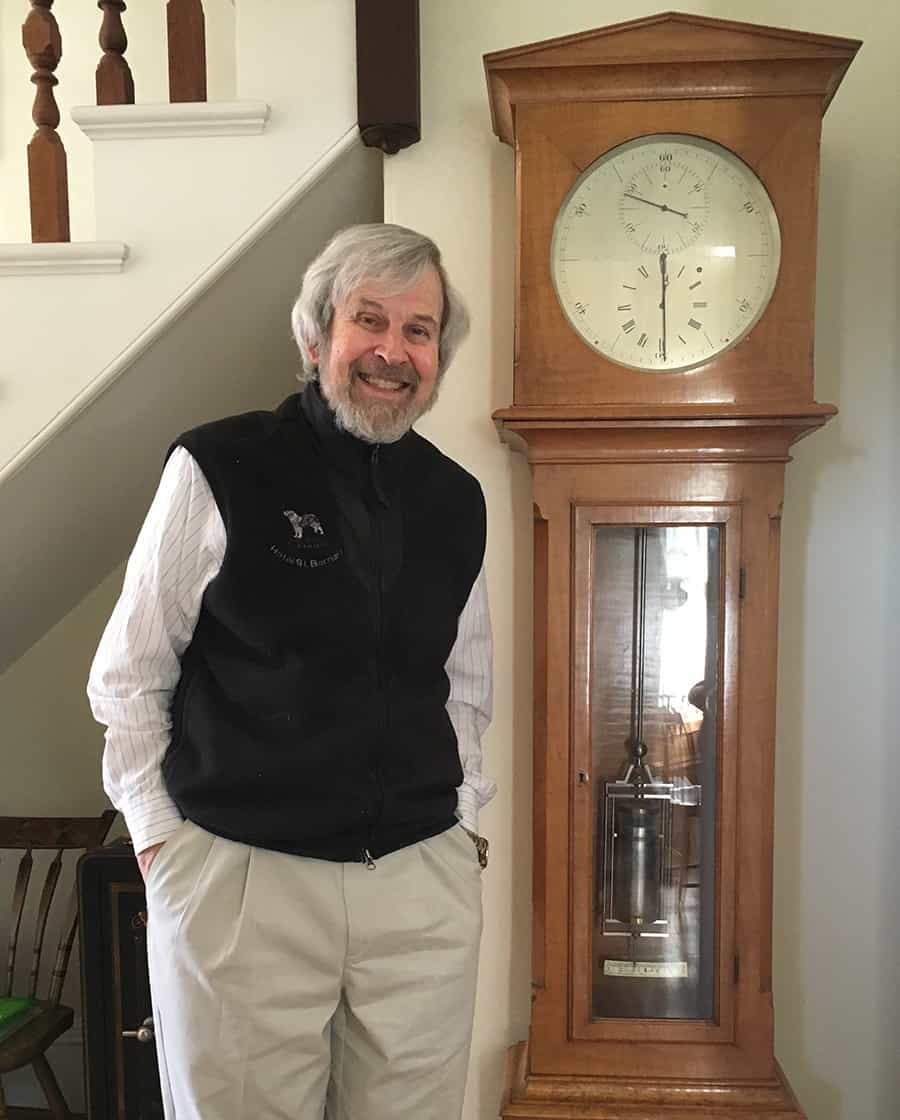
Awe struck Linn Hobbs’ fascination with materials extends to those used in the many clocks at his home on the outskirts of Boston, Massachusetts. (Courtesy: Robert P Crease)
If you want to get a sense of the power and beauty of materials science, there are few better places to start than a chat with Linn Hobbs. Living in retirement in a three-storey house on the outskirts of Boston, US, Hobbs spent most of his career at the nearby Massachusetts Institute of Technology and retains an almost child-like enthusiasm for materials science. “[It] explores the kinds of things you can find in an average 10-year-old’s pocket,” he claims. “Salt, sand, string, rust and bone.”
Born in 1944 in Detroit, Hobbs’ interest in science started early. He was a licensed ham-radio operator at 11, later built his own communications equipment and these days still broadcasts from the top floor of his house. In 1962 Hobbs went to study engineering science at Northwestern University in Illinois, where the first book he read was on metals. “I was astounded that so much science could be applied to so much of the world around us,” he told me when I visited last summer. “I read the book like a novel, cover to cover in three days.”
Hobbs tried to major in materials science, but was informed that Northwestern had no major in the subject. All it had was a graduate materials-science department, which had been set up by the Advanced Research Projects Agency (ARPA) just two years earlier. ARPA itself had been established as a panic measure by the US government, which realized it lagged the Soviet Union in missile technology following the launch of Sputnik – the world’s first artificial satellite – in 1957. ARPA was designed to foster the study of metal alloys, ceramics and other compounds that could withstand the extreme pressures and temperatures of missiles and spacecraft.
When Hobbs arrived at Northwestern, even the university’s grad students in materials science had little prior training in the new discipline.
So when Hobbs arrived at Northwestern, even the university’s grad students in materials science had little prior training in the new discipline. Undeterred, Hobbs learned alongside them, despite his status as a lowly undergraduate. With his electronics experience as a radio operator, he ended up building equipment to study the motion of dislocation defects in lithium fluoride, which has a crystal structure identical to that of table salt.
After graduating from Northwestern in 1966, Hobbs went to the UK on a Marshall Scholarship, winding up at the University of Oxford, which then was a world-leader in the electron microscopy of materials. Hobbs’ subsequent career leant heavily on this tool, using it in a constantly widening set of applications that were turning materials science into an ever more unified field.
Hobbs’ hobbies
During his decade at Oxford, Hobbs took up a series of life-long hobbies, each involving different facets of materials science and reflecting a growing fascination with historical artefacts.
One hobby was antiquarian horology, instigated by his purchase of an old clock in an Oxfam charity shop for £10. On my visit to his house, our conversation was periodically punctuated by bright, high-timbre chimes from many of his subsequently acquired clocks. Hobbs explained the role of various clock materials – wood, iron, steel, brass, bell-metal, even sapphire – and discussed how they drove the history of these devices. Material behaviour was also critical for the forte pianos (an early kind of piano) in Hobbs’ collection of these instruments.
At Oxford, Hobbs became the wine steward at Wolfson College. This was a prestigious gig: Oxbridge colleges take their wine very seriously and Hobbs now owns a 2500-bottle wine cellar. As he showed me round it, Hobbs stopped to explain the structure of Champagne bottles; the indent at the bottom, called a punt, is mechanical reinforcement. He also explained the different colours of glass bottles – their silicate glass deriving from sand – with green being so common as the iron impurities causing it are the hardest to remove. A stack of skis next to the wine cellar then led Hobbs to explain why his knowledge of their component materials saw him becoming a consultant for a ski company.
Linn Hobbs is keenly aware just how integral materials science is to different fields of research.
Hobbs is keenly aware just how integral materials science is to different fields of research. He told me how he once listened to a young biologist describing difficulties with in vitro studies of cellular processes in bone formation. These studies were being carried out in Petri dishes, prompting Hobbs to enquire as to the nature of the glass substrate. Looking at him blankly, the biologist exclaimed in frustration: “Glass is glass!” Hobbs replied, “No, it’s not,” and explained that there are many types of glass, whose composition, surface chemistry, topography and defects all affect biological processes.
During his career, Hobbs has also studied the materials science of natural polymer fibres used by ancient cultures – the “string” in the child’s pocket – as well as metal corrosion processes, such as the rusting of iron. As for bone, Hobbs and his research group used electron microscopes to study bone biomineralization processes. In collaboration with the Royal National Orthopaedic Hospital in London, he also examined how bone bonds to orthopaedic implants, discovering that bone mineralization can begin after just three days.
The critical point
Few American materials scientists, it is safe to say, have been awarded an OBE (Order of the British Empire), which Hobbs received for his long involvement with the Marshall Scholarship programme and other Anglo-American educational initiatives. As for our conversation, it left me with an appreciation for two aspects of the history of materials science. One is that it is driven by society’s demands for better materials in everything from housing, clothing and transport to defence, art and medicine. The other is the role of scientific instruments in advancing our understanding of materials. This has simultaneously extended materials science into previously separate fields – notably metallurgy, ceramics and glass, polymers, biology and medicine – and unified much scientific research in these varied disciplines.
Supporting talent Cinzia Casiraghi was one of the guest editors of a focus issue of JPhys Materials on female contributions to 2D materials. (Courtesy: Cinzia Casiraghi)
Female scientists make some amazing contributions to advanced materials, with almost a third of all researchers on the European Union’s €1bn Graphene Flagship project, for example, being women. But when it comes to, say, positions of leadership or invitations to speak at conferences, we’re far from reaching gender equality.
We wanted to celebrate the work of women investigators at different stages of their careers – not only to recognize their excellent science, but also to show the next generation of women scientists that there are lots of female role models out there. We wanted to fix the “leaky pipeline” by helping and supporting the progression of other women scientists. Long term, we are also planning a series of focus issues in all areas of advanced materials, which would provide a list of top women researchers who could be used for subject reviews, invited talks and so on.
What’s been your impression of the overall quality of the papers published so far?
The issue currently has 13 research papers, one review article and one perspective, with a further three papers under peer-review. The quality has been very good, as shown by the incredible number of downloads – on average 850 each – in the first five months. The work on “Spectroscopic properties of few-layer tin chalcogenides”, led by Zeila Zanolli from the Catalan Institute of Nanoscience and Nanotechnology in Barcelona, has already been cited five times. Indeed, no matter what the gender of the author, quality has been always the number one criterion for whether a paper gets accepted in JPhys Materials.
By publishing this focus issue, did you come across talented women scientists whose work you personally hadn’t been aware of before?
Yes, and, unfortunately, I am sure there are many other talented women I am also not aware of. I have been very impressed by the collaborative and supportive network that this special issue has initiated. Women are often accused of not being able to build networks, but for this issue, that has certainly not been true.
What’s been the reaction from the authors and the wider community to this focus issue – did they find it a worthwhile exercise?
In general, both female and male scientists have enthusiastically supported this focus issue, often saying how very much needed it was. Remarkably, it wasn’t just the women we’d invited to write for the issue who suggested other female contributors; we also received names from male colleagues, showing they’re also sensitive to the subject too. Only in a very few cases were our invitations turned down because of the gender angle to this project. This feedback was very useful as it gave rise to interesting discussions with the scientists around gender issues and how to improve female representation in science.
Have any of the women who contributed papers found it has helped their research careers, raised their profiles, or led to new opportunities?
I hope so. Zeila Zanolli, for example, became an associate professor after publishing her work, while Lucia Gemma Delogu from the University of Padua, Italy, on the back of her JPhys Materials paper, used this opportunity to write a review article on the results obtained from the two European projects she has co-ordinated, by providing visibility to her role in the projects.
Yes, several. First, although I have been involved in 2D materials ever since the discovery of graphene, it’s remarkable how fast the field’s been growing, and, as consequence, how many people, especially emerging scientists, are struggling to get visibility. Second, it has been interesting to see how female scientists from different geographical areas or at different stages of their careers reacted in different ways to the special issue. There are lots of data we could extract from this exercise that could be used to improve gender equality in this field.
You’ve published 15 papers so far – are there any more in the pipeline?
We expect a total of 16 papers.
What progress has been made on plans for further JPhys Materials focus issues covering the work women scientists in other areas of advanced materials?
The next focus issue – on “materials for energy” – is already open for submissions. But as it’s on a different topic, I personally am not involved in it, although I am delighted that JPhys Materials is following up on our exercise. It’s being guest-edited by Giulia Grancini (University of Pavia, Italy), Serena Corr (University of Sheffield, UK), Deli Wang (Huazhong University, China), Claudia Schnohr (University of Leipzig, Germany) and Mari-Ann Einarsrud (Norwegian University of Science and Technology). More than 40 confirmed submissions have already been received, which is amazing. I am looking forward to that focus issue being published and comparing the reactions between the two communities.
Pushing the limits of silicon electronics to make smaller, faster and more efficient devices is getting harder. One possible way forward is to take advantage of the many phenomenal properties of nanocarbon structures that have been discovered over the past 40 years. However, amongst this suite of carbon nanomaterials there has been a conspicuous lack of wire-like materials with metallic properties that can be reliably customized and produced. This poses a challenge because connecting nanocarbon semiconductor components with the same old copper or silver wires can take the shine off their appeal.
While carbon nanotubes with metallic properties can be made with relative ease, the snag is that a heap of nanotubes with semiconducting properties are inevitably produced at the same time. Unfortunately, separating metallic nanotubes from semiconductor nanotubes en masse remains something of a dark art. Another option is graphene; with the unprecedented mobility of its π-electrons, doping can push graphene’s conductivity well above copper. But if graphene is cut into strips called graphene nanoribbons (GNRs) to make wires for interconnects, the material’s delocalized electrons become confined by the width of the nanoribbon. This opens a bandgap that makes the material a semiconductor or even an insulator.
Now, researchers in the US have shown how to produce graphene nanoribbons with robust metallic properties to order, providing the missing link for all-carbon electronic devices. The work was led by Daniel Rizzo, Gregory Veber and Jingwei Jiang, who are researchers in the labs of Michael Crommie, Felix Fischer and Steven Louie at the University of California, Berkeley.
Synthesis of theory and experiment
“The search for metallic GNRs has been a long-standing question in this field,” says experimental physicist Crommie. Not only is demand high for metallic GNRs, but for quite a while now people have known what is needed to make them. Somehow, nanoribbons must be decorated with features that have an electronic state right in the middle of the bandgap – a so-called zero-mode state. Then, it must be ensured these zero-mode states are effectively equidistant from each other so that electrons can hop with equal ease between these states along the length of the nanoribbon.
“That idea is actually a century old,” says theoretical physicist Louie, adding that pioneers right at the beginning of quantum mechanics were contemplating similar ideas. The catch, as Louie points out, is that this is very hard to do in practice. Just getting the zero-mode state at all is far from trivial, not only to design in the first place but then to synthesize. What often happens is that the states end up paired so that electrons can hop about within each pair much more easily than they can hop to the adjacent pair, making the material a semiconductor. Conversely, when the building blocks are made evenly spaced, there is typically no zero mode.
Despite these challenges, the possibility of producing metallic GNRs retains its allure because of the atomically tailored features of GNRs that can be reliably and selectively produced when stitching GNRs together from specific precursor molecules. This is something that is simply not possible for carbon nanotubes. The hunt was on not just for the right precursors but also the reaction conditions to make them assemble into nanoribbons with equidistant zero mode states.
“Cross your fingers”
“You can’t go in there with a pair of tweezers to guide their assembly,” says chemist Fischer. “Once you put the molecular precursors on the surface and provide the energy they need to form a nanoribbon all you can do is cross your fingers and hope that all the planning and design that went into the structure of the precursor pays off and channels the self-assembly to the one desired GNR.”
The whole scenario may sound like a parent hopelessly trying to nudge their wayward offspring back onto the straight and narrow, but in this case the group’s collective expertise in materials physics, precursor design and synthetic chemistry paid off. The researchers used a precursor made of linked strips of aromatic rings, which stitch into the graphene nanoribbon backbone, and a methyl group that adds an extra carbon atom along the edge. They then stitched them together head-to-tail on a gold surface to produce nanoribbons where the extra carbon atom resulted in a tiny promontory alongside a sheltered cove of atoms, thus producing equally spaced zero mode states stemming from alternating sides of the nanoribbon. Sure enough, scanning tunnelling spectroscopy maps of the nanoribbons revealed the much hoped for metallic behaviour due to hopping between the zero mode states.
The inbuilt fix
But that is where the plot thickens. When the researchers further explored the properties of their system with ab initio calculations, they realised the underlying gold surface was inducing doping and surface fields that were actually helping to hold together the extremely narrow bandwidth of metallic behaviour in their GNR. Take the gold surface away and a bandgap would open up again.
To get around this, one must remember that the honeycomb structure of carbon atoms in graphene can also be described by two superposed overlapping triangular lattices. This symmetry affects the electron behaviour because mobility along the metallic zero-mode states is confined to one sublattice as opposed to hopping also between two sublattices. However, hopping along just one sublattice is harder because an electron must hop to its second nearest neighbour, and so has further to go.
The problem was easy to fix in theory, as Louie points out, because it involves breaking the symmetry of the sublattices. Achieving this in practice is quite another matter, but fortunately the researchers were able to exploit the tiny cove structures on the edges. Using further chemical reactions to join the carbons on either side of the mouth of these coves they formed five-membered carbon rings, thus directly connecting two sites from one sublattice (something not allowed in regular graphene). This broke the symmetry, increasing the metallicity by a factor of twenty. “We got lucky with our design,” says Fischer. “The fix was built-in essentially.”
Having demonstrated a procedure for producing graphene nanoribbons with a robust broad metallic mode, Crommie is confident that with “all the tricks of organic chemistry,” the process could be improved to make these materials widely available. Possible next steps include creating electronic devices with the nanoribbons to evaluate their performance directly, and exploring their behaviour on different surfaces and in heterostructures.
“The wobbling shadow of the M87* black hole” is a video made by astronomers working on the Event Horizon Telescope (EHT), which in 2019 found the first direct visual evidence of a black hole and its “shadow”. The video above is an animation of three years in the life of the black hole M87* as its shadow wobbles. It shows extremely hot and turbulent gas swirling around the event horizon of the black hole before plunging into the abyss.
The animation is done in the wavelength of radio waves that EHT is sensitive to and towards the end of the video the image is blurred to simulate the limited resolution of the EHT.
Baking a pie
One of the most remarkable things about the plethora of known exoplanets is that many of them orbit extremely close to their stars while travelling at extremely high speeds. K2-315b is no exception, moving at a blistering 81 km/s in a tight orbit around a cool star that is about one fifth the size of the Sun. The exoplanet itself appears to be about the same size as Earth, but is expected to have a surface temperature of about 180 °C. That is about the right temperature for baking a pie. But that is not why K2-315b has been dubbed the “π planet” – it takes just 3.14 Earth days to orbit its star.
K2-315b was studied by Prajwal Niraula and colleagues using the NASA Kepler Space Telescope’s K2 mission and the SPECULOOS network of ground-based telescopes. They described their observations in The Astronomical Journal.
Science is a human endeavour, so it is not surprising that the scientific literature can sometimes reflect social attitudes that were prevalent when the papers were written. In “Science journals are purging racist, sexist work. Finally”, Adam Marcus and Ivan Oransky of Retraction Watch applaud the removal of papers published less than a decade ago “that have been called out for both their content and their lack of scientific rigour”. They also call for the more aggressive retraction of papers that although not based on repugnant ideas, are nonetheless wrong and pose threats to the credibility of the scientific literature.
The first ultrasound imaging device that converts acoustic signals directly into light has been created by Hyeonggeun Yu and colleagues at North Carolina State University in the US. The device was fabricated by depositing an organic LED onto a piezoelectric crystal and it can produce real-time images without the need for signal processing. This approach could significantly reduce the costs of advanced ultrasound imaging techniques, making them far more accessible in applications including engineering and medicine.
Ultrasound is an increasingly popular technique for imaging the interiors of objects – animate and inanimate. It works by transmitting sound waves into an object using a piezoelectric transducer, then using the same transducer to pick up sound reflected from structures within the object. The resulting voltages in the transducer are then converted into electrical signals, from which images can be reconstructed by a computer. Since ultrasound achieves low-cost, real-time imaging without the need for powerful magnets or ionizing radiation, it offers significant advantages over approaches such as magnetic resonance imaging and X-ray scans.
Recently, improvements to ultrasound technology have replaced single, moveable transducers with large arrays of more than 10,000 devices, allowing for scans with far higher resolutions and image-production speeds. However, these newer techniques involve far more sophisticated hardware, and require far more complex approaches to signal processing compared with previous approaches. This significantly drives up the cost of ultrasound imaging, making it inaccessible to many groups that would benefit from the technology.
OLED on PZT
In their study, Yu’s team found a way of avoiding signal processing by fabricating a layered organic LED (OLED) on top of a lead zirconate titanate (PZT) piezoelectric crystal. In the resulting “p-OLED”, the voltages induced by ultrasonic waves are converted directly into light, which is displayed on a screen built into the transducer itself – eliminating the need for signal processing.
Yu and colleagues demonstrated this concept with an 8×8 array of p-OLED transducers immersed in water – each of which produced a 1×1 cm pixel in the final ultrasound image. With a total thickness of just 120 nm, the organic LED screen had a high luminescence and low turn-on voltage, while demonstrating a high conversion efficiency between mechanical and optical energy.
The team now hopes that through relatively simple improvements to their setup, they will be able to produce images with resolutions as high as 500×500 pixels. They also believe that they will soon be able to manufacture their p-OLED for just around $100 – a remarkable improvement on the price tags of previous transducer arrays, which could exceed $100,000. Ultimately, these improvements could make advanced ultrasound imaging accessible for those providing high-demand medical services such as foetus imaging, drug delivery, and early cancer detection. Elsewhere, it could lead to far more cost-effective ways for engineers to monitor the structural health of buildings and infrastructure.

 In recent years, the pharmaceutical industry has been interested in biocatalysis (enzymes) for the production of drugs and drug intermediates due to the high selectivity of enzymes. However, many of these enzymes require expensive cofactors. Electrochemistry provides an opportunity to do cofactor regeneration to decrease the cost and the sustainability of biocatalysis for synthesis.