A recent electromagnetic signal from a distant quasar could have been created by merging black holes, according to an international team of astronomers led by Matthew Graham at the California Institute of Technology. The researchers made the connection after modelling the aftermath of such a merger in an active galactic nucleus (AGN) and their calculations predict a second related flare from the quasar in the future.
Over the past several years, the LIGO–Virgo observatories have detected gravitational waves from merging black holes. So far, these observations have largely originated from ancient clusters of stellar remnants and no electromagnetic signals from the mergers have been observed. However, Graham’s team believe that electromagnetic signals could be generated when black-hole mergers occur in AGNs including quasars. An AGN is the central core of a galaxy and contains a supermassive black hole that is surrounded by hot, gas-filled accretion discs. AGNs are thought to have dense populations of black holes.
When two black holes merge, they create a black hole and the team’s models suggest that this black hole would shoot off at a high velocity through the accretion disc, leaving behind a hot, glowing shockwave. This would briefly increase the brightness of the quasar, until the new black hole eventually left the accretion disc.
Bright flare
To find evidence for such an event, they searched for any coincidence between gravitational wave measurements made by LIGO/Virgo, and archival data from the Zwicky Transient Facility (ZTF) in California – which searches for objects that rapidly change in brightness at visible wavelengths. Graham’s team found that a possible gravitational-wave signal (dubbed S190521g) spotted by LIGO–Virgo in May 2019 was followed 35 days later by a bright flare from a distant quasar. Such a delay would be consistent with their predictions because light travelling to Earth would be slowed down within the quasar’s opaque accretion disk.
If the team’s calculations are correct, it would mean that a pair of black holes, with a combined mass of around 150 times that of the Sun, merged within this quasar’s disc. The resulting body would have then been kicked away from the disk at high velocity, creating a strong electromagnetic signal.
Graham and colleagues calculate that the black hole would have then orbited the disk for about 1.6 years before plunging back in to be swallowed by the central supermassive black hole. This, they say, would create a second bright shockwave, which should soon be picked up by the ZTF. The team is monitoring ZTF observations closely for signs of a second flare. If it is spotted, it will give them a high degree of confidence that their model is correct.
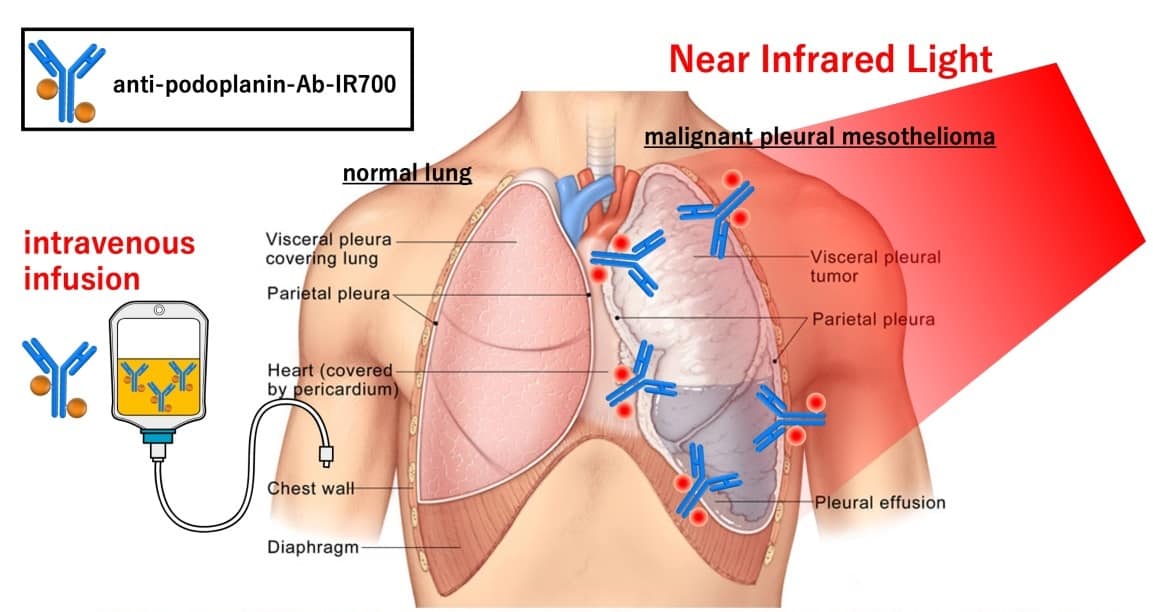
An antibody–drug complex can be targeted and activated in a cancerous lung, leaving the normal lung healthy. (Courtesy: Kazuhide Sato)
A rare type of cancer affecting the lining of the lung, malignant pleural mesothelioma (MPM) has always been very difficult to treat. That may be about to change, though. Kazuhide Sato and his colleagues at Nagoya University have found a potential new treatment that combines light and a targeted antibody, according to their latest research published in Cells.
MPM, most often caused by exposure to asbestos, is often diagnosed late and has a very poor prognosis, with almost no options for treatment. To overcome this, Sato adapted a strategy recently developed for treating other cancers: near-infrared photoimmunotherapy (NIR-PIT).
Illuminating the target
NIR-PIT combines two key methods together into one treatment. First, an antibody targets the tumour with precision. In this case, the antibody targets a protein called podoplanin, which is found in the membranes around the outside of cells. Whilst podoplanin is found on many cell types, certain cancers have a particularly large number of podoplanin proteins on their surface. MPM is one such cancer.
Next, the researchers attached to the antibody a drug molecule that is activated by exposure to NIR light. This light can be directed specifically at the site of the tumour.
Together, this antibody conjugate delivers and activates only the drug molecules in the targeted location – killing cancer cells with reduced damage to the rest of the body. This means fewer side-effects for the patient. The same method has already been given fast-track approval in the USA for treating a head-and-neck tumour. Thanks to this latest study, it may now also be adapted for MPM.
Seeing the light
The lung is a particularly good target for NIR-PIT, according to Sato. “The lungs and chest cavity contain a large amount of air and are thus very good at effectively transmitting near-infrared light,” he explains. That light is absorbed by the drug molecule – IR700 – attached to the antibody, which sticks to the outer membrane of the cancer cells. These cells then break apart and die.
Compact NIR light sources allow light to be easily targeted to the desired treatment site. (Courtesy: Kazuhide Sato)
In this study, the team showed that the antibody conjugate will bind to its target protein on the surface of cells. When exposed to NIR light, the team saw those cells swell, burst and die. This approach killed isolated cancer cells and, in mice with MPM tumours, caused a reduction in tumour volume compared with a control group of mice. Importantly, without the NIR light, the antibody conjugate caused no damage to the cells, showing how the treatment can be accurately targeted.
Whilst Sato and his team say that further work is required to ensure other, healthy cells with the same protein on their surface are not adversely affected, they envision that the method will be a promising anti-cancer strategy. It still needs extensive testing to prove its safety and performance in humans before use in the clinic. If successful, though, it may represent an important step forward towards treating an aggressive and often incurable cancer.
As the complexity of treatment options continues to increase, it is critical that the plan second check process improves to ensure patient safety.
MIM SureCalc MonteCarlo® provides the means for physicists to administer fully automated, independent 3D Monte Carlo calculation instead of manual point dose checks, improving the scope and accuracy of dose calculation without sacrificing time.
Developed in partnership with Radialogica, MIM SureCalc MonteCarlo provides dose calculation solutions for plan second check and adaptive assessments by utilizing an advanced Monte Carlo algorithm powered by SciMoCa™.
MIM SureCalc MonteCarlo improves multiple areas of the QA workflow to ultimately help physicists ensure that they are delivering the best plan possible for patients. Monte Carlo accuracy for dose calculation and automation is leveraged to provide a single platform that serves multiple purposes and centralizes data.
This webinar will walk through MIM SureCalc MonteCarlo and how it can help ensure the highest quality plan for each patient.
The webinar presented by Neha Dwivedi will talk about:
Current realities in pre-treatment and plan second check QA.
Catching plan inaccuracies with plan second check.
Treatment progress tracking with automated adaptive assessments.
Neha Dwivedi is currently a senior clinical support engineer in the radiation oncology department at MIM Software. She has been with the company for five years. Neha has a BS in biomedical engineering from Case Western Reserve University.
As well as floating balloons and making your voice sound funny, helium plays a crucial role in science and medicine – thanks to its cryogenic properties. Although the second most abundant element in the universe, helium is rare on Earth because it is much lighter than air.
Helium is produced deep underground by the radioactive decay of uranium and thorium. As it rises to the surface it can become trapped in the same geological features that contain natural gas – from which helium is extracted.
For various reasons, the helium industry is currently in a state of flux and medical and scientific users of the gas are concerned about future supply problems – which some worry could be exacerbated by “frivolous” uses of helium such as in balloons.
As a result, some in the helium user community are celebrating today as “Helium Conservation Day”. Introduced last year by the company Quantum Design, it falls on the anniversary of the first liquefaction of liquid helium, which was done in 1908 by the Dutch physicist Heike Kamerlingh-Onnes. The above video from Quantum Design explains the importance of helium conservation.
Long shot
You’ll be delighted to know that researchers have revisited a classic problem in the animal kingdom: why can penguins poo such large distances? The discovery was made in 2003, when researchers from Germany, Finland and Hungary – who bagged an IgNobel prize in 2005 for their efforts – found that some penguins can fire their excreta as far as 40 cm, allowing these aquatic birds to continue to nurture their eggs without sitting in a sea of faeces.
Now researchers in Japan have modified this pooping model to calculate the maximum distance that a penguin could manage to fling their dung when at a certain height. Thanks to the penguins’ “strong rectal pressure”, which they calculate to be higher than previous work, the team finds that the maximum distance is 1.34 m, beyond which lies the “safety zone”. The team says this information could be “useful” for zookeepers, who want to avoid getting hit by the firing faeces. So next time you visit the penguin enclosure – do remember to keep your faecal distancing.
Spaghetti has long fascinated physicists. In 2005 French physicists used computer modelling and high-speed photography to explain why dried spaghetti usually breaks in two places when bent. The next big pasta breakthrough came in 2018, when researchers in the US came up with a way of ensuring that a bent piece of dried spaghetti only broke in one place – by twisting it.
Cantilevered pasta
Now, Fathan Akbar and Mikrajuddin Abdullah of the Bandung Institute of Technology in Indonesia have shown that bending dried spaghetti in the presence of steam reveals a smorgasbord of information about the pasta’s material properties. The duo cantilevered horizontal spaghetti strands over a pot of boiling water and observed their sagging using a video camera. They also did a similar experiment using vertical columns of the pasta.
They found that the Young’s modulus (a measure of stiffness) of the spaghetti decreased exponentially in time with steam exposure. This was linked to the rate at which water diffused into the spaghetti and the duo suggest that their technique could be used as a general way of measuring the rate of diffusion of vapour molecules into materials.
In any normal year, the American Association for Physicists in Medicine (AAPM) would be preparing to welcome around 4000 attendees to its annual meeting and exhibit, which in 2020 was due to take place in Vancouver, Canada, as a joint meeting with the Canadian Organization for Medical Physicists (COMP). But this year will be different: the bustle of the traditional conference venue will be replaced by a virtual meeting platform that will allow delegates to attend scientific presentations, take part in live Q&A sessions, and interact with leading equipment vendors in an online exhibit hall.
The scientific programme for the Joint AAPM | COMP Virtual Meeting will feature six tracks of specially curated presentations, along with a series of interactive online ePosters. Delegates can opt to attend the live presentations, which will each be followed by an interactive Q&A, or catch up a via an on-demand service that will be available for six weeks after the event.
The virtual meeting platform will also host networking and social events, such as an online fitness challenge that will enable delegates to earn points for completing a daily 30-minute activity. Meanwhile, the online exhibit hall will be open for delegates to browse company and product information, view video presentations, and schedule meetings with almost 60 exhibitors. A vendor-focused seventh track of presentations offers delegates a deeper dive into specific products and services, with experts available to answer questions immediately after their talks. As a taster, some of the latest innovations from the vendor community are highlighted below.
Robotic radiosurgery system delivers precision, speed and motion synchronization
The CyberKnife S7 System from Accuray is the first robotic and fully automatic system for stereotactic radiosurgery (SRS) and stereotactic body radiation therapy (SBRT), enabling clinicians to deliver personalized treatments to more patients. The system delivers non-surgical stereotactic treatments in any part of the body with sub-millimetre precision, with powerful motion tracking capabilities and adaptive treatment planning to enable high doses of radiation to be targeted directly at the tumour.
The CyberKnife system offers real-time motion synchronization to automatically adapt the treatment in response to any movement of the target or patient. It exploits Accuray’s proven Synchrony technology, which uses artificial intelligence to continuously synchronize the radiation beam to the target location.
Such real-time adaptation allows clinicians to escalate the radiation dose delivered to the tumour while minimizing the exposure of healthy tissue, and also enables more precise and effective hypofractionation treatments. The fully automated system ensures there is no impact on standard workflows or delivery speed, while also avoiding unnecessary manual tasks and reducing the potential for error.
The CyberKnife S7 is the first SRS/SBRT system to have a linear accelerator mounted on a robotic arm, which provides flexibility and freedom of motion for delivering complex beam profiles to any part of the body. An integrated stereoscopic X-ray system provides continual image guidance and real-time tracking information to maintain accuracy and precision during treatment.
The fully automated system is designed for operational efficiency, providing personalized treatment planning while also maintaining patient throughput. Treatment plans can be optimized in as little as 60 seconds, in some cases enabling same-day planning and treatment delivery, while hypofractionated treatments can be delivered in just 15 minutes.
More information about the CyberKnife S7 System can be found on the Accuray website.
The CyberKnife S7 system from Accuray combines artificial intelligence with a linear accelerator mounted on a robotic arm (Courtesy: Accuray)
Seeing is believing: a new way to visualize radiotherapy
The BeamSite video imaging system from start-up company DoseOptics enables clinicians to visualize in real time how the radiation beam is being delivered to the patient. The system captures video-rate images of both the entry and exit beams during treatment, and also provides record and playback capabilities for sharing observations and investigating any anomalies with the clinical team.
The BeamSite system offers direct video imaging for most common radiotherapy modalities, as well as total skin electron therapy. Such real-time visualization is particularly useful for monitoring any stray radiation during treatment, checking that the patient remains in the correct position while the beam is being delivered, and identifying and understanding any errors or near misses.
The system works by detecting the faint Cherenkov radiation that is emitted by human tissue when it is irradiated with high-energy electrons or photons. Unique time-gating technology ensures that each pulse of radiation delivered by the linac contributes to the image recovered, and time-integrating software accumulates the Cherenkov emission to create an image that overlaid in real time on the area being irradiated. The camera and software operate remotely to provide an independent check and measurement tool for beam shape and delivery.
The team from DoseOptics will be presenting several talks and posters during the virtual AAPM meeting to explain the BeamSite system in more detail. They will be:
A recent study by medical physicists at the UT Health San Antonio Cancer Center reveals that patients with simple lung or liver lesions could be treated more efficiently – and just as effectively – with optimized dynamic conformal arc therapy (DCAT). While traditional DCAT generally does not achieve the same plan quality as volumetric modulated arc therapy (VMAT), optimizing the DCAT delivery by varying the dose rate and gantry speed delivers highly conformal dose distributions.
“At our institution, this would mean that 40–60% of lung and liver patients who would normally be treated using VMAT could be treated more efficiently using optimized DCAT, while maintaining the same plan quality,” conclude Sotiri Stathakis and Niko Papanikolaou. “These patients could benefit from significantly shorter treatment times, which are easier to tolerate and reduce the risk of intrafraction movement.”
Stathakis and Papanikolaou exploited the Monaco treatment planning system from Elekta, which offers a variable dose-rate feature for DCAT delivery. It also includes segment shape optimization for DCAT, which optimizes the beam weights and shapes to improve conformality and prevent damage to healthy organs.
Comparing the plans produced for 19 patients using both VMAT and optimized DCAT revealed that both techniques achieved the same plan quality when the targets were located away from other critical organs. But DCAT could be delivered 2.5 times faster than VMAT – which could be particularly beneficial for treatments exploiting the deep-inspiration breath-hold technique.
“Based on the results of this study, lung and liver patients with simple, spherical lesions that are not close to organs-at-risk are ideal candidates for optimized DCAT,” conclude Stathakis and Papanikolaou. “The potential for application of optimized DCAT to other treatment sites, such as pancreas, brain and prostate, is of great interest and the subject for future investigation.”
Optimized DCAT (top) can deliver a similar treatment plans to VMAT for lesions on the liver, as shown in this example, and the lung. (Courtesy: Elekta)
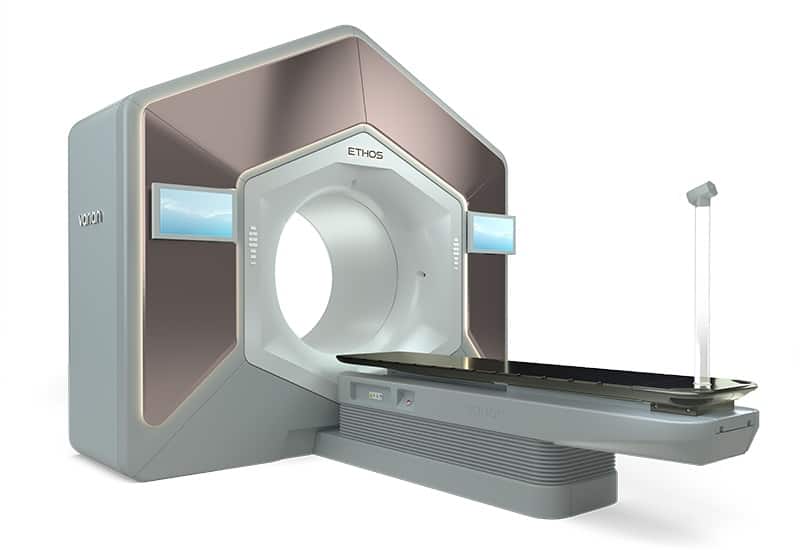
The new Ethos™ therapy system from Varian enables clinicians to adapt radiotherapy treatments to daily changes in the patient’s anatomy. It has been designed to help deliver treatments that better target the tumour, reduce the dose to healthy tissue, and achieve the clinical objectives for each treatment plan.
Ethos incorporates artificial intelligence (AI) to increase the capability, flexibility and efficiency of radiotherapy. It has been designed to allow physicians to assess and adapt treatment plans daily, enabling them to deliver more personalized cancer care and offering the potential for improved patient outcomes.
Ethos therapy integrates Iterative cone-beam CT and multimodality images on the treatment console, providing clinicians with an up-to-date view of the patient’s anatomy to help make better informed treatment decisions. The streamlined workflow of Ethos therapy is enabled by its AI-driven planning and contouring capabilities, making it possible to better visualize daily changes and enabling physicians to make any adjustments to the treatment within minutes.
The Ethos radiotherapy system from Varian offers the flexibility to adapt treatment plans on a daily basis. (Courtesy: Varian)
Optimization technique enables more effective treatment planning
RaySearch Laboratories explains in a new white paper how multi-criteria optimization (MCO) helps clinicians select the optimal treatment plan for their patients through a streamlined and intuitive workflow. The company’s RayStation treatment planning system supports MCO for intensity-modulated radiotherapy, as well as volumetric modulated arc therapy, tomotherapy and proton pencil-beam scanning.
Such MCO techniques start with a series of ideal clinical objectives, such as delivering a uniform dose to the target volume and zero dose to critical organs, from which a series of possible plans are generated. Clinicians can explore these different plans through RayStation’s interactive interface, which exploits slider controls to alter the dose distribution and examine in real time the impact on the defined clinical goals.
As well as such manual navigation, the module also offers an automatic option that optimizes the treatment plan based on a prioritized list of clinical objectives. The dose distribution selected through the chosen navigation process can be converted to machine parameters through a dose mimicking optimization that minimizes discrepancies between the navigated dose and the deliverable plan.
A number of research studies have shown how MCO can speed up the treatment planning process, while also delivering higher quality plans than standard inverse planning. MCO has also been shown to enable novice dosimetrists to create treatment plans of comparable quality to more experienced planners using traditional techniques, while a proof-of-concept suggests that MCO could enable treatment plans to be created in a single meeting between a physician and a planner to save time and improve clinical decision making.
In this example, the MCO module in the RayStation treatment planning system was used to reduce the dose delivered to the right parotid for a head-and-neck patient. (Courtesy: RaySearch)
Autocontouring delivers equivalent results in half the time
DLCExpert is an automatic contouring system from Mirada Medical that exploits artificial intelligence and deep learning to delineate organs-at-risk and other anatomical structures, which is an important but time-consuming element of radiotherapy treatment planning. An expert assessment by oncologists at MAASTRO Clinic in the Netherlands suggests that the time needed for thoracic contouring can be reduced from 20 minutes when done manually using existing clinical routine to just 10 minutes with the deep-learning approach, while validation results available from Mirada show that DLCExpert produces OAR contours of a similar quality to those drawn by professional clinicians.
DLCExpert exploits Mirada’s Zero-Click Contouring platform, which uses background processing to deliver contours before planning gets underway. Contours can be validated using any treatment planning software, or the RTx imaging workstation available from Mirada.
DLCExpert supports all major anatomical sites including breast, lung, head and neck, and prostate. To test out its capabilities, visit www.autocontouring.com to take Mirada’s modified version of the Turing test to try to identify which contours are drawn by radiation oncology professionals and which have been automatically drawn by Mirada software.
More information about the deep-learning techniques used in DLCExpert can be found in a technical white paper from Mirada Medical.
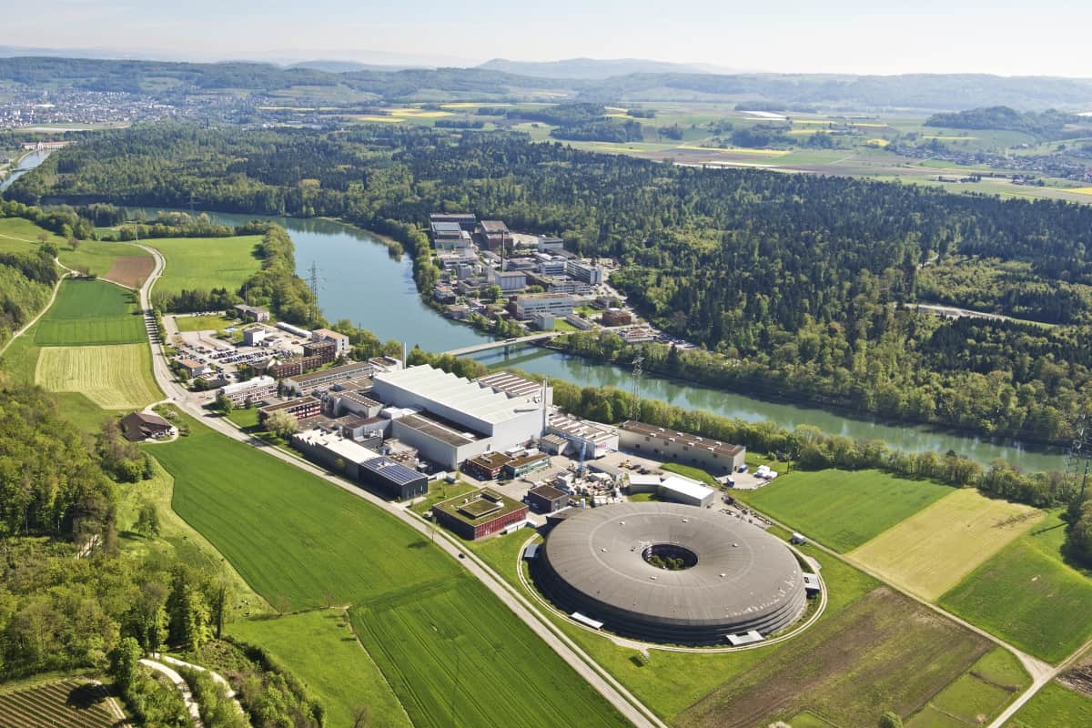
Tony Lomax, Chief Medical Physicist at the Paul Scherrer Institute in Villingen, Switzerland, offers an insight into the different imaging modalities that are needed for proton therapy in a new white paper for Siemens Healthineers. Most important for making initial clinical decisions are the offline imaging techniques used to diagnose the disease, which determines whether the patient will be treated with protons or conventional radiotherapy, and to map out the extent of the tumour and any critical structures and organs nearby.
Such anatomical imaging is generally achieved with computed tomography and magnetic resonance imaging, which offer excellent spatial resolution and good anatomical contrast. MR imaging is also used to image the activity and function of the tumour, often in combination with positron-electron tomography (PET). Lomax points out that it can be difficult to match these two datasets, since PET has a lower spatial resolution and does not provide anatomical information, which means that imaging systems such as PET-CT and PET-MRI are now emerging to improve the precision with which the tumour can be defined.
For proton therapy it is particularly important to understand how the tumour or surrounding organs might move during treatment, which requires time-resolved images to be captured before treatment gets underway. By far the most popular is 4D CT, although major artifacts in the reconstructed data can be caused by any variability in the patient’s breathing. Interest is therefore growing in other techniques, such as 4D MRI, that can reduce these motion artifacts and also capture data over longer periods of time.
Lomax also reviews the options for online imaging during proton therapy. Commercial proton therapy machines are now equipped with cone-beam CT (CBCT) systems for in-room 3D imaging, which is widely used to ensure that the patient is positioned in the same way for each treatment. But some proton centres have opted instead for in-room CT systems that provide the same diagnostic quality as pre-treatment CT, and which also offer useful information for adapting the treatment in response to any anatomical changes, such as the size and shape of the tumour.
While MR-guided systems are now emerging for real-time imaging in radiotherapy, Lomax points out that integrating MR with a proton machine presents several practical challenges that have yet to be overcome. Online 4D imaging is also not yet available for proton therapy, which means that real-time motion tracking must instead rely on time-resolved 1D or 2D imaging. Lomax highlights two recent studies in which such 2D surrogates based on X-ray fluoroscopy or ultrasound monitoring have been used to reconstruct 3D movements using motion models derived from 4D MRI imaging.
A new flexible material for passive cooling that was inspired by a volcano dwelling beetle has been developed by scientists in China, the US and Sweden. The film reflects around 95% of solar irradiance, and can reduce the surface temperature of objects by around 5 °C. It could be used to cool everything from buildings to electronics, the researchers say.
There are some 30,000 species of longhorn beetle, often characterized by having antennae that are much longer than the insect’s body. In southeast Asia one bright golden species, Neocerambyx gigas, is often found living on the slopes of active volcanoes, particularly on the Indonesian islands of Java and Sumatra. In these extreme environments summer temperatures often top 40 °C, while ground temperatures can exceed 70 °C. When it gets really hot, the beetles stop moving and foraging to help shed excess heat and reduce heat absorption.
Han Zhou, a materials scientist at the Shanghai Jiao Tong University in China, told Physics World that this ability to survive in extreme climates and the beetles “very brilliant appearance” attracted her team’s interest. Zhou and colleagues in Shanghai, the University of Texas at Austin and Sweden’s KTH Royal Institute of Technology wondered if the beetle had microstructures that increased its light reflectance to help it regulate its body temperature.
Keeping cool
“We measured their temperature under the light irradiation, and we found that this beetle can lower their surface temperature by 1.5 °C in air and by about 3 °C in vacuum, Zhou explains. “We also measured their optical properties. We found that these beetles have very high optical reflectivity in the visible and the near-infrared light region.”
Using scanning electron microscopy, they discovered that the hardened forewing – called the elytra – of these longhorn beetles is covered in tiny fluffs. These hair like-structures are triangular in cross section and reach densities of around 25,000 per square centimetre.
The researchers found that these forewings reflect 65% of solar irradiation in the visible to near-infrared range, but only 30% if the triangular fluffs are removed, they report in the Proceedings of the National Academy of Sciences.
Pleated fluff
In cross section, the fluffs have two smooth sides and one corrugated side with frills, the scanning electron microscopy revealed. The pleats created by the corrugation and frills are around 1 micron in width and 0.18 micron in height. The team modelled how light interacts with the fluffs and this has shed light on the origins of the forewings’ optical properties.
The study reveals that the pleats create a strong scattering effect that increases reflectivity, no matter the incident angle of the light. The corrugated edge also adjusts the angle of transmitted light, thereby increasing the chance that it experiences total internal reflection inside the fluffs. And light that passes through the smooth sides at most angles also experiences total internal reflection when it hits the inside of the corrugated edge.
“We used this beetle as a model to synthesize some bio-inspired materials,” Zhou explains. They developed a polymer film with triangular surface undulations to mimic the reflective properties of the fluff-covered forewings. To recreate the performance of the fluffs’ pleats they embedded spherical aluminium oxide ceramic particles into the polymer. These are ideal, the team says, because they exhibit a strong light scattering affect and have negligible absorption of visible to near-infrared light, so generate very little heat under direct sunlight.
Pyramid power
Experiments showed that pyramid structures on the surface of the film produced the highest reflectance, compared with cones and prisms. The final film was 500 micron thick, embedded with randomly distributed 2 micron aluminium oxide particles, and covered in an array of 8 micron wide and 5.7 micron high pyramids.
The film has a reflectance of around 95% and real-world tests highlighted its cooling ability. A mobile phone case made from the material reduce the temperature of the device by as much as 4.5 °C, compared to a case without the film. And on a sunny day, sheets of the material on the bonnet of a car reduced the surface temperature by an average of almost 4 °C and a maximum of more than 7 °C, compared with patches of white paper of a similar size and thickness. The researchers say that this demonstrates that the flexible film is a promising passive cooling material for electronic devices and vehicles.
Zhou believes that the material could have many uses, from cooling fabrics to helping cool buildings. Next, she says, they would like to work out how to manufacture the film on much larger scales, and they are looking to see it can doped with other materials to give it new properties, such as high conductivity.
Tooth cracks that are not detected in X-ray CT scans (left) can be seen using indocyanine green near‐infrared fluorescence (ICG-NIRF) imaging. (Courtesy: Jian Xu and Zhongqiang Li)
Cracked teeth can be identified in their early stages using near infrared fluorescence (NIRF) imaging, researchers in the US have demonstrated. The approach – which can distinguish between different types of crack and reveal their depth – is more reliable than existing modalities and may help better diagnose the source of otherwise inexplicable toothache.
Cracked teeth are a common condition and, thanks to their potential to allow bacteria across the enamel–dentin junction, also the third highest cause of tooth loss. Despite this, the condition is often overlooked in its early stages. “Cracked teeth can be difficult to diagnose clinically as patients’ symptoms often aren’t reproducible and cracks can be barely visible to the naked eye,” explains oral surgeon James Allison of Newcastle University, who was not involved in the present study.
At present, there is no dependable clinical method for detecting the presence of cracks in tooth enamel. Visual and surgical-microscope-aided inspection is an unreliable approach, and dye-staining is time consuming and cannot reveal cracks beneath the surface of teeth. Common imaging modalities like X-ray and cone-beam CT, meanwhile, do not offer a high enough resolution. MicroCT scanning, with its higher resolution, can reveal larger cracks – but is only viable on extracted teeth, rendering it useless in a clinical setting.
Conventional near-infrared imaging – in which light is passed through dental structures and scattered to produce image contrast – has, like X-ray imaging, been shown capable of detecting some cracks in teeth. But it cannot distinguish between crack types or provide further information on the extent of the damage.
In their study, engineer Jian Xu of the Louisiana State University and colleagues have instead turned to NIRF – in which image contrast is generated by the differential accumulation within teeth of a fluorescent dye (here indocyanine green) which is excited by infrared light. To demonstrate the potential of the technique, the researchers compared the images of 16 extracted cracked teeth produced by NIRF with both those from near-infrared transillumination and X-ray imaging.
They found that the fluorescence approach was consistently able to reveal cracks in enamel that were not visible in the X-ray images – and was able to highlight more cracks than conventional near infrared imaging. They also report that an angled exposure gave better image contrast, as it created shadows under each crack. From these, one can determine crack depth and whether the crack is in enamel alone or has also reached the dentin.
Furthermore, the team noted that cracks could be revealed by immersing teeth in the fluorescence agent for only one minute – although longer periods produced clearer images. In practice, the dye could be applied to patient teeth via a mouthwash. In fact, indocyanine green has the benefit of being entirely safe to swallow – although has been known to cause allergic reactions.
“We use indocyanine-green-assisted near-infrared imaging to address the major drawback of the current state-of-the-art dental imaging: failure to detect some critical dental diseases, for example, early stage cracks and caries,” Xu tells Physics World. Alongside this, he adds, the new technique does not rely on the use of bulky imaging sensors and avoids the ionizing radiation-based health risks associated with X-ray techniques.
“The idea of checking all teeth for cracks would be unlikely to be cost effective as a health intervention,” notes dental radiologist Keith Horner of University Dental Hospital Manchester. Enamel cracks are not normally treated, he explains, but the approach could be useful for diagnosing patients with toothache where decay or restoration is not an obvious cause of the pain.
Manipulating atoms into quantum entangled states is hard enough, but then proving that you have achieved that entanglement is harder still. But that is the goal of an experiment at the Institute for Quantum Optics and Quantum Information (IQOQI) in Vienna, as explained in this interview with physicist Michael Keller – recorded before the COVID-19 pandemic.
Keller’s team is seeking to create momentum-entangled particles by cooling helium atoms into a Bose-Einstein condensate and “kicking” them with lasers. As observing entangled states directly tends to destroy the entanglement, the researchers have devised indirect methods for study the helium atoms’ properties. The group’s long-term goal is to carry out tests on these momentum-entangled states to probe the foundations of quantum physics.
Launched three decades ago, the Hubble Space Telescope is a triumph of the modern scientific era. In this episode of the Physics World Weekly podcast we hear from David Leckrone, who began working on Hubble before it launched and was the mission’s senior project scientist in 1992–2009.
Leckrone, whose new book Life with Hubble is an insider’s view of the Hubble mission, talks about the shock of discovering that the telescope’s optics were blighted by spherical aberration and how scientists and engineers banded together to work-out a solution. He describes how the Space Shuttle programme played a crucial role in maintaining Hubble and he also pays tribute to the people who made the mission such a success.
The COVID-19 pandemic has changed the very nature of the scientific conference and physicists have responded by using various online platforms to keep the lines of communication open. IOP Publishing’s Tim Smith talks about a Twitter poster conference that he is helping to organize for 15–16 July and how you can get involved – and even win a prize for best poster.
In a separate audio clip on the Physics World blog, David Leckrone talks about some of the outstanding science that has been done by the Hubble Space telescope over the past 30 years. His highlights include the 1994 impact of fragments of the comet Shoemaker-Levy 9 with Jupiter, and the landmark 2001 measurement of the Hubble constant by Wendy Freedman and colleagues.
Recently, I had a fantastic conversation with David Leckrone, an astrophysicist who spent 33 years working on NASA’s Hubble Space Telescope. Much of that interview is featured in today’s episode of the Physics World Weekly podcast, where Leckrone explains what it was like when he and his colleagues realized that the telescope suffered from spherical aberration –and how they saved the mission from disaster.
Unfortunately, the interview is too long for all of it to be included in the podcast, so I have decided to present Leckrone’s scientific highlights in a standalone clip. Enjoy this insider’s view on the comet Shoemaker-Levy 9 impact of Jupiter and other Hubble triumphs of discovery.



 As the complexity of treatment options continues to increase, it is critical that the plan second check process improves to ensure patient safety.
 Neha Dwivedi is currently a senior clinical support engineer in the radiation oncology department at MIM Software. She has been with the company for five years. Neha has a BS in biomedical engineering from Case Western Reserve University.