Ministry of Recovery and Discovery

Am I the only one who thinks that perhaps some of our technology has come too far, too fast? And are we really better off as a result of this? Whenever I bring up the subject among friends and colleagues it invariably results in my being told to “get back to the dark ages” (which I’m old enough to remember). If I have interpreted the current general opinion correctly, it’s that all progressive technology is good, and that rather than eventually making us redundant and supine, the world without it would be a very dark place indeed.

So, when I become World Leader, my first task will be to establish a Ministry of Recovery and Discovery (MoRD). This novel ministry will continue to produce inventions of benefit to mankind, but with the restriction that they do no harm to the planet – for example, by leaving a trail of pollution. Simultaneously, it will tackle the removal of those insidious evolutions that threaten our well-being and that of our planet.

One of the first challenges for the MoRD will be to remove all traces of the internal combustion engine. As a major source of pollution worldwide, its demise would greatly help the environment and would herald a new era of opportunity and challenge. Companies manufacturing things such as bicycles and horseshoes would receive a boost, and what a joy it would be to hear the soothing clatter of hooves making their way along the smooth motorway surfaces, mingled with the soft purr of the electric vehicles developed to fill the needs of emergency services and public service providers. Of course, a new breed of highly efficient, high-capacity batteries would be developed to replace the lithium cell, and the positively archaic lead-acid museum piece.

An important follow-up task for the MoRD would be the removal of any residual legacy left by petrol and diesel cars – perhaps by removing carbon dioxide from the atmosphere using the relatively cheap and efficient direct air capture method, which is currently being pioneered at a few locations, including Cambridge in the UK. At the same time, the ministry’s botany team would be striving to perfect a new breed of trees and plants with enhanced powers of photosynthesis, giving at least twice the capacity for oxygen production.

Further strong candidates for removal include plastics, mobile telephones and nuclear power. Both nuclear power and plastics share the same virtually insoluble problem of waste disposal. The indestructible nature and consequent build-up of long-half-life nuclear waste, the potential for accidental radiation hazard and the proliferation of nuclear weaponry no longer makes nuclear power an attractive option. The current estimate of high-level waste held worldwide is in excess of a quarter of a million tonnes and rising, which is a lot of dangerous material to have around, whether deeply buried, launched into space, submerged beneath the oceans, or elsewhere.

Plastic, with its indestructible nature and ability to insinuate itself into the very fabric of our planet at an alarming rate, has more than outlived its initial usefulness. The diverse scale of its use – from the majority of packaging, to low-friction bearings, to polypropylene used to ensure the physical integrity of the humble tea bag even – leaves a formidable plastics legacy of pollution and destruction. Research at the MoRD to earnestly develop a truly recyclable, biodegradable replacement will be a priority. In the interim period, tea drinkers will need to resort to the faithful tea pot – and with growing sales the emergence of a new, attractive, heat-retaining, truly non-drip tea pot would be welcomed by tea drinkers everywhere.

Of the various clean forms of energy, wind, solar and wave seem to have the greatest potential, especially if their efficiency continues to improve. Vertical axis wind turbines (VAWTs) are particularly versatile, without the need to “track the wind” by yawing the rotor or pitching the blades. Easier maintenance than the horizontal equivalent is assured as generator and gearbox can be positioned at ground level. This also makes them an attractive proposition for the domestic situation, inviting initiatives such as collaboration with rotary clothesline manufacturers. VAWTs are generally longer-lasting, cheaper, can be situated closer together and have higher power outputs than horizontal-axis versions.

Research into the biological effects, if any, of microwave radiation associated with mobile telephones has continued to be minimal and inconclusive since the 2000 Stewart report by the Independent Expert Group on Mobile Phones (IEGMP). Biologist William Stewart concluded, quite correctly, that there was no evidence that such microwave radiation had any harmful effect on humans – much to the relief of mobile phone manufacturers – although, interestingly, he advised that the use of mobiles by children should be discouraged. Stewart’s conclusions, of course, offer no guarantee of safety and one recalls how asbestos, X-rays and cigarettes were retrospectively condemned after considerable damage had been done.

With the current world population racing towards eight billion, it is clear where we are heading, and the destination isn’t that attractive. Facing dwindling resources such as air, water, food and space, the MoRD will have to take the initiative to meet this challenging situation if we are to survive, although it will be difficult to come up with a humane and ethical solution.
Droplets created by speech could contribute to COVID-19 spread, new study suggests

Droplet clouds emitted during 1 min of loud speech by an individual infected with the SARS-CoV-2 virus could contain more than 1000 virus particles – according to new calculations done by scientists in the US. This work, alongside observations that these speech-generated droplet clouds persist in a confined space for 8-14 min, supports suspicions that COVID-19 may be transmitted when infected individuals speak.“This direct visualization demonstrates how normal speech generates airborne droplets that can remain suspended for tens of minutes or longer and are eminently capable of transmitting disease in confined spaces,” write the team in a paper in PNAS Brief Report describing the work.

The piggybacking of viral particles (virions) in droplets generated by coughing and sneezing is a recognized route of respiratory virus transmission. However, the role of fluid droplets emitted during speaking is less-well known. Given the current COVID-19 pandemic, this spoken transmission route is attracting considerable attention, particularly in relation to spread from asymptomatic carriers.

Philip Anfinrud and Adriaan Bax’s biochemical physics teams at the National Institute of Diabetes and Digestive and Kidney Diseases (NIDDK) (part of the National Institutes of Health) in Bethesda, Maryland recently made high resolution visualizations of droplets produced when a person speaks. They described this work in the New England Journal of Medicine.

Visualizing saliva spray

To observe droplets after they are produced by speech, the NIDDK teams used a laser and optics visualization system. The droplets were created by having a person repeat the phrase “stay healthy” several times (with short delays in between) into the “speaker port” of a cardboard box and then capturing the crossing of the droplets through a sheet of intense laser light. The high sensitivity of the system enabled the teams to detect small droplets, 20-100 micron in diameter, and so record a higher volume of droplets than previous studies.

Now the researchers have quantified those experiments to assess the potential for speech droplets to transmit SARS-CoV-2 viral particles in an enclosed environment. In work described in a PNAS Brief Report, Anfinrud and colleagues used an improved experimental setup to derive quantitative estimates for how long the smallest droplets remain airborne and how many there are. where the researchers write.

Careful estimations

The researchers described the droplets using two exponential decay functions, with the top 25% in scattering brightness and the dimmer 75% fraction having exponential decay constants of 8 min and 14 min, respectively.

They used the weighted average decay rate of all droplets to calculate the terminal velocity of the droplets and, given smaller droplets fall at a slower rate (staying airborne for longer), from the velocity they were able to estimate the average fallen droplet size.

The probability a droplet contains a viral particle depends on the initial droplet volume, but evaporation causes a rapid decrease in volume after release from the mouth. The team made shrinkage assumptions based on the relative humidity and temperature of the experimental settings, estimating the initial droplet size to be 12-21 micron in diameter.

The researchers were unavailable for comment, but the NIDDK responded to questions on their behalf.  The NIDDK  told Physics World that the researchers were “surprised to find the enormous number of particles in the 12-20 micron range [as this was far higher than suggested by previous research] that, after drying out and shrinking to about 4 micron, remain airborne for many minutes”.

Finally, to determine the probability a droplet encapsulates at least one virus, the team estimated the total volume of emitted saliva in their experiments, and combined this with data from recent research that shows saliva from a COVID-19 patient contains on average about seven million virus particles per millilitre.

They found that a second of speaking could be expected to generate 17-90 virion-containing droplets.

Substantial probability of transmission

In their latest paper the authors conclude, “These observations confirm that there is a substantial probability that normal speaking causes airborne virus transmission in confined environments.”

Julian Tang, a virologist at the University of Leicester, UK, who has expertise in respiratory viruses and their transmission, comments that this work is a “nice visualization method”. But points out that it would be “even more powerful” if the researchers combined their expertise with other disciplines to quantify live virus transported by speech droplets from COVID-19 infected volunteers.

The NIDDK says that Anfinrud and Bax’s team are building a third-generation laser-scattering apparatus to further address questions regarding speech droplet transmission.

The mystery of missing marine plastic

In the May 2020 issue of Physics World, science journalist Marric Stevens wrote about the problem of the missing plastic in the world’s oceans. Although we are starting to see large amounts of plastic in the oceans, the quantity is far smaller than we expect to see – based on the quantities of plastic being released into the oceans every year. In the latest episode of the Physics World Stories podcast, Andrew Glester digs deeper into the mystery to find out where the plastic might be ending up.

To learn about the threat of plastic to marine wildlife, Glester meets Lucy Quinn, a seabird ecologist with the British Antarctic Survey. Quinn was the researcher who captured public awareness of the plastics issue when she appeared in the BBC’s Blue Planet 2 showing the horrific impacts of ingested plastic on an Albatross colony in Bird Island, South Georgia.

Also in the podcast, physical oceanographer Erik van Sebille outlines the extent of the missing plastics issue. He explains how his research on ocean flow at Utrecht University in the Netherlands can help to better understand where the plastics are ending up. While Alethea Mountford from Newcastle University, UK, describes how oceanographers combine physical measurements with modelling to get a handle on the issue.

To find out more, read ‘The search for the missing plastic‘, a feature originally published in the May 2020 issue of Physics World – a special edition on plastic waste.

This podcast is sponsored by Teledyne Hastings Instruments.

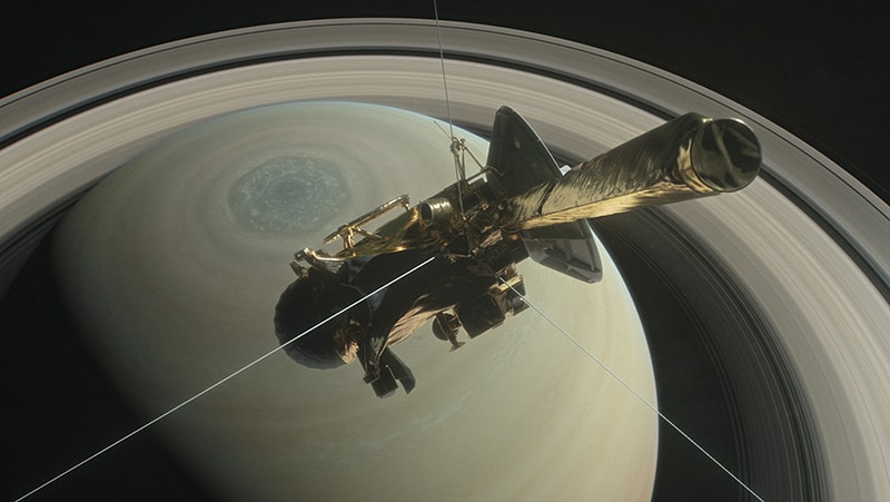
The Cassini spacecraft mission at Saturn

Want to learn more on this subject?

In September 2017, the NASA/ESA Cassini-Huygens spacecraft mission ended its 20 years in space by burning up in Saturn’s atmosphere. The end-of-mission orbits was designed to better understand the interior of Saturn and its magnetic field.

ICL webinar

This webinar will describe these end-of-mission results as well as some of the other surprising discoveries made during the orbital tour at Saturn, including water-vapour plumes at the small moon Enceladus and implications that this has for potential habitability.


Want to learn more on this subject?

Michele DoughertyMichele Dougherty is professor of space physics at Imperial College London. She is leading unmanned exploratory missions to Saturn and Jupiter, and was the principal investigator for the magnetometer instrument onboard the Cassini mission to Saturn as well as being the principal investigator of the magnetometer for the JUpiter ICy moons Explorer (JUICE) of the European Space Agency due for launch in June 2022. She is head of the physics department, is a fellow of The Royal Society, was awarded the Royal Astronomical Society Geophysics Gold Medal in 2017, was awarded a CBE in the 2018 New Year Honours list, and was awarded the Institute of Physics Richard Glazebrook Gold Medal and Prize.


Tipsy sludge worms simulate active polymers

Clumps of wriggling – and sometimes drunken – sludge worms have been used by physicists in the Netherlands to simulate the behaviour of self-moving polymers. Antoine Deblais and colleagues at the University of Amsterdam used the creatures to gain new insights into the properties of poorly understood “active polymer” materials by measuring the viscosity of worm clusters as they were subjected to shear forces.

Polymers such as silk and polyester are some of the most familiar and most studied materials.  However, scientists know much less about active polymers, which use stored energy or energy in the surrounding environment to move and change shape. When active polymers interact with each other and the fluid surrounding them, new structures and dynamics can emerge – something that could be useful for developing new technologies. However, studying this in the lab or using computer simulations is extremely challenging.

Now, Deblais’ team has used tangled clusters of living sludge worms as an analogue system for studying active polymers. These long, slender animals closely mimic the behaviours of active polymer molecules, and are also widely available in many pet shops, making them ideal for simple, inexpensive experiments. Furthermore, their activity can be varied by placing them in water at different temperatures, and they can even be temporarily incapacitated when exposed to alcohol – allowing them to resemble inactive polymers more closely.

Disentangling freely

In their experiment, Deblais and colleagues studied the physical properties of the worms by putting them in a shallow cylindrical container of water. They then pressed a rotating plate onto the surface of the mixture, subjecting it to a shear force. In a typical polymer, tangling between molecules would resist these forces, increasing the viscosity of the mixture. In contrast, the movements of the worms caused any coils to disentangle more freely. This meant the warmer mixtures, which contained the most active worms, were up to 100 times less viscous than those containing inactive, alcohol-infused specimens.

The team also looked at an effect called“shear thinning”, which occurs when higher shear forces force the polymer strands to align themselves, thereby lowering the viscosity. In contrast, Deblais’ team found that mixtures containing more active worms displayed less shear thinning than their inactive counterparts when the plate was spun at several rotations per second. They believe that this occurs as the bending, stretching, and contracting motions of the wriggling worms works against the alignment process.

Deblais and colleagues now hope that their research will pave the way for a new experimental research field they call “polymer-like worms”, which could lead to much better models of similar systems on microscopic scales. Their research could also lead to sophisticated new techniques for modelling a variety of microscopic biological systems.

The research is described in Physical Review Letters.

Nano-optomechanical resonator detects low-frequency bacteria vibrations

Researchers in Spain and France have measured the vibrations of individual bacteria by coupling them to a nanomechanical device with a similar resonance frequency. This new optomechanical spectrometry technique could offer an alternative to current methods of detecting and classifying bacteria and other biological particles.

Proteins, viruses and bacteria all vibrate at frequencies in the terahertz and gigahertz range. Their vibrations carry valuable information about their structure and mechanical properties, but efforts to study these using optical inelastic scattering techniques are extremely challenging because the bioparticles change shape and deform as they vibrate.

The new method, developed by a team led by Javier Tamayo and Eduardo Gil-Santos from the Instituto de Micro y Nanotecnología (IMN-CSIC) in Madrid, involves coupling the mechanical vibrations of bacteria to an ultrahigh frequency (UHF) nano-optomechanical resonator made from a GaAs microdisk. Such coupling is only possible when the resonance frequencies of the disk and the bacteria are similar, Tamayo explains. In contrast, previous experiments relied on the biological particles vibrating much faster than the micro- and nanomechanical resonators or microcantilevers used to measure the particles’ mass and stiffness.

Going beyond mass and stiffness measurements

The Madrid team’s disks support two different types of vibration: radial breathing modes (RBMs), which correspond to a radial expansion and contraction of the disk (and therefore depend on its diameter), and so-called optical whispering gallery modes, which correspond to resonances within the disk’s structure. In both cases, the disk vibrates at frequencies in the GHz range, which can actually surpass the low-frequency vibration modes of bioparticles.

In their experiments, Tamayo and colleagues deposited a single S. epidermis bacterium onto their optomechanical microdisks using an electrospray ionization technique. The bacteria are round in shape and have a radius of roughly 400 nm. The researchers measured the fundamental RBM frequencies of the disks before and after depositing the bacteria, then used a general theoretical framework to describe the coupling between the bacteria and the disks. This framework allowed them to calculate the resonant frequencies of a bacterium’s low-frequency vibration modes based on their “before and after” measurements of the disks’ RBM frequencies.

To determine the mechanical coupling between a bacterium and their nanomechanical resonator, the researchers had to measure very tiny fluctuations – a few picometres (10-12 m) – at ultrahigh frequencies. Such measurements were only possible thanks to the strong optomechanical coupling of the disks to the bacterium. Indeed, the coupling is so strong that these devices can measure displacements of just attometres (10-18 m) – a value similar to the precision of the kilometre-sized interferometers used to measure gravitational waves, Tamayo says.

The work was done within the framework of the EU FE VIRUSCAN project, which aims to use optomechanical resonators to detect viral particles based on their physical parameters. The idea is to build up a “library” of the mechanical and vibrational properties of different viruses and bacteria.

Members of the Madrid team, who report their work in Nature Nanotechnology, are now planning to use their technique to measure the vibration modes of viruses, which are much smaller than bacteria. “This future work will be more challenging,” Tamayo tells Physics World.

Dual-layer spectral CT proves feasible for routine practice

© AuntMinnieEurope.com

Reduced-dose dual-layer spectral CT (DLCT) is feasible in routine practice, despite the required higher tube potential, researchers from Heidelberg University Hospital, Germany, reported in a study published in European Radiology.

DLCT can deliver comparable objective and subjective image quality to that of reduced-dose single-layer CT (SLCT), reported Thuy Duong Do, senior physician at the Clinic for Diagnostic and Interventional Radiology, and colleagues. Also, further dose reduction in the thorax might be possible by adjusting mAs thresholds.

Conventional CT acquires images in a single broad energy band, but spectral CT separates energy into two or more narrow energy bands. Because different types of energies are absorbed differently by tissues, they can provide insights into the different chemical compositions of tissues.

“DLCT acquisitions allow material decomposition (virtual non-contrast, iodine-only imaging and effective atomic numbers) as well as the calculation of virtual monoenergetic images,” noted the authors, adding that several clinical studies have shown the benefits of DLCT for head CT to image intracerebral lesions and haemorrhage, for thoracic CT, for vertebral CT to differentiate bone lesions, and for abdominal CT angiographies to improve delineation of visceral arteries.

“For the image acquisition of such data, a tube potential of either 140 kVp or 120 kVp is necessary to allow for spectral decomposition under the exploitation of the energy-specific X-ray absorption of different materials,” they explained. “In contrast to changes in tube current, changes in tube potential have a nonlinear effect on radiation dose.”

Study details

The Heidelberg group’s overall aim was to quantitatively and qualitatively evaluate image quality in DLCT compared with SLCT in the thorax, abdomen and pelvis in a reduced-dose setting.

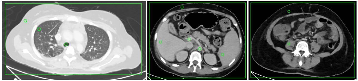
Example images

The researchers performed intra-individual, retrospective comparisons in 25 patients who received at least one acquisition of all three acquisition protocols – SLCTlow (100 kVp, iCT, Philips Healthcare), DLCTlow (120 kVp) and DLCThigh (120 kVp, IQon Spectral CT, Philips Healthcare) – covering the venous-phase thorax, abdomen and pelvis with matched volumetric CT dose index between SLCTlow and DLCTlow.

All examinations were conducted in the craniocaudal direction and supine position, with automatic exposure control, using an iohexol contrast agent (Accupaque 350, GE Healthcare). Contrast agent application was performed using a power injector with an injection rate of 3 ml/s.

Reconstruction parameters were identical for every scan. Image quality was assessed quantitatively at 10 measurement locations in the thorax, abdomen and pelvis by two independent observers, and subjectively with an intraindividual forced-choice test between the three acquisitions. The authors extracted dose–length product (DLP) and volumetric CT dose index (CTDIvol) for dose comparison.

The main findings were as follows:

  • Despite matched CTDIvol in acquisition protocols, CTDIvol and DLP were lower for SLCTlow compared with DLCTlow and DLCThigh (DLP of 408.58, 444.68, 647.08 mGy·cm, respectively; p < 0.0004), as automated tube current modulation for DLCTlow reached the lower limit in the thorax (mean 66.1 mAs vs limit 65 mAs).
  • Noise and contrast-to-noise ratio (CNR) were comparable between SLCTlow and DLCTlow (p values, 0.29–0.51 and 0.05–0.20), but CT numbers were significantly higher for organs and vessels in the upper abdomen for SLCTlow compared with DLCTlow. DLCThigh had significantly better image quality (noise and CNR). Subjective image quality was superior for DLCThigh, but no difference was found between SLCTlow and DLCTlow.
  • DLCTlow showed comparable image quality to SLCTlow, with the additional possibility of spectral postprocessing. Further dose reduction seems possible by decreasing the lower limit of the tube current for the thorax, the researchers noted.

The team was surprised to see that the transition from 100 kVp to 120 kVp tube potential worked out so well in terms of both image quality and patient radiation exposure, according to corresponding author Stephan Skornitzke, a medical physicist at Heidelberg University Hospital.

“I would have expected that we would lose some contrast with the increase in tube potential, but the contrast-to-noise ratio turned out to be very similar before and after the switch to the new scanner and new protocol,” he told AuntMinnieEurope.com in an email. “Another surprise for me was to see how well the automatic exposure control is able to adjust the tube output to the patient anatomy.”

For the 120 kVp protocols, the exposure control hit the lower threshold for the tube current–time product in the thorax for a number of patients, where it wanted to regulate even lower but could not, he continued.

“This shows us that we can still optimize our acquisition protocols with an attention to detail and reduce patient radiation exposure,” Skornitzke explained. “The automatic exposure control was hitting the lower threshold for the tube current–time product in the thorax for a number of patients. We are in the process of evaluating an adjustment of this lower threshold, which may allow to further reduce patient radiation exposure, especially in smaller patients. However, we will have to carefully consider any potential impact on diagnostic image quality.”

Looking to the future

For follow-up studies based on the spectral imaging capabilities of the CT scanner, the researchers are investigating the potential clinical benefit of the large number of available spectral postprocessing applications. For example, they are evaluating calcium-suppressed imaging, which could allow for more accurate evaluation of bone marrow and fractures.

Also, they are planning to further investigate the connection between automated exposure control, image quality and patient radiation exposure.

“In the context of appropriate imaging, the automated exposure control is a very important tool, as it serves to ensure that the radiation is applied exactly where it is needed,” Skornitzke noted. “Today, with 3D-modulation, online-adaption and specific technologies like ‘Liver Boost’, we have a large number of methods available that help us to guarantee adequate image throughout the scan.”

More research is necessary to evaluate the complex interaction between these technologies, radiation exposure, and image quality, he stated.

The COVID-19 pandemic has provided new challenges for the radiological community regarding the fast and reliable assessment of the associated changes in the lung, and the Heidelberg group is involved in the research of these topics.

“Of course, the pandemic has affected all of our lives. However, as researchers in radiology, we are in a comparatively privileged situation, where our research often involves only a small number of people and a lot of work can be performed digitally, so that our research has so far only been affected minimally,” Skornitzke pointed out.

  • This article was originally published on AuntMinnieEurope.com ©2020 by AuntMinnieEurope.com. Any copying, republication or redistribution of AuntMinnieEurope.com content is expressly prohibited without the prior written consent of AuntMinnieEurope.com.

New material could be used to make a liquid metal robot

A liquid metal lattice that can be crushed but returns to its original shape on heating has been developed by Pu Zhang and colleagues at Binghamton University in the US. The material is held together by a silicone shell and could find myriad uses including soft robotics, foldable antennas and aerospace engineering. Indeed, the research could even lead to the creation of a liquid metal robot evoking the T-1000 character in the film Terminator 2.

The team created the liquid metal lattice using a special mixture of bismuth, indium and tin known as Field’s alloy. This alloy has the relatively unusual property of melting at just 62 °C, which means it can be liquefied with just hot water. Field’s alloy already has several applications – including as a liquid-metal coolant for advanced nuclear reactors.

Zhang and colleagues combined the alloy with a silicone shell through a complex hybrid manufacturing process that combines 3D printing, vacuum casting and so-called “conformal coating” – a technique normally used to coat circuit boards in a thin polymer layer to protect them against the environment. The silicone shell is what allows the lattice to “remember” a desired shape and restore such when the alloy is melted.

Shell skeleton

“Without the shell, it won’t work, because the liquid metal will flow away,” Zhang said. “The shell skeleton controls the overall shape and integrity, so the liquid metal itself can be confined in the channels.”

To illustrate the potential of the lattice technology, Zhang and colleagues made several demonstration structures – including honeycombs, the letters BUME (for Binghamton University mechanical engineering), a spider web-like mesh and a lattice in the shape of a human hand. When crushed and reheated, all eerily return to their original shape.

When solid, Field’s alloy is very strong and stable and is far stiffer than most shape-memory polymers, according to Zhang. A crucial benefit of the new material is that an object can easily be crushed down into a much smaller spaces for transport or storage before being restored to its usual shape. The researchers think this would make the material ideal for use in space missions, where it could be used to make antennas or building superstructures that could packed tightly on spacecraft ship and then expanded on arrival on the Moon or another planet.

Space cushions

The material could also be used to make cushions because it can absorb a considerable amount of energy when crushed. Zhang suggests that this could be useful for building reusable spacecraft. “Normally, engineers use aluminium or steel to produce cushion structures,” he says. “After you land on the Moon, the metal absorbs the energy and deforms. It’s over – you can use it only once.”

In contrast, a spacecraft with landing cushions built using a liquid metal lattice could be reused over and over again. “Using this Field’s alloy, you can crash into it like other metals, but then heat it up later to recover its shape,” Zhang said.

“There’s been growing interest in machines and structures that can change their shape, stiffness, and ability to bear load. These architectures have potential use in emerging applications like soft robots that mimic biological organisms, wearable computing systems that can conform to the body’s natural motion, or wearable robotics that can assist in human motor tasks,” says Carmel Majidi, a mechanical engineer from the Carnegie Mellon University. “[This work] nicely builds on past research in stiffness tuning and shape memory materials,” he says, adding it. “is an excellent demonstration of how low melting point metals can be used for creating smart and adaptive structures”.

Enormous changes

Michael Dickey —a chemical engineer at North Carolina State University agrees, adding, “This work nicely takes advantage of the capabilities of 3D printing, the elastic energy of elastomers, and the enormous changes in modulus that occur when low melting point metal alloys melt”.

With their initial study complete, the researchers are now working to improve the durability, strength and energy absorption capacity of their liquid metal lattice materials – alongside scaling up and refining the manufacturing process for such.

Zhang also has another goal in mind. “Our dream is to build a liquid metal robot,” he said. “Now we have a hand, so we’re one step further.

The research is described in the journal Additive Manufacturing.

Proton CT system lines up for small-animal studies

SIRMIO proton CT system

Small-animal irradiation studies are an essential component of cancer research, bridging the gap between cell experiments and clinical realization of emerging radiotherapy techniques. But while photon-based preclinical radiation research platforms have been commercialized, the same does not hold true for protons.

The SIRMIO project, established by Katia Parodi at Ludwig-Maximilians-Universität München (LMU), aims to bring together proton therapy and preclinical research by developing a dedicated platform for small-animal proton irradiation. To achieve high-precision placement of the Bragg peak within the target, the team is equipping the platform with a proton CT (pCT) system.

“So far, image guidance on small-animal photon radiation research platforms, which are being adopted for proton therapy, uses X-ray cone-beam CT,” Parodi explains. “But the relationship between X-ray attenuation and proton relative stopping power (RSP) is uncertain. Moreover, experience from human tissue cannot be easily translated to calibration curves for murine tissue. Hence, we decided to rely on proton imaging to provide low-dose pre-treatment alignment, along with the possibility of tomographic imaging for more accurate stopping power information.”

Parodi and colleagues have now performed a Monte Carlo (MC) study of their proposed pCT system, to assess its feasibility and optimize the design of the detector components.

Detector designs

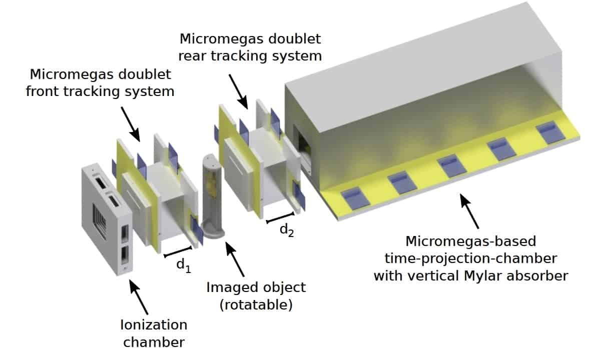
The SIRMIO pCT is a single-particle tracking system, comprising tracking detectors on either side of the imaged object and a residual range detector placed downstream. The tracking detectors, which estimate particle trajectories through the object, each contain a doublet of Micromegas planar gaseous detectors. The residual range detector, which measures the energy lost by each proton (expressed as water-equivalent path length, WEPL), is a Micromegas time-projection-chamber with vertical Mylar absorbers.

“Low-energy proton imaging is challenged by scattering in the detector,” says co-author Jonathan Bortfeldt. “Hence, we chose gas-based Micromegas to realize the lowest possible material budget, while offering high spatial resolution and high count-rate capability. The proposed technology is scalable and could be tailored in future to clinical application.”

The spatial resolution of a reconstructed pCT image is determined by the precision of the estimated proton trajectories. For Micromegas detectors, this depends on two fixed factors: the pitch of the readout strips (500 μm) and the detector-to-object distance (4 cm); plus two adjustable parameters: the material budget and the spacing between planes in each doublet. Using MC simulations of a 75 MeV scanned proton beamline, the researchers aimed to optimize these latter two parameters.

Conventional Micromegas use three layers of copper strips in the readout structure. To reduce the material budget, first author Sebastian Meyer simulated two redesigns: removing the last layer of readout strips from the active area; and replacing the 33 μm copper strips with 9 μm thick aluminium strips. Simulations of the three structures in a water phantom showed that, for the aluminium strip design, the average RMS path estimation deviation was around 0.29 mm, compared with 0.36 and 0.39 for the two- and three-layer copper strip designs, respectively.

The researchers also varied the distance between the two planes in the tracker doublets, from 1 to 10 cm. They found that a spacing of at least 7 cm maximized the accuracy of the estimated proton trajectories, giving an average RMS path deviation of 0.18 mm. To avoid spatial resolution degradation, they chose 10 cm spacing as optimal.

Simulated pCT images of a slanted edge phantom revealed spatial resolutions of 1.9, 2.2 and 2.8 mm1, for the two- and three-layer copper and aluminium strips designs, respectively. The team notes that this performance is comparable to that of X-ray cone-beam CT systems commonly used in preclinical research.

To optimize the residual range detector, the researchers quantified RSP accuracy for Mylar absorber thicknesses of between 250 and 1000 μm, by simulating pCT of a water phantom with tissue-equivalent inserts. An absorber thickness within 500–750 μm gave the best trade-off between WEPL resolution and detector complexity, providing sub-0.5% RSP accuracy.

Small-animal studies

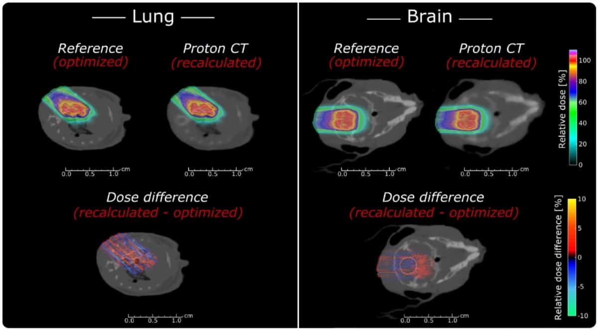
Using their optimized pCT design – aluminium detector layers spaced by 10 cm and a residual range detector with 500 μm thick absorbers – the researchers simulated pCT images of a mouse head. The pCT images clearly resembled the reference anatomy at a low noise level, but with blurring due to the limited spatial resolution.

Treatment plans

To investigate whether such pCT images are suitable for treatment planning, they used a MC-based proton treatment planning system to plan brain and lung tumour treatments in a mouse model. Comparing optimized reference plans with plans recalculated on the pCT images showed that pCT enabled sub-millimetre accuracy. The average relative range errors were -0.02±1.42% and +0.87±0.98%, for the lung and brain cases, respectively. The corresponding absolute water-equivalent range differences were -0.01±0.20 mm and +0.09±0.10 mm.

Parodi notes that both detector components of the pCT system are already under construction and being tested. “A full-size tracking Micromegas prototype with aluminium strips, as well as a reduced version of the full-scale time-projection-chamber range telescope with Mylar foils as absorbers, have been already realized in-house and successfully commissioned in low-energy proton beams from the Munich Tandem accelerator,” she explains. “Currently, larger scale prototypes are being realized and the full system pCT realization is expected for fall 2020.”

The study is published in Physics in Medicine & Biology.

Ask me anything: Carole Mundell

What skills do you use every day in your job?

I think the key quality that I use in both my academic role and as chief scientific adviser is curiosity. That may sound a bit imprecise, but actually being able to question, challenge the status quo, and not being afraid to contest assumptions I think is really important. And my curiosity really comes from my background as a physicist.

What do you like best and least about your job? 

What I like best about my job is the people, and the fact that wherever I go and whatever I do, I see that science makes a difference – whether it’s inspiring young children, or scientific breakthroughs that really help human health, or applications that improve people’s lives and livelihoods and make the world a better place. When I give public lectures on astrophysics, I always get amazing questions from the youngest children. You can see their curiosity sparked and their passion for understanding how things work. I think that always progressing that knowledge is one of the best parts of my job. This kind of curiosity and connection is very human, and so it doesn’t matter what country I’m in – it’s a consistent factor.

People are afraid of getting things wrong. I think that limits our ability to be creative and innovative

Although I consider it a privilege to travel for my job, I do miss my family when I am away from home – this is something that many people experience in their careers. I think it’s natural in today’s world, and it’s not specific to my job, or even just science. But now with modern technology, it’s so much easier than it would have been for previous generations. I still write a handwritten letter to my children whenever I travel, but obviously we’ve got Skype and FaceTime and any of the technical devices that we can connect to, so that does make it easier to be away from home.

What do you know today that you wish you knew when you were starting out in your career?

I wish that I had been more confident and relaxed about taking risks and trying new things. Sometimes people worry about that – asking questions and making mistakes. I see this particularly in academia, where a combination of competition, individualism and insecurity, plus the measurement and reward culture, can create an unhelpful atmosphere of perfectionism and judgement. This makes people risk-averse and afraid of being seen to get things wrong. I think that is unfortunate as it limits our ability to be creative and innovative and actually to make new discoveries. Instead, I say – have a go at it, have an experience and if it doesn’t work out how you expected, learn from it. You’ll be a better scientist and enjoy your work more. In turn, be the positive, encouraging voice in the room and you might be pleasantly surprised by how others respond. I enjoyed my time as PhD student because I had the right combination of support and encouragement from my supervisor and other senior scientists, as well as some space to try things out for myself. That is important as you develop your independence – and don’t be afraid to ask questions.

It’s also good to take new opportunities when they arise – even if beyond your comfort zone –  they will stand you I good stead in the future in ways you cannot plan. Some years ago, I was fortunate to be selected to attend the BBC Women Experts training course. It was fascinating and I met incredible women from a range of backgrounds. It gave me the confidence to accept media engagements that I would otherwise likely have turned down. That has been valuable in helping shape my ability to communicate with diverse audiences.

Copyright © 2026 by IOP Publishing Ltd and individual contributors