My physics degrees covered a wide range of topics, from quantum mechanics to electronics. I decided to specialize in geophysics, specifically remote sensing, which makes use of sensors, wave propagation and signal/image analysis. My PhD was about radar imaging of Venus, and afterwards, I worked on sonar imaging of the Earth. Having worked with a multitude of people over several decades, I have realized that everybody needs a physicist. The technical skills we learn, such as numeracy and programming, are useful to work with all other disciplines. As physicists, we are not afraid of equations or complex instruments, and have the tools to adapt to many different things. Today, I am a Chartered Geologist, as well as a Fellow of the Institute of Acoustics.
Soft skills will vary with the jobs, and they are not always taught in a degree, but I was fortunate to be able to catch up on the job. Communication skills are extremely important: it is crucial to explain and “reach out” to other disciplines. People management is key when working in multidisciplinary teams: respecting each other and understanding we might have different ways of looking at the world but still all want the best outcomes. Time management becomes increasingly important as we progress, and keeping a healthy work–life balance (making us more productive in the end).
What do you like best and least about your job?
Discovery is the best part of the job: every day, you learn new facts, new skills and you can participate in very innovative projects. Right now, I am working on acoustic signatures of climate change in the Arctic, on measuring and mitigating the impacts of human noise on marine life, as well as working with some world-leading industries. It means I work with a variety of researchers and end-users, but I also need to interact with policy-making and help design the international standards of the future. Second best is the possibility to encourage younger scientists to pursue their own ideas, helping them connect across the field, and then step back and see how well they are doing. Teaching is a great opportunity for that: I love the “light bulb” moments, when students realize they can do something hard by themselves.
Every job has its downsides, of course. For me, it is having to justify how we meet particular targets, fill in audit forms with unclear guidance (and sometimes ridiculously short deadlines). But it is more than balanced by the pleasure in seeing our students thrive, keeping in touch after they graduate and seeing how they develop. There is also the thrill of watching the results of our research being used around the world.
What do you know today that you wish you knew when you were starting out in your career?
“Follow your heart” is a common answer, but it needs to be modulated with a scientific approach to the possible outcomes, and you need to remember the future is unpredictable. Originally, I wanted to become an astronomer and look at planets and stars. Job prospects in France were limited at the time I chose my undergraduate options, and I focused on other skills. Be open to possibilities, and be aware that not all dreams are possible, but don’t abandon them either. Remember that there will be new types of jobs in the future that we cannot expect right now. Some authority figures can be very assertive about what needs to be done and how. My personal advice would be never to take these forceful statements as the gospel truth. It is good to listen to different voices, and to make your own, justified choice. Finally, linear careers are no longer the norm, and you might need to change jobs along the way. The skills we learn as physicists are useful for all sorts of things, and Physics World has plenty of very good role models and personal stories to show how useful physics is everywhere.
At this stage of my career, most of what I use is actually my connection with physics as a community and as a culture. Though most of my job is about emphasizing careers outside of academia, I have to interact very closely with academics to help them become more effective career mentors – so a deep understanding of the challenges and concerns they have is very important. Since I also give lots of advice directly to students, understanding what they are going through from a first-hand perspective really helps.
I also use all kinds of communication skills (writing articles, writing proposals, creating and giving presentations), an understanding of statistics (when analysing and communicating about career and employment data), project management skills and leadership skills.
What do you like best and least about your job?
What I like the most about this job is that it gives me an opportunity to create a far broader positive impact on the physics community than I would probably have as an individual academic working in a single institution. At the APS, I get a “birds-eye” view of all kinds of efforts that are going on to make physics as a discipline more relevant to the aspirations, interests and identities of 21st-century physics students. Because of this, I get to help make connections between change-makers in the community so that their efforts are more impactful.
The down side is that I really don’t get to do much “physics” anymore – which is something I definitely miss. I loved both teaching physics and doing physics research, and they aren’t really part of my daily activity anymore.
What do you know today that you wish you knew when you were starting out in your career?
I wish I had thought more broadly about the kinds of activities that really make me excited and happy, apart from the physics research I had been doing. There are lots of ways in which your career path can scratch a particular “itch” (for example, a technical sales and marketing career is actually great for people who enjoy teaching physics, because you have to learn to explain the science behind your product to people with lots of different backgrounds so that they understand it). My advice would be to pay special attention to your passion, believe that what you do with your life can align with that, and start finding ways to use your skills and knowledge to pursue that dream.
Physicists at the Moscow Institute of Physics and Technology along with colleagues at the Kurnakov Institute of General and Inorganic Chemistry and the Tretyakov Gallery have solved a longstanding mystery surrounding a famous painting.
“The Portrait of FP Makerovsky in a Masquerade Costume” was painted by Dmitry Levitsky in 1789. The work appears to have been done in three sections and it had not been clear whether all three had been painted by Levitsky or had been later additions.
The team used infrared and Raman spectroscopy, scanning electron microscopy, energy dispersive X-ray spectroscopy and other techniques to work out that the entire work was indeed done by Levitsky.
From art to nature, researchers at Lehigh University in the US have been studying how whales and other cetaceans propel themselves with relative ease through the water. They say that they are the first to create a model that can quantitatively predict how that shape and motion of a fin can be tailored to maximize propulsion efficiency.
“We’re studying how these animals are designed and what’s beneficial about that design in terms of their swimming performance, or the fluid mechanics of how they swim,” explains Keith Moored.
Honeywell says that it will release the world’s most powerful commercial quantum computer by mid-2020. The US-based manufacturer of scientific and commercial equipment says that the device is based on trapped ions, which is a different technology than that being pursued by most other commercial developers including Google and IBM. Honeywell researchers have published details of a smaller version of the machine that has a “quantum volume” of 16 and say that it should be straightforward to scale this up to 64.
The fundamental requirement for quantum computation is a set of quantum bits (qubits) that can interact to form quantum logic gates that process quantum information. In principle, quantum computers can perform certain computational task much faster than conventional computers. However, qubits tend to be very fragile so creating practical quantum computers is a significant scientific and technological challenge.
Some experts use the concept of “quantum volume” as a figure of merit for a quantum computer. Developed at IBM, it considers the number of qubits, the degree of connectivity between qubits and the qubits’ coherence times (how long they survive). So far, IBM has created a system with a quantum volume of 32, but Honeywell says it can do better.
Long tradition
While most commercial quantum computers use superconducting circuits as qubits, the first ever qubits were made from trapped ions in 1995 by David Wineland, Chris Monroe and colleagues at NIST in Colorado. “Back then, we were thinking, ‘Somebody smart in solid state physics is going to figure out how to scale this up, because to scale you have to have a solid-state system,’” recalls Monroe – who is now at the University of Maryland. Superconducting circuits have since become popular because they are solid-state systems that can be built using lessons learned from the semiconductor industry,
Recently, however, the superconducting platform has encountered problems. “Superconducting systems had so many advantages they went through in years what had taken decades of work in trapped ions and rapidly surpassed them,” says quantum computing expert Barry Sanders of the University of Calgary in Canada. “The question is whether the golden era is gone,” he adds.
While Monroe was an early proponent of solid-state quantum computers, he is now a sceptic. “We don’t know how to make perfect little solid-state qubits and replicate them to be absolutely identical,” he says. “I’m turning completely around on this: I don’t think any solid-state system will ever scale,” referring to the need to integrate relatively large numbers of qubits to create useful quantum computers.
External errors
In 2015, Monroe co-founded IonQ, which in 2019 produced the first commercial quantum computer made from trapped atomic ions. “With atomic qubits, each qubit is an atomic clock: it is by definition a perfectly replicable system. All the errors are from the outside world – laser beams, microwave fields, imperfect vacuums,” he says. While these errors can be difficult to deal with, he points out that the semiconductor industry overcame formidable challenges in order to produce modern computer chips.
Now, Honeywell has released details of a trapped-ion quantum computer that it intends to launch commercially this year. A subsidiary called Honeywell Quantum Solutions claims to have developed a new ion trap that allows the qubits to remain coherent for significantly longer than in competing systems. The researchers say their system uses ytterbium-171 ions as qubits and barium-138 ions for sympathetic cooling.
Honeywell says that unlike competing designs, its system simultaneously meets several important requirements for a commercially viable ion-based quantum computer. While the researchers were unavailable for comment, they claim in a preprint on arXiv that the near-term obstacles to the scalability of their design are not severe. The four-qubit device described in the preprint has quantum volume of 16 and but Honeywell this will be boosted to 64 in a new device released this year.
Sanders describes the preprint as “very honest”. “They’re saying ‘this group did this, this group did that, and that group did that, but we’ve integrated all these advances into one system,’ he says.
Monroe says: “It’s not going to be easy for trapped ions to scale, but we can predict with confidence that we’ll have better lasers, better integration with optical chips and ion traps themselves, better detectors, all these things, and I think having a big company like Honeywell involved is a big deal because they have a rich history of this type of engineering.”
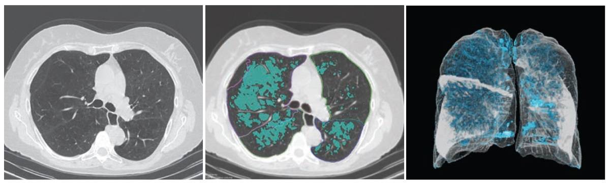
An artificial intelligence (AI) algorithm can provide fully automated quantification of emphysema, offering potential as a tool for image-based diagnosis and quantification of emphysema severity, according to research published in the American Journal of Roentgenology (AJR 10.2214/AJR.19.21572).
After testing prototype AI software on over 140 patients, a multinational team of researchers found that the algorithm showed very strong correlation with traditional pulmonary function tests.
“The ability of an AI-based system to analyse high-dimensional image data and generate valuable clinical information without input from human observers should thus hold considerable potential for granular quantitative imaging approaches to provide patients with accurate diagnoses and steer treatment,” write the team, led by Andreas Fischer of the Medical University of South Carolina (MUSC) and the University Medical Centre Mannheim in Germany.
Although several visual, semiquantitative and quantitative techniques are used to assess emphysema, these methods are subject to increased interobserver and interpatient variability, according to the authors.
A 65-year-old man with severe lung emphysema (light blue on images) seen on axial CT image (left), CT image including AI-based calculation (centre) and 3D model (right). (Courtesy: AJR)
To see if AI could accurately quantify emphysema, the researchers used a deep image-to-image network – a multilayer convolutional neural network that had been trained and tested on more than 10,000 CT datasets acquired on scanners from three vendors at over 20 clinical sites in the US and Europe. Approximately 25% of these datasets were from patients with emphysema.
The algorithm was retrospectively evaluated on 141 patients who had received unenhanced chest CT and spirometry measurements within six months of each other at MUSC between August 2017 and July 2018. All CT exams had been acquired on one of three scanners from Siemens Healthineers: Somatom Definition Flash, Force or Emotion.
To determine if reconstruction methods would impact performance, the researchers applied the algorithm to two reconstruction kernels. The first method used a section thickness of 1.5 mm with a long kernel, while the second utilized a section thickness of 1.5 mm with a soft-tissue kernel. Emphysema was quantified using spatial filtering and a threshold of -950 Hounsfield units.
The patients had a mean spirometry-based Tiffeneau index (TI) of 0.57. The first reconstruction method showed a mean percentage of emphysema of 9.96 ± 11.87%, while the second method had a mean percentage of 8.04 ± 10.32%.
The algorithm correlated very strongly with the TI on both the first (Spearman correlation coefficient = -0.86) and the second (Spearman correlation coefficient = -0.85) CT reconstruction methods. Both results were statistically significant (p < 0.0001).
The results indicate that AI-based emphysema quantification meaningfully reflects clinical pulmonary physiology, according to the researchers.
“Further investigation is needed to establish quantified AI-based emphysema analysis as a potential biomarker for patients with COPD, thus improving diagnostic performance for a specific outcome and maximizing information retrieval to better understand the causes of disease,” the authors write. “Thus, AI-based pulmonary emphysema diagnostics could contribute to complementary phenotyping as part of an imaging biomarker for patients with COPD to shape the individual therapy of patients as a common diagnostic tool, in combination with pulmonary function tests.”
As an undergraduate student at the University of St Andrews from 2015 to 2019, my thirst for knowledge, and fine beverages, was quenched by two part-time jobs. The first was as a tour guide at Kingsbarns whisky distillery, and the second was as a retail assistant in a local drinks shop. Indeed, I would frequently joke that I was actually paid to drink, but both jobs gave me a plethora of skills profoundly useful for physics – I gained confidence in public speaking, learnt time management, and got used to dealing with challenging people. What’s more, I always know which beverage to choose for any occasion.
One evening in the shop during our organic and vegan wines month, my boss – a sponge of booze information – set about educating us on the world of “biodynamic” wine-making. The idea is that wine-makers plant, grow and even advise consumption depending on the phases of the Moon with respect to the Sun, constellations and the planets. Biodynamic agriculture was pioneered by Austrian philosopher and esotericist Rudolf Steiner, in the 1920s. Its application to the wine industry is credited to German enthusiasts Maria and Matthius Thun, who practised biodynamic gardening. They eventually applied their 50 years of research and experimentation to publish the first “biodynamic wine calendar” in 2010.
Days are split into four distinct categories: fruit days, root days, leaf days and flower days. In brief, fruit days are those on which the Moon rises through “fire” constellations – they correspond to both good harvesting and optimal drinking days. Flower days – when the Moon rises through “air” constellations and plants should be left to their own devices – benefit aromatic wines. Leaf days are those on which the Moon rises through “water” constellations, and plants should be watered on these days. Finally we have root days, when the Moon rises through “Earth” constellations and all the “lunar energy” is concentrated in the roots of the plant – these are the worst of all for drinking wine.
I was sceptical and craved some scientific evidence – surely, given that large supermarket chains, and even some Grand Cru Bordeaux vineyards, are coming out as biodynamic converts, there must to be some credibility to these biodynamic claims. My scientific intuition was telling me that it was as much a genuine prospect as homeopathy or astrology, and was perhaps evidence of a large-scale placebo effect. Still, as an open-minded scientist, I was prepared to give biodynamic wine the benefit of the doubt.
To my surprise, there was only one legitimate peer-reviewed paper on the subject published in PLOS One in 2016, alongside many biased individual studies. The paper, entitled “Expectation or sensorial reality? An empirical investigation of the biodynamic calendar for wine drinkers”, tested 19 professional New Zealand wine critics, who tasted 12 pinot noirs on two fruit and two root days. The study found that while the wines tasted different on each day, there was no correlation between a more negative experience and root days, or vice versa, as suggested by the biodynamic calendar.
Critics of the paper within the biodynamics community claimed that pinot noir was too dull a grape to have any marked differences, and that the study would have benefited from an aromatic white that would “sing” on fruit days; or a tannic red to taste astringent on root days. The biodynamic enthusiasts’ studies were entirely dependent their preconceptions: there was a suspicious correlation between calendar followers tasting a difference and sceptics not; there was even some divide within the followers themselves with some admitting “wine doesn’t taste bad on root days, it just tastes better on fruit days” (with no evidence provided). Additionally, there are some obviously fundamental flaws in both the experiment and the calendar itself: are four tasting days in total sufficient? How were uncontrollable variables accounted for? Moreover, a fruit day can change into a root day at any minute; would that mean that in an instant a wine would turn from tasting of blackcurrants to bell pepper?
In essence, there are many different variables that can influence the drinking of a wine. Due to the volatility of alcohol, factors such as poor weather and low atmospheric pressure temperatures have a significant impact on taste, such that aromatics taste stiff, and flavour is subdued. Also, drinking even the best of wines in a foul mood never bodes well for true appreciation. Furthermore, bottle storage and wine temperature will affect aromatics: a riesling might taste like burnt plastic when warm, like honey blossom at optimal temperature, and like acid when chilled. Most importantly, drinking should be done in good company and in a comfortable environment in order for you to feel most at ease; being anxious, irate or distracted will hurt the drinking experience.
Perhaps we should transfer our robust scientific methods from exoplanets and lasers to wine – if nothing else, these studies would make for an interesting social activity at conferences
With so few scientific studies, perhaps we should transfer our robust scientific methods from exoplanets and lasers to wine – if nothing else, these studies would make for an interesting social activity at conferences. Until the biodynamic way of drinking has been rigorously tested scientifically, I encourage you to drink whatever you want, whenever you want, and however you want (safely). It’s your wine and you shouldn’t have to wait until Jupiter’s four moons are visible and form a direct line to Orion’s belt to enjoy it.
Physicists in New Zealand have used optical tweezers to combine three atoms, with two of the atoms forming a molecule in the presence of the third. They were able to measure the rate at which this “three-body recombination” occurs and found it to be much lower than had been expected. The technique could be used to provide important information about how atoms combine to form molecules.
In atomic physics three is a crowd because it is notoriously difficult to calculate how three or more atoms will interact with each other to form a molecule. “Fundamentally, we think that, if we write up the equations of motion from quantum mechanics, these will describe our system,” explains atomic physicist Mikkel Andersen at the University of Otago in Dunedin. “But you can only do that and get an exact solution for a very, very simple system. The question then often becomes finding out, under different conditions, what’s important and what we can throw out of the equations.”
The question is not just of academic interest, as everything around us results from atoms combining. Previous researchers have studied atomic combination using Bose–Einstein condensates, but these contain many atoms, which makes it difficult to disentangle various effects. “We’re trying to develop the capability to build small quantum systems such as molecules atom by atom,” explains Andersen.
Scattered photons
In the new work, Andersen and colleagues at Otago and Massey University in Auckland cooled three atoms of rubidium to 17.8 μK in three separate optical traps positioned about 4.5 μm apart. They then carefully merged the three traps, allowing the atoms to interact. After about a second, the researchers irradiated the combined trap with light, counted the number of photons it scattered and inferred from this how many atoms it contained.
By repeating the experiment multiple times with the same time interval between merging and measurement, the researchers measured the probability of the trap containing various numbers of atoms after that time interval. By varying the time interval, the researchers measured how these probabilities evolved in time.
The results provide the first-ever observation of three-body collisions and combinations at an atom-by-atom level. Andersen explains that the results provide important information about how molecules form: “If you put two atoms in an optical tweezer they don’t usually form a molecule by themselves because something else needs to take away the binding energy But if you put in three, two of them can form a molecule, with and the third taking away the binding energy.”
Lower rate
The researchers were surprised that the rate of this “three-body recombination” was more than ten times lower than theoretical predictions and previous experimental work in Bose–Einstein condensates had suggested. Indeed, Andersen was so perplexed by the results that he had an independent group of people repeat the measurements. “I was convinced it could not be true!” he recalls.
Since describing the research in Physical Review Letters, he says, “We have been contacted by a number of people from the scientific community with suggestions as to what could be going on so, in the next year, we will be conducting a number of experiments to verify whether or not these suggestions are actually true.”
Cold-atom experimentalist Hans-Christoph Nägerl of the University of Innsbruck in Austria, who works with Bose–Einstein condensates, is impressed by the work. “I think this bottom-up approach to looking at few-body processes really has a future,” he says. “I think we’ll see a lot of results with similar systems.”
He is less convinced by the team’s conclusions regarding the recombination rate, however. “First of all, the temperature is not really ultracold, so I’m not too surprised that the rates are different from the zero-temperature expectations,” he says. “The second point – which they raise – is the role of confinement. A few years back my group published a paper – which they don’t cite – looking at what happens to correlations in confined dimensions. We only looked at the repulsive case, but the evidence clearly showed that confinement strongly modifies the three-body correlations.” His conclusion is that “it’s a very nice addition to what’s been done previously. Will one see new physics? It’s hard for me to judge.”
The novel coronavirus responsible for the current pandemic has only been known for a few months but scientists have already gained a vast amount of information about it. Some of this knowledge has been gained by structural biologists who use techniques first developed by physicists. In this podcast episode, the science journalist Jon Cartwright explains what X-ray crystallography and other techniques can tell us about how the virus reproduces.
Also featured in this episode is the philosopher Vanessa Seifert, who is fascinated by the relationships between chemistry and quantum physics. We also travel to exotic exoplanets and chat about our fantastic student contributors.
We are all working at home here on Physics World, so this podcast was recorded using laptop microphones rather than the usual professional equipment.
It probably originated in one of the several species of horseshoe bat found throughout east and south-east Asia. Possibly, a pig or another animal ate the bat’s droppings off a piece of fruit, before being sold at a wet market in Wuhan, China, and subsequently infecting one of the stallholders. Or maybe the first transmission to a human occurred elsewhere.
There is a lot we don’t know about the novel coronavirus now called SARS-CoV-2 and its resultant disease, COVID-19. What we do know is that Chinese authorities alerted the World Health Organization (WHO) to the first known cases in Wuhan at the end of last year. Less than a fortnight later, one of those infected people was dead. By the end of January, with more than 10,000 diagnosed cases and 200 fatalities in China alone, and with the virus cropping up far beyond the country’s borders, the WHO declared a global emergency.
As of this article’s publication (19 March), the WHO reports that the virus has spread to 166 countries, areas and territories, with over 205,000 confirmed cases worldwide and the number of deaths exceeding 8500. The status of “pandemic” was officially designated on 11 March and many countries have introduced social distancing, travel restrictions and quarantine methods to try to curb the spread. Festivals, sports events, parades and conferences are being called off due to the front-line support services they require and the concern that large gatherings of people could help spread the virus. The American Physical Society, for example, axed both its annual March meeting in Denver, Colorado, and April meeting in Washington DC.
When it comes to viruses, there is good reason to worry about novelty. Throughout its history, humanity has had to contend with new diseases springing up seemingly out of nowhere, spreading like wildfire and leaving scores of dead in their wake. In ages past, bacterial plagues were often the source of that terror. Since the birth of modern medicine, however, novel viruses have assumed the mantle of doom. Take Spanish flu for example, which killed up to 100 million people a century ago, and then more recently, HIV, which has led to around 32 million deaths to date. It is only a matter of time before another devastating pandemic, and though epidemiologists do not know what type of virus it will be, they do know that it will be different from anything witnessed before.
Whether or not SARS-CoV-2 is the next “big one”, there is something else epidemiologists are grimly aware of: today, disease travels fast. The Black Death that ravaged Europe, as well as parts of Asia and Africa, in the mid-14th century spread at an average of just 1.5 km a day – hardly surprising, since this was before ships could reliably cross oceans and the fastest mode of transport was by horse. Contrast that with the 2015 outbreak of Zika virus in South America, where the daily dispersion was on average 42 km, peaking in the densest-populated parts of Brazil at 634 km. Faced with more populous cities, more mobile people and more international travel, scientists must respond to the threat of viral pandemics faster than ever.
Structural biology has reached the stage where it’s fast enough for almost anything
Fortunately, those scientists now have much more efficient tools at their disposal. Structural biology – the study of the structure and function of biological macromolecules – has come a long way since it was first used as the basis of rational (as opposed to trial-and-error) drug design 30 years ago. Back in the early 1990s, viral structures deposited in the Protein Data Bank – an international repository for structures of biological macromolecules – numbered just a few dozen annually, but by the mid-2010s, there were well over 500 new additions a year. Modern techniques, such as automation and cryo-electron microscopy (cryo-EM), mean that viral structures can be identified almost instantly in many cases. “Structural biology has reached the stage where it’s fast enough for almost anything,” says Alexander Wlodawer, chief of the macromolecular crystallography laboratory at the US National Cancer Institute in Frederick, Maryland.
But is it fast enough to halt a pandemic?
The speed of physics
Physics-based techniques play a huge role in the field of structural biology. The vast majority of biological macromolecule structures are obtained by X-ray crystallography, going back to 1934, when John Desmond Bernal and Dorothy Hodgkin recorded the first X-ray diffraction pattern of a crystallized protein, the digestive enzyme pepsin. Their work stemmed from that of physicists such as Wilhelm Röntgen, who discovered X-rays; Max von Laue, who discovered that X-ray wavelengths are comparable with inter-atomic distances and are therefore diffracted by crystals; and William Henry and William Lawrence Bragg, who showed how to use a diffraction pattern to analyse the corresponding crystal structure. Hodgkin went on to win the 1964 Nobel Prize for Chemistry for her determinations by X-ray techniques of the structures of important biochemical substances.
Single biological molecules also diffract X-rays, but only very weakly. Crystallization, as Bernal and Hodgkin employed for pepsin, is helpful because it results in the repetition of huge numbers of molecules in an ordered, 3D lattice, so that all their tiny signals reinforce one another and become detectable – by photographic plates in the early days and by active pixel detectors today. These signals are not images of the molecules, for there are no materials that can substantially refract, and thereby focus, scattered X-rays. Rather, the signals are merely the sum of the contributions of X-rays diffracted from different parts of the molecule. To pick apart these contributions, structural biologists rely on a mathematical tool – the Fourier transform. The calculated contributions are then equated with possible atomic structures by a lot of careful (and now largely computer-driven) interpretation.
Of course, to obtain the signals in the first place requires X-rays. Nowadays, synchrotron radiation sources – large facilities that accelerate electrons in a continuous ring – are ideal for macromolecular crystallography because they produce high-intensity X-rays with a very narrow spread of wavelengths. At these machines, according to Wlodawer, diffraction datasets that would have taken months with X-rays from traditional rotating anode generators take just seconds to compile.
Technological developments such as these spurred the first forays into rational drug design, in which scientists study the structure and function of molecules in order to work out what drugs might bind to them – and in the case of viruses, prevent them from replicating. Antiviral drugs for HIV were an early success. When HIV protease was identified in 1985 as an essential enzyme – and therefore a potential drug target – in the virus’s life cycle, it took four years for its first crystal structures to be determined, and a further six years for the first licensed drugs to inhibit it. “That’s probably one of the best-documented cases of how quickly rational drug design can go,” says Wlodawer, who contributed to the international effort.
Today, it might have gone faster. The four-year delay in obtaining the structure of HIV protease was primarily due not to the brilliance or quality of X-rays, but to the lack of sizeable crystals of the enzyme. Current synchrotrons and ever newer free-electron lasers – which extract diffraction data from molecular crystals in the few femtoseconds before they are annihilated – employ techniques such as serial crystallography to build up a complete diffraction dataset from numerous partial datasets of crystals that would otherwise be too small. Moreover, both the crystallization and data collection are now automated, so that structural biologists need not even visit a light source themselves: they simply post their proteins to a facility and download the dataset when it is ready.
The analysis of SARS-CoV-2 is a prime example of this type of modern pipeline in action. On 5 February this year, a little over a month after the Chinese authorities disclosed the existence of the new coronavirus, a research team led by Zihe Rao and Haitao Yang at ShanghaiTech University in China uploaded the structure of the virus’s main protease to the Protein Data Bank (DOI: 10.2210/pdb6lu7/pdb), having obtained the dataset using X-ray crystallography at the Shanghai Synchrotron Radiation Facility. “A decade ago, that would have taken a year,” says Wlodawer. “At least.” The structure is already helping pharmaceutical companies to explore potential drugs, such as those used to tackle HIV.
The protein pipeline
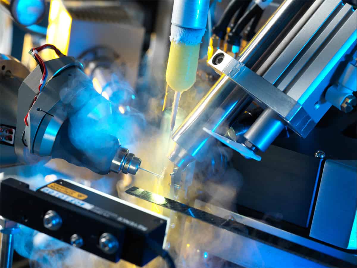
Even if molecules refuse to be crystallized, there is still the chance of obtaining structures using cryo-EM, a technique pioneered by Jacques Dubochet of the University of Lausanne in Switzerland, Joachim Frank of Columbia University in New York City, US, and Richard Henderson of the MRC Laboratory of Molecular Biology in Cambridge, UK, for which they shared the 2017 Nobel Prize for Chemistry. In a cryo-EM experiment, a solution containing the biomolecule or complex of interest is applied to a sample holder, or “grid”, as a thin layer. The grid is flash-frozen in liquid ethane to vitrify the sample, which is then imaged by the electron microscope with low doses of electrons to minimize radiation damage. Because single molecules or complexes are imaged directly, there is no need for crystallization.
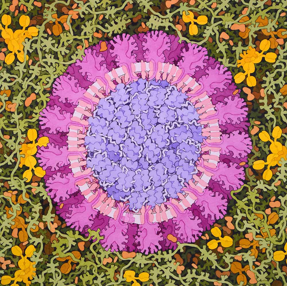
Not pretty in pink This painting depicts a coronavirus just entering the lungs, surrounded by mucus secreted by respiratory cells, secreted antibodies, and several small immune system proteins. (Courtesy: David S Goodsell, RCSB Protein Data Bank)
Thanks to cryo-EM, Daniel Wrapp and Nianshuang Wang of the University of Texas at Austin, US, and colleagues were able to obtain the structure of an outer “spike” protein of SARS-CoV-2 that is believed to enable the new virus to weasel its way into host cells. From harvesting the protein to submitting a paper to the journal Science on 10 February, the entire process took just 12 days (10.1126/science.abb2507). “Without cryo-EM,” says the University of Texas’s Jason McLellan, an author on the paper, “it may not have been possible at all.”
The structure of the external spike is more useful for creating coronavirus vaccines than drugs. If host cells are exposed to virus-like particles that brandish the same external features, while being hollow inside, those cells can still help the body build an immunity but without the risk of being exposed to a dangerous, fully fledged virus. David Stuart – a structural biologist at the University of Oxford in the UK and director of life sciences at the Diamond Light Source, a “third-generation” synchrotron – has used this synthetic trick to create a new vaccine for foot-and-mouth disease. This virus, which is still devastating livestock in large parts of Africa, the Middle East and Asia, is in a family of single-stranded “positive sense” RNA viruses that also includes polio and human rhinovirus – the latter being behind most cases of the common cold. “Only in the past few years have we been able to exploit structural biology to understand immunity to disease,” he says. The knowledge of viral structures can even be used to design synthetic “therapeutic antibodies” to directly attack diseased cells, he adds.
Stuart obtained the structure of the foot-and-mouth virus itself back in 1989 at the (now defunct) Synchrotron Radiation Source in Daresbury, UK. Indeed, it was one of the first viral structures ever deposited in the Protein Data Bank. For that reason, he knows first-hand how much the techniques have progressed. “Getting those first structures, that was a big deal!” Stuart recalls.
Beware the unknowns
It is too early to predict how long it will take to develop drugs or vaccines for SARS-CoV-2. A US biotechnology firm, Moderna Therapeutics, has already begun human trials for a vaccine candidate, but even if it successful, it could still take up to 18 months for it to be available to the public. Borrowing the infamous terminology of the former US defence secretary Donald Rumsfeld, Stephen Cusack – the head of the European Molecular Biology Laboratory (EMBL) in Grenoble, France – puts the virus in a category of formidable “unknown unknowns”, which covers viruses that break out without precedent, such as HIV, Zika and the 2002 coronavirus, SARS-CoV. But Cusack says we should still beware the more familiar “known unknown” pandemic influenza, which has struck three times since the Spanish flu of 1918. Its most recent incarnation in 2009, swine flu, is believed to have infected up to a fifth, and killed up to half a million, of the world’s population – although that, relatively speaking, was not such a bad case. Similar numbers are met every year with seasonal flu – in Cusack’s terminology, the “known known”.
Though he graduated as a physicist, Cusack has spent much of his career studying influenza as a structural biologist, and in particular its polymerase – the enzyme behind the virus’s transcription and replication. Like other viruses, in order to replicate, influenza has to produce a code for its proteins known as messenger RNA (mRNA). This needs to match the mRNA of the healthy host cell that the virus is invading in order to trick the cell to produce more of the virus. Some viruses have their own enzymes to synthesize the matching mRNA from scratch; influenza does not, and instead steals a “cap” from the host-cell mRNA as a primer. Biochemists have known of this influenza “cap snatching” for many years, but in 2014 Cusack’s group used structural-biology techniques to understand its basic mechanism at an atomic level. In their most recent work, yet to be published, the researchers have employed cryo-EM to snapshot different stages of the entire polymerase transcription – in effect creating a molecular movie – in order to uncover weaknesses that can be targeted by drugs. “If you can stop this mechanism from working, you can stop the virus from replicating,” Cusack says.
All of which suggests that structural biologists are well-equipped to tackle the next pandemic, be it a known-unknown or unknown-unknown. Whether their techniques are sufficiently advanced to prevent some of the huge death tolls humanity has suffered in the past, however, is still an open question. Those who study complex networks believe they can now predict the rate at which pandemics spread in the modern world (see box below), although these only stress the shortness of the deadlines on which scientists must act. Moreover, finding a drug is only the first step in a long regulatory process involving fabrication, initial toxicology testing and clinical trials.
Even then, there is no guarantee of success. In 2018 Xofluza was the first antiviral drug for influenza approved by regulators in Japan and the US for decades, billed by the press as a “miracle cure” that was able to stop the virus dead in its tracks just 24 hours after a single dose. A year later, the Japanese company that developed it, Shionogi, discovered that, far from being killed off, the virus in patients taking the drug spontaneously mutated into a more resilient form. Working with Shionogi, Cusack and colleagues used structural-biology techniques to show that although the mutated virus bound to the drug less tightly, it was also less fit to replicate, leaving a question mark over the drug’s efficacy. “No-one knows whether the drug will be useless in a year or two,” he says.
And therein lies a lesson. “The virus is always cleverer than you are,” Cusack says. “But we knew that anyway.”
The global village
International effort Experts from the World Health Organization novel coronavirus pneumonia investigation group visit Wuhan to conduct field research. (Courtesy: Top Photo Corporation/Shutterstock)
Back in the 14th century, when the bubonic plague known commonly as the Black Death was advancing through Europe, geographical proximity to an infected town or village was a reliable indicator of how likely a neighbouring settlement’s inhabitants would contract the disease. In today’s globalized world, however, with the ease of long-distance travel, that is no longer true – at least according to the physicist Dirk Brockmann of Humboldt University Berlin in Germany and the physicist-turned-social-scientist Dirk Helbing of ETH Zurich in Switzerland. In 2013, based on an analysis of airports, the pair showed that computer models could better describe the progress of previous epidemics, such as the swine flu of 2009, if they spread through an “effective” distance between points – one that is dependent not on miles or kilometres but the density of traffic flow between them (Science342 1337).
Although their model – and others like them – could be important for predicting the spread of current viruses and targeting disease-control measures, population flow between cities is not always known to high accuracy. In a preprint posted online in February this year, however, Piet Van Mieghem and colleagues at the Delft University of Technology in the Netherlands showed that it is possible to make short-term predictions about the rate of progression of the latest coronavirus, SARS-CoV-2, from Wuhan to other cities in the Chinese province of Hubei if population interactions between cities are inferred from initial observations of the spread, rather than relying on prior knowledge. Comparing their model with real data, they found that its predictions for infections in Hubei cities three days ahead were within 7.5% of the actual numbers (arXiv:2002.04482). “The coronavirus pandemic has been a good case to demonstrate the power of our method,” says Van Mieghem.
Researchers at Imec, a Leuven, Belgium-based centre for nanoelectronics and digital technologies, have developed a wireless receiver and transmitter small enough to fit inside a millimetre-scale capsule. The transceiver, which was presented at the International Solid-State Circuits conference in San Francisco, US, last month, is 1/30th the size of today’s state-of-the-art systems and could be used in a broad range of so-called “ingestibles” – sensors that monitor health conditions from inside the human body.
Like their external, wearable cousins, ingestible sensors are designed to measure and track health parameters over a period of time. Unlike them, however, ingestibles need to be able to transmit data autonomously to a receiver station outside the body. “The development of such devices brings along a specific set of challenges,” says Imec’s Christian Bachmann. “They have to be extremely small, consume very little power and be able to connect wirelessly.”
A miniaturization revolution
Bachmann, who serves as programme manager for the Sensitive Networks project at Imec’s laboratory in Eindhoven, Netherlands, explains that these goals are only achievable thanks to an ongoing “miniaturization revolution” in nanoelectronics. This revolution has enabled researchers to develop smart, small and lightweight devices that combine minimal power consumption with maximal patient comfort.
Imec’s wireless transceiver occupies a volume of less than 55 mm³, with areas of 3.5 and 15 mm2 on its sides. It supports transmissions at the medical 400MHz frequency band and contains an on-chip tuneable antenna. According to the Imec team, the most significant achievement is that the module does not require a crystal-based oscillator. Such oscillators are commonly used to precisely stabilize the frequency of radio signals and network protocol timing, but the Imec researchers instead created an on-chip mechanism that, in effect, uses the wireless network to calibrate itself. The lack of an off-chip crystal device made it possible to achieve the extremely small “form factor” needed for an ingestible.
According to Bachmann, on-chip tuning for the miniaturized antenna does more than just guarantee reliable data transmission. It also offers a large flexibility in impedance tuning – meaning that it works over a wide range of impedances in the body, equivalent to a filled and empty stomach. “We foresaw a tuneable impedance that can ‘tune’ itself to conditions seen in the digestive tract, so that the transceiver always makes a reliable link,” he says.
A range of uses
While ingestible devices are in their infancy, their potential benefits are considerable. As well as helping to monitor digestive processes and diagnose gastrointestinal diseases, ingestibles could also replace procedures like endoscopic inspections and stool sample analyses – which can be very uncomfortable and provide only one-time observations. Bachmann says the Imec transceiver has several potential use cases, including ingestibles designed to monitor health, nutrition or biomarkers that indicate the presence of disease. However, he cautions that clinical applications will require a further integration of electronics at the nano level.
“For the moment, there are some camera-integrated pills that can be used to study the digestive tract,” he says. “But these pills are still quite big today.” More advanced applications will probably arise as early demonstrators in the healthcare domain. Apart from cameras, ingestibles could also carry other sensors, which – like the transceivers – will need miniature batteries capable of powering them for several weeks.
Despite these obstacles, Giovanni Traverso, who studies ingestibles and implantable robotics at the Massachusetts Institute of Technology in the US, calls the Imec device “a welcome contribution to the field”. Traverso, who was not involved in the Imec project, adds that a smaller transceiver “certainly boosts the miniaturization of ingestibles” by making more room for other components such as sensors and batteries. “It’s key to make the devices smaller and smaller, as this maximizes the safety while transiting the gastrointestinal tract,” he says.
The end result
That raises an important question: what happens after an ingestible has done its job? Does it just follow the natural way out of the body? Bachmann’s response is that Imec is currently exploring ways of fixing ingestible sensors at certain locations along the gastrointestinal tract. “This would enable longer recordings in specific places of interest and keep patients comfortably outside the hospital while their health data is collected and sent in real time to a doctor,” he says.
Traverso notes that this question is particularly important in his work, which focuses on using ingestibles for drug delivery. For this application, it may be important to keep ingestibles in place for days or even weeks. “We’ve made a lot of progress during the past five years, keeping devices in place in the stomach or in the gastrointestinal tract,” he says. For example, a device could be swallowed as a pill, unfold in the stomach, and then dissolve in the acid environment once it has delivered the required drugs.