The rise and rise of ‘new big science’

“They do nuclear work there and jam your GPS,” my taxi driver warned me as we drove from the airport in Knoxville to the Oak Ridge National Laboratory (ORNL). Nestled in a lush green area between the Cumberland and the Great Smoky Mountains in the middle of Tennessee, it was here during the Second World War that the US created fissionable material for the Manhattan atomic-bomb project. Informing me he’d have to drop me off at the gate, I asked my driver to go in anyway and leave the device on. He did – and the GPS worked fine.

With its huge, federally funded scientific facilities, the Manhattan project helped to usher in a phase change in physics from small-scale efforts to what became known as “big science”. The term in fact was popularized by ORNL’s then director Alvin Weinberg in an article he wrote for Science in 1961 (134 161). Big science was his term to describe how particle accelerators and reactors were fostering a seemingly unstoppable rise in the scale of scientific facilities, instruments and budgets, with increasingly specialized researchers working in ever-larger collaborations.

Many physicists began to embrace the moniker “big science” to enhance the prestige and funding of high-energy physics. But Weinberg felt that physics as a whole did not share that culture. ORNL and other national labs in the US and elsewhere, he pointed out, had a more varied character, focusing as they did mostly on materials science and condensed-matter physics. For this reason, he lamented the enthusiasm with which many of his fellow scientists were greeting big science.

In “new big science”, large facilities and labs are increasingly supporting a “research ecology”, consisting of relatively small instruments, experiments and collaborations.

There is, however, another phase change going on in science that shows that Weinberg’s instincts were sound. In this latest phase change, which historians are calling “new big science”, large facilities at ORNL and other labs are increasingly supporting a “research ecology”, consisting of relatively small instruments, experiments and collaborations. Research is growing not so much bigger as more complex.

Small teams interact in ever more interactive and networked ways. The change manifested itself first in the 1970s and 1980s at light sources, where one beam port often began to serve many different research projects and one project often took place at several different ports. But the flexible collaborative research of new big science is now also apparent at neutron facilities, such as at Oak Ridge.

Spalls and thrills

I had come to Tennessee to see the new big science in action at the Spallation Neutron Source (SNS), the largest facility to have been built at Oak Ridge since the Manhattan Project. Ground was broken for the $1.4bn project in 1999, and it began operating in 2006. As I toured the SNS, instrument scientist Bryan Chakoumakos explained how its linear accelerator runs pulses of protons up to 1 GeV before slamming them into a mercury target, which splits or “spalls” into many small pieces.

This spallation process creates sprays of neutrons, which spread down instrument pipes that surround the target, like the gaps between slices of half a pie. The SNS has 18 beam pipes, nine on each side, sometimes with more than one instrument on each pipe. Consisting of different kinds of diffractometers and spectrometers, those instruments are placed a dozen or more metres from the target, partly because the resolution increases with distance and partly to fit in blocks that shield users from radiological hazards.

Spallation sources, Chakoumakos told me, have given neutron research additional scope and flexibility, making it useful not just for condensed-matter physics but for chemistry, biology and engineering too. Industrial participation is growing as well. Automotive companies have inserted batteries and even entire engine blocks into the SNS’s neutron beams, while aviation and engineering companies use them to study new alloys.

Industrial participation – small-scale research for applied purposes at a large facility – is one hallmark of the new big science.

Industrial users at the SNS range from large corporations whose researchers are experienced with the machine and how it works, to small firms for which the SNS staff has to perform “concierge service”, helping them set up and use the instrument and interpret the result. This new industrial participation – small-scale research for applied purposes at a large facility – is one hallmark of the new big science.

A further sign is the increasing interaction between research at facilities such as the SNS and work at light sources elsewhere. One reason for this cross-disciplinarity is the ability of neutrons to investigate the magnetic structures of materials. Another is that neutrons can easily locate hydrogen and other light elements in, say, proteins, which is hard or even impossible with X-rays.

“The large fraction of users at the powder diffractometers collect data at both X-ray and neutron sources,” says ORNL’s associate director Paul Langan. The use of cold neutrons – with long wavelengths – has further expanded neutron research into fields such as chemistry and biology.

The critical point

In the new big science, the practice of research is not necessarily getting bigger in scale – it’s also becoming more flexible and interactive. Growing institutional interactions are developing between facilities – with, in the case of the SNS, five other labs building different major parts of it. This kind of co-operation has been increasing among the US national labs and is now far bigger than when the SNS itself was built. Furthermore, small research groups, many involving industrial participants, are thriving without needing ever-narrower specialization.

Another key sign of the new big science is the increasing collaboration between neutron researchers and those using X-ray instruments at light sources, such as the Advanced Photon Source and the National Synchrotron Light Source at the Argonne and Brookhaven national labs respectively. Such interactions indicate a big change in the way materials scientists do research at user facilities. Those interactions, in turn, also create new challenges for scientists, science educators, managers and those who synthesize the knowledge and information that are generated.

Quasicrystalline Bose–Einstein condensate provides a glimpse of physics in higher dimensions

A Bose–Einstein condensate (BEC) of ultracold atoms has been created on a 2D quasicrystalline optical lattice by Ulrich Schneider and colleagues at the University of Cambridge in the UK. The pioneering study could pave the way for quantum many-body simulations in fractal systems and systems with higher dimensions.

Quasicrystals are materials with structures that are not periodic in space but have some long-range order. Quasicrystals have the fractal quality of self-similarity and can be related to crystals that exist in higher dimensions.

Optical lattices are created using standing waves of laser light that trap atoms at regular intervals. Physicists have already studied ultracold atomic gases within optical quasicrystals that were created by overlapping two or more optical lattices with different lattice spacings.

Unwanted scattering

One of the main goals of these studies has been to simulate the behaviours of quantum many-body systems within quasicrystals, and potentially, in higher dimensions. So far, however, these efforts have been hindered by laser-induced resonances in some of the trapped atoms, which scatter the laser light in unwanted directions.

In their study, Schneider’s team formed an optical lattice from a planar arrangement of four intercepting 1D optical lattices, separated by a 45° angle. They then fine-tuned each lattice to create a quasicrystal with eightfold rotational symmetry. By tuning their optical lattice far from the resonant frequency of the atoms, they could ensure minimal unwanted scattering.

Their experiments began with a pre-formed BEC composed of potassium atoms that is created in the absence of an optical quasicrystal. Then, the optical lattices were switched on in a pulse lasting few microseconds – creating the quasicrystal.

Forbidden momentum

Schneider and colleagues found that during the brief time their quasicrystal was turned on, atoms in the BEC scattered photons from one beam to another. This put the atoms in distinctive, time-varying series of momentum states. While lower momentum states are forbidden for atoms in periodic lattices, those in the researchers’ quasicrystal-like BEC moved to successively lower, and more closely-spaced momentum states through a series of small photon-induced velocity boosts.

The physicists say that this behaviour constitutes a “quantum walk” in their quasicrystal’s momentum space, resulting in a diffraction pattern that appeared as an infinite series of progressively larger octagons. This is similar to the discovery of quasicrystals by Dan Shechtman and colleagues in 1984, who observed electron diffraction patterns that displayed rotational symmetries beyond the two- three- and six-fold symmetries allowed in periodic crystals.

From their observations, Schneider’s team interpreted their 2D quasicrystal as a projection of a 4D lattice onto a 2D plane, demonstrating the possibility of simulating higher dimensions with quasicrystal-like optical lattices. Their work could soon allow for controllable simulations of quantum many-body systems over a wide range of new quasicrystal environments; potentially allowing for explorations of exotic new 4D phenomena.

The research is described in Physical Review Letters.

Mangroves deliver research surprise

­

Rates of growth and loss in mangrove forests are higher than previous estimates, according to satellite measurements of the Rufiji, Zambezi, Ganges and Mekong deltas. And natural expansion and growth of new mangrove forests could partially compensate for declines caused by human activity.

“What we see for these types of mangrove delta regions is that there is rapid expansion and growth — in about 20 years, forest canopies can reach a mature height of over 20 m,” says David Lagomasino from the University of Maryland and the NASA Goddard Space Flight Center, both in the US. “In some places that fast growth can help to offset the losses, both in terms of extent of the forests and carbon density.”

Mangrove forests make up a relatively minor part of the Earth’s total forested area, but their importance exceeds the scale of their footprint. They sequester proportionally more carbon than almost any other ecosystem — their maximum storage potential above and below ground is more than 1000 tonnes per hectare – and they provide food, shelter, fuel and coastal protection.

Mangrove extent is decreasing due to urbanization and expanding agriculture and aquaculture but natural coastal processes like erosion and deposition also play a role.

By measuring changes in mangrove area and height at four major river deltas, Lagomasino and colleagues discovered that, although there was a net loss of carbon across the study areas, this was partially balanced by the growth and spread of forests elsewhere in the same regions.

The researchers examined satellite images of the Rufiji delta in Tanzania, the Zambezi delta in Mozambique, the Ganges delta in Bangladesh, and the Mekong delta in Vietnam, captured between 1995 and 2000. They identified pixels corresponding to forest using the normalized difference vegetation index (NDVI) — a method based on the ratio of red and infrared wavelengths characteristic of plant leaves. Then they took the change in forested area as the difference between this reference NDVI signal and that for the years 2000 to 2016.

The amount of carbon that an area of forest stores depends on its height and density, but in situ studies are prone to bias because of access and logistics issues. To get a more representative view, the researchers used canopy-height measurements from satellite radar observations, matching them to the changing mangrove coverage.

Although radar measurements do not yet characterize mangrove forests completely, the researchers are confident about their conclusions overall.

“We have compared our remote sensing-based models with the growth from individual field plots and have had very similar values,” says Lagomasino. “Though there are differences in the density and species of trees between young and mature forests, two forests of the same height – a young one with thousands of thin trees, and an old one with a couple hundred thick trees – may have similar amounts of carbon.”

Follow-up research will focus on what the future might hold for these regions. “Changes in the rate of sea level rise will certainly have an impact on the erosion and progradation of mangrove forests,” says Lagomasino. “Under certain conditions with enough sediment, a mangrove forest may expand with sea level rise, but other conditions may lead to the collapse of the ecosystem.”

Lagomasino and colleagues from the Universities Space Research Association and the Forest Service in the US, the World Wide Fund for Nature in Germany, and the University of Dar es Salaam, Tanzania reported the findings in Environmental Research Letters (ERL).

Sticky biomaterial could repair corneal injuries in patients

Vision loss is a serious and common symptom of corneal injuries, with more than 1.5 million cases of corneal blindness reported every year. Corneal injuries typically result from a trauma or an infection to the cornea. These can cause scarring and stromal thinning, which can lead to visual impairment and even blindness.

Researchers at the University of California Los Angeles (UCLA), Harvard Medical School and Northeastern University have now developed a biomaterial named GelCORE, which aids the sealing and repair of corneal tissue. The biomaterial is comprised of a gelatin-based adhesive and photoinitiators. Published in Science Advances their research shows how the biomaterial hardens when exposed to visible light, producing a transparent hydrogel with similar biomechanical features to that of the cornea. This adheres to the cornea and encourages regeneration of cells and tissue to repair any damage at that site.

The GelCORE advantage

This technology provides an alternative solution to the current standard treatment options available, which include the use of synthetic glues, corneal transplants and surgery to graft tissue onto the cornea. Both surgery and transplantation risk post-transplant complications such as infection or rejection, and the glues in current use are associated with low biocompatibility, poor transparency, and are undesirably rough and difficult to handle.

Other adhesive materials developed for eye injuries use ultraviolet light, which can introduce toxicity issues. The use of visible blue light to activate the functionalized gelatin adhesive is unique to GelCORE, giving it an important advantage.

Preclinical trial success

The researchers trialled their GelCORE product in preclinical rabbit models of a corneal injury. Here, they made a 3mm long partial cut in the rabbit’s cornea and applied GelCORE concentrations of 20% to the wound, which was then exposed to visible light for 4 minutes.

They found that GelCORE adhered firmly to the damaged tissue in the cornea. Observations over the subsequent day found no inflammation at the site, and a smooth surface across the cornea. After one week of application, they were still able to observe GelCORE at the site on the cornea they had damaged and the product remained transparent. Over a few weeks, the researchers were able to identify the formation of new cell tissue forming, indicating tissue regeneration.

The group additionally showed that by adjusting the concentration and the amount of time exposed to light, the properties of the GelCORE material could be altered. This versatility in the properties of the GelCORE is exciting as it enables product modification and the ability to tailor the product to different patient needs.

Nanotechnology is neither saviour nor destroyer of the world, it’s just very useful

Historically, nanotechnology has provoked its fair share of hype, but also some fears around possible impacts on human health and the environment. In reality, nanotechnology is neither saviour nor destroyer of the world – it is an exciting emerging research field with the potential to help with a variety of global challenges.  Today’s nanoscience community is busy with such worthwhile tasks as developing methods of water purification, producing nanoparticles for bioimaging and developing new coatings to prevent corrosion to steel structures.

Find out more by reading the Physics World Focus on Nanotechnology and Nanomaterials, which you can access in our digital magazine or via the Physics World app for any iOS or Android smartphone or tablet.

Topological physics pioneer and Nobel laureate David Thouless dies at 84

The theoretical physicist David Thouless, who shared the 2016 Nobel Prize for Physics, has died age 84. In the 1970s Thouless pioneered in the study of the topological states of matter, which has since blossomed into a thriving branch of condensed matter physics with potential applications ranging from electronic devices to quantum computers.

Thouless was born in 1934 Bearsden, Scotland – which is near Glasgow. He studied physics at the University of Cambridge before travelling to the US to complete a PhD at Cornell University in 1958 under the supervision of Hans Bethe. He worked at several universities in the UK and US before becoming professor of physics at the University of Birmingham in 1965.  Following stint at Yale University, Thouless settled at the University of Washington in 1980. He remained there until 2014 when he returned with his wife Margaret to live in Cambridge.

Thouless share the Nobel prize with Duncane Haldane and Michael Kosterlitz. Working at the University of Birmingham in 1972, Thouless and Kosterlitz identified a completely new type of phase transition that can occur in 2D materials, where topological properties play a crucial role. As a result, they were able to show that superconductivity or superfluidity can occur in 2D layers – something that had not been expected prior to their work

According to the Nobel committee, the pair’s work “resulted in an entirely new understanding of phase transitions, which is regarded as one of the 20th century’s most important discoveries in the theory of condensed-matter physics”.

Thouless is survived by Margaret and their three children.

Shear forces help make stretchable hydrogel

Shear forces can convert a sol into a gel. This surprising new behaviour, observed in an aqueous solution of a pseudopolyrotaxane with copper ions added to it, is similar to that in dissipative, far-from-equilibrium, biological systems capable of self-healing.

“Usually supramolecular hydrogels, like the ones we studied in this work, are destroyed by shear forces and convert into a sol,” says Wei Jiang of the Southern University of Science and Technology in China, who led this research effort. “This makes our result surprising and exciting.”

Molecular self-assembly is common in the biological world and leads to large and complex architectures that have very specific functions. Bioinspired synthetic versions of these have been built in recent decades, but they are usually stable in thermodynamic terms, unlike their biological counterparts, which work in the far-from-equilibrium state. Far-from-equilibrium means they constantly require an input of energy (or fuel) to continue functioning.

Dissipative self-assembly

This mechanism, known as dissipative self-assembly, would come in very useful in materials science because it could produce properties that exist in biological systems, such as self-healing and adaptability. The problem is that most synthetic dissipative self-assembly structures made to date are soft and have poor mechanical properties, something that limits their applications.

Jiang’s team has now self-assembled a supramolecular hydrogel under shear force. The researchers made their gel by simply adding copper ions to a solution of pseudopolyrotaxanes, which themselves are formed by threading molecular tubes on polyethylene glycol chains. When the solution is shaken vigorously for up to 30 seconds it transforms into a gel that gradually relaxes back to the sol state over time (hours to days at room temperature depending on the concentrations of the gelators) It can be converted back into a gel, however, by reapplying a shear force (the fuel). This process can be repeated many times.

Intrachain to interchain coordination

The mechanisms at play in the sol to gel transition rely on a shear-induced transition from intrachain to interchain coordination with the Cu(II) ions, explains Jiang. “The initial mixture is in a solution state because Cu(II) ions form intrachain coordination and there is no, or very little, cross-links among the pseudopolyrotaxanes. Shear forces destroy this intrachain coordination though and this results in kinetically fast interchain coordination, which leads to more cross links and thus gelation (the material becoming thicker).”

The resulting transient hydrogel has good mechanical properties, even when compared to permanent hydrogels, in that it is highly stretchable (it can be stretched up to 30 times its original length). This is in contrast to previous shear-thickening systems. It is also self-healing under a shear force as mentioned.

“The high extensibility comes from the fact that the molecular tubes are loosely packed on the polymer chains and can slide ‘frictionally’ when stretched,” explains Jiang. “This means that it might be used to prepare mechanoresponsive materials, as a first type of application, by incorporating the supramolecular coordination unit into other systems,” he tells Physics World.

The team, reporting its work in Nature Chemistry 10.1038/s41557-019-0235-8, says that it is now busy further improving the mechanical properties of its dissipative material.

A new approach to 3D brain visualization

© AuntMinnieEurope.com

Researchers from Spain have developed a technique that allows users to visualize and interact with head MRI and CT scans as 3D models using virtual reality (VR) and augmented reality (AR). The technique relies on proprietary software applications specifically designed for examining radiology images.

The group’s visualization approach features three different software applications that can convert standard 3D anatomical models typically viewed on a 2D screen into files compatible with VR and AR technologies (J. Med. Syst. 10.1007/s10916-019-1239-z).

The study shows how clinicians can integrate VR and AR into the radiology workflow and have access to a more realistic, comprehensive display of brain and skull anatomy, first author Santiago Izard, a doctoral student at the University of Salamanca and CEO of VR/AR start-up ARSoft, told AuntMinnieEurope.com.

“In this moment, it is not usual to create 3D models [for VR and AR] due to the long processing time and additional costs it demands,” he said. “The key clinical implication of the system we are implementing is to allow doctors to work with 3D models created from radiological results cheaply and very easily.”

Cranium model

Beyond traditional imaging

Technological advancements in recent years have led to major improvements in graphics processing and rendering capabilities, paving the way for the visualization of medical images as complex 3D models, according to the authors. Although many computer programs can generate 3D models from 2D medical images, few are exclusively geared toward the examination of radiology images using VR and AR.

One of the major challenges with integrating high-quality 3D anatomical models into VR and AR is that the models generally consist of too many polygons for VR and AR devices to handle, they noted. Most computer programs reduce the number of polygons that make up a model to be able to visualize it in VR and AR, but this comes at the cost of image quality.

Seeking to address this barrier, Izard and colleagues obtained head MRI and CT scans and used segmentation software to select regions of interest on the scans and turn them into 3D meshes. Next, they reduced the complexity of the 3D meshes by applying the marching cubes algorithm.

After simplifying the 3D meshes, the researchers input them into one of three distinct software applications they designed using computer software (Unity3D, Unity Technologies):

  • VR Viewer: Produces 3D models compatible with VR headsets (Oculus and Samsung Gear VR, Oculus)
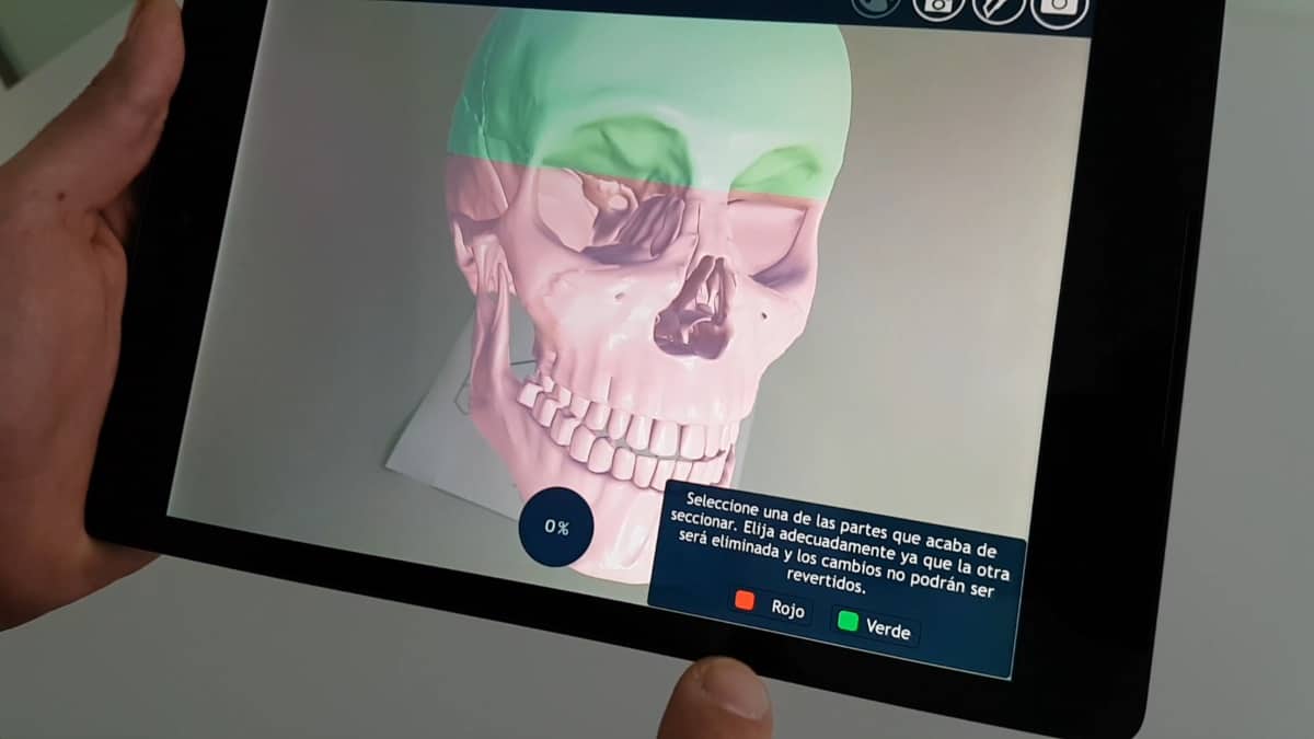
  • AR Viewer: Allows for the visualization and manipulation of 3D models with Android and Apple devices
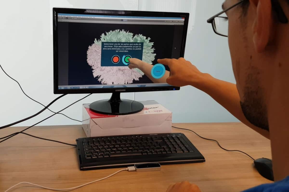
  • PC Viewer: Works with a USB motion sensor (Leap Motion controller, Leap Motion) to allow users to interact with 3D models on a monitor using hand motions

These software applications help improve the final step in the process of examining radiology images using VR and AR, making complex 3D models compatible with VR and AR devices without compromising image quality, the authors wrote. Ultimately, the technologies offer “fast and efficient interaction, including rotating, scaling or cutting the 3D models [to view] complex internal structures. In addition, this system can be used by clinicians to store and explore clinical neuroimages from different locations.”

3D brain model

Automated segmentation

Looking ahead, the researchers proposed a way to overcome yet another barrier to using VR and AR for viewing radiology images: the time-consuming process of image segmentation. Their proposal involved using artificial intelligence (AI) algorithms to automatically segment regions of interest on MRI and CT scans.

“We are currently working on creating and implementing advanced AI algorithms for image segmentation,” Izard said.

The algorithm they are developing relies on a cellular neural network — a computing paradigm similar to neural networks — to detect key areas for automated segmentation.

“Our tool fully integrates AR and VR technology with radiological imaging and is specifically designed to study radiology-based results and even plan surgeries,” he said. “These technologies allow [users] to interact with 3D models in a realistic way without having to print them, saving time and money.”

  • This article was originally published on AuntMinnieEurope.com ©2019 by AuntMinnieEurope.com. Any copying, republication or redistribution of AuntMinnieEurope.com content is expressly prohibited without the prior written consent of AuntMinnieEurope.com.

Making graphene by the tonne

Your scientific background is different from that of most people who work on nanomaterials. How did you get into this field?

Essentially, I’m a prospector: someone who finds things and then brings them to light. I have a lot of experience working on mineral-resource projects, but I’m also self-educated in science, particularly geology and palaeontology. I discovered dinosaur palaeontology when I was in my 30s and on a break from my career in minerals, and I had some success in that field. One of the dinosaurs I helped find was dubbed “the world’s best-preserved dinosaur” by Guinness World Records, and I also discovered one that could be a new species. It’s currently being studied at the Royal Ontario Museum in Canada.

At first, I tried to apply a mineral-exploration approach to palaeontology, bringing modern technology into an old-world business. That didn’t work, but lab-based techniques for investigating fossils have improved massively. We can now sift through the guts of a dinosaur to study its fossilized last meal, look at the pollen and work out the species of plants it was eating 77 million years ago. That sort of clarity wasn’t available before.

Eventually, though, I came back to the world of minerals, created Talga and floated it on the stock market in Australia in 2010. We were planning to look for gold, but we quickly moved towards graphite instead. That became our chief focus in 2011.

Why did you shift away from gold? I mean, for a prospector, gold is sometimes worth thousands of dollars an ounce, whereas graphite…

Gold prices go up and down, usually in cycles. But graphite is used in the anodes of lithium-ion batteries, and the outlook for growth in that market was fantastic. You’re talking about a 25–50% combined annual growth rate, and there’s 10 times more graphite than lithium in a battery. That made graphite a more interesting commodity than gold – at least to us.
What got you interested in graphene?

At a graphite conference in late 2013, I got talking with someone who was working on graphene. He’d been involved with liquid crystal displays (LCDs) from their inception, and he said that for LCD technology, the process from invention to commercialization took about 20 years. I asked, “Well, why be in graphene now, when commercial applications could still be a long way away?” His response was that improvements in computing power mean that we can now model the impact of different materials better, and the way we finance R&D in materials has also matured – with the result that the commercialization process is a lot faster than it was.

That drove me to investigate how we could get graphene from our graphite. The method we now use, electrochemical exfoliation, is usually employed to make graphene from very high-purity synthetic graphite, in the region of 99.9% carbon. But when we tested it on our graphite ore – straight from the ground, costing in the region of hundreds of dollars a tonne to extract – we found to our surprise that we could liberate graphene directly from the rock. With that sort of advantage, we started putting a lot of weight behind scaling up.

Who did you bring in to help you as Talga evolved?

At first we relied on relationships at universities where academics were publishing a lot of papers on these techniques. But very quickly we saw the limitations of this, so beginning in May 2014 we hired chemists and physicists to do our own graphene-processing development and our own graphene product development. We still collaborate with universities, particularly the University of Cambridge in the UK, but predominantly we employ our own team and we now have about 35 people spread across four operations centres.

We’ve followed a vertical integration model. We started with the mineral assets, but to extract the maximum amount of value from them we needed a process that would give us a cost and volume advantage. The next step, bolting on our own R&D, came after we learned that graphene doesn’t like to play well with a lot of materials. It’s probably the world’s ultimate additive (after water), but you have to functionalize it and there’s a lot of chemistry and know-how that goes into making it fit for purpose. That meant we needed an in-house scientific team.

What are the advantages and disadvantages of that model?

The advantages are that in the long term you are almost unbeatable on a cost basis, because you’re not paying a middleman at any stage. You can optimize your production systems to make best use of your natural materials. You do not have the strategic supply-chain problems associated with buying from someone who may go broke or get taken over by another company. And we can keep our intellectual property levels high, because we own everything from the mine up to the customer.

On the downside, our management team is busier. Essentially, we’re developing three businesses instead of one, and each of them has different skillsets. We have a mineral-resource team working in Sweden; our processing team is in Germany; the R&D and product team is in the UK; and the admin and capital comes from our Australian base. That is quite a lot to handle. But we think it is the best way to go and we are very happy with that strategy so far.

Talga senior scientist Karanveer S Aneja

Going back to the graphene-commercialization timeline, we’re now 15 years into the hypothetical 20-year development period. Where do you see the field heading in the next few years?

It’s maturing, but it’s easy to be disappointed by the difference between how fast you think things will happen and how fast they really go. We get weekly cold calls from companies, some with multi-billion-dollar annual revenues, that are looking for a graphene supplier or R&D provider, but the products themselves are still in development. Most companies that buy graphene are using samples to test or develop a product – there’s no really large-scale orders yet.

In the next couple of years, though, I think we’ll see a breakthrough. Some of these test programmes will finish, commercialization decisions will happen, and people will start to need graphene in volume. Eventually, we believe that graphene additives will be a much larger industry, both in value and in tonnage, than the global battery industry is for Talga’s battery products. But the pathway to commercialization is bumpy, and I think that’s why we’re seeing a lot of graphene companies struggling in the capital markets. There will probably be a fair amount of M&A (merger and acquisition) activity in this sector.

Ultimately, though, I think we’re following the same path as carbon nanotubes (CNTs). You had the “valley of despair”, as it were, after a lot of production capacity was built, but today, CNT production is greater than ever. Certainly, the tonnage is far bigger than for graphene, and the prices are still very high. So with the potential for producing graphene at a lower cost than CNTs, and with possibly a much greater range of products, I think the commercialization of graphene is inevitable. It’s just that the focus will be on more basic industrial products rather than the hi-tech stuff that gets all the media attention.

What types of products might create a step change in the market?

I think in the short term it’s going to be coatings, thermal materials and thermoplastics, and maybe also batteries. These products already contain some amount of carbon, and this is where graphene can, I think, be commercialized first – not by creating an entire new class of product or a revolution in performance, but by incremental performance gains from small amounts of additives making a bigger difference and replacing other types of carbon. For example, graphene may replace carbon black in some products and traditional graphite in others. These are maybe not the most exciting applications, but they are certainly very large ones and they will sustain considerable profit margins.

Turning back to Talga, is there anything that you know now that you wish you knew when you started?

That’s a real toughie. I guess maybe I would have moved a little faster to our vertically integrated model. You do get led down a few stray pathways when you’re learning about nano products. Probably, with our own in-house team, we would have learned some of those things a bit faster. But there’s not a heck of a lot I’d change apart from that.

Coming from a mining perspective, we bring a lot of practical efficiencies and scale to what we do. For example, we own the largest natural graphite deposits in Europe, and the Earth’s crust supplied the energy required to crystallize that carbon. We don’t have to do it ourselves. Another way of looking at it is in terms of density. Compared with, say, chemical vapour deposition, where you have to precipitate the carbon out of a gas, we are dealing with rocks that contain up to 50% graphite. You don’t need to move much raw material to produce many, many tonnes of product. We also operate in Sweden, which has super-low power costs and some of the least CO2-emitting power on Earth, so we have a very clean process. And our processing technology is scalable. If, ultimately, someone wanted hundreds of thousands of tonnes of graphene in a pipeline, flowing onto ships somewhere, we could provide that.

We carry this culture into our product development too. We’ve been working for a while on incorporating graphene into concrete, which is seen as a low-value product, but there’s a lot of sustainability and performance benefits in adding graphene to concrete. We didn’t hear much about this before we started talking about it, but there are now other companies interested and university research groups publishing work on it. We feel we helped pollinate that idea because of our large-scale, practical outlook.

Any advice for people working on commercial applications of graphene?

Well, I wouldn’t like to go head to head with us in the future on the production side, so I guess my advice would be to specialize in something. Find a niche. There are many great graphene applications that have been worked on, but haven’t quite made it because, after an initial bout of excitement, there’s not been the right follow-up. Specialize in something where you can follow through and don’t let it go.

  • Enjoy the rest of the 2019 Physics World Focus on Nanotechnology & Nanomaterials in our digital magazine or via the Physics World app for any iOS or Android smartphone or tablet.

Excitement grows about the Event Horizon Telescope, physics of bent rulers, could you be the next director general of CERN? 

Here at Physics World we are getting very excited about an upcoming announcement from astronomers working on the Event Horizon Telescope, which will be made on 10 April. The aim of the telescope is to take the first image of a black hole, so fingers crossed!

To whet your appetite, you can watch the above video of astrophysicist Avery Broderick who explains how the telescope (which is really several telescopes worldwide) works. There is much more about the Event Horizon Telescope in the feature article “Portrait of a black hole” by Tushna Commissariat.

For something a little more down to earth, there is a fascinating paper in Physics Education called “The curve shape of a bent ruler—analytical, numerical and experimental studies”. It is a comprehensive theoretical, numerical and experimental study of how rulers bend by Anders Johansson, who is an adult education teacher in Sweden. “The experimental investigations, the algebraical expressions and the numerical simulations can be useful in high school teaching and at undergraduate university level,” he writes in the abstract of his paper.

Could you be the next director-general of CERN? The world’s most famous physics lab is looking for a leader as outlined in this recruitment notice.

If you need some tips for polishing your CV, check out “Leadership required”, which offered advice to potential candidates when the job was last up for grabs in 2014. And if you get the job, here is what to expect on a typical day “A day in the life of CERN’s director-general”.

Copyright © 2026 by IOP Publishing Ltd and individual contributors