Researchers have moved closer to the real-time verification of hadron therapy, demonstrating the in vivo accuracy of simulations that predict particle range in the patient. The new Monte Carlo tool is a key component of a system that measures particle range during treatment and compares it with the predictions.
Housed in the synchrotron facility at the National Centre of Oncological Hadrontherapy (CNAO) in Pavia, the INSIDE system is being developed by an Italian collaboration (See INSIDE in-beam PET monitors proton range). It combines a PET scanner that maps positron emitters generated during irradiation and Dose Profiler, a new tracking detector that detects signals from secondary charged particles produced by heavy ion beams.
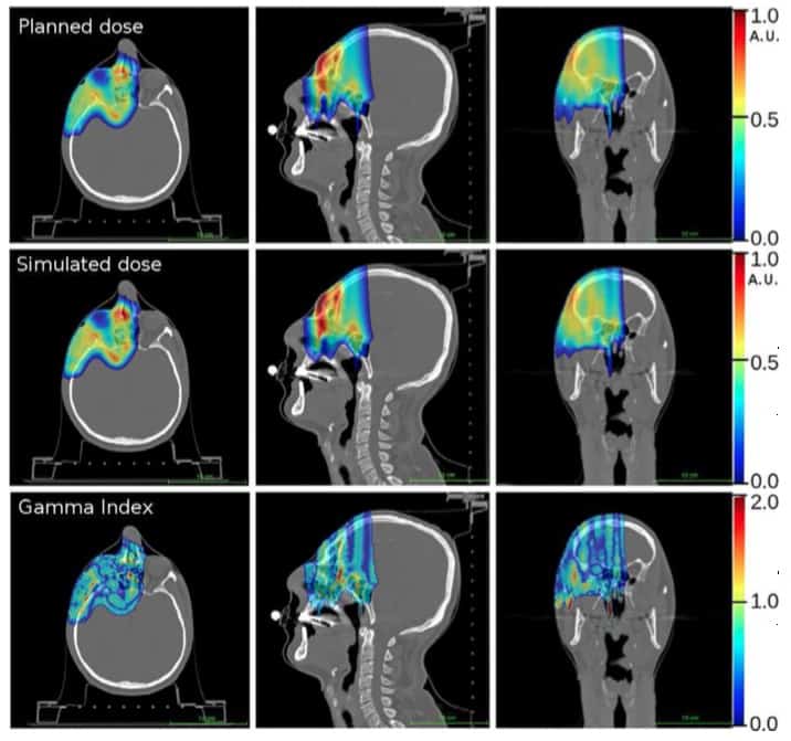
In their latest study, the researchers used their simulation tool in the first analysis of in vivo PET data, acquired from a single patient in December 2016 (Physica Medica 10.1016/j.ejmp.2018.05.002).
By monitoring particle range during treatment, clinics can identify when changes in anatomy produce unacceptable deviations from a patient’s planned treatment. For example, tumours may shrink as they respond to treatment, while specific anatomy such as the paranasal sinuses can contain air or higher density mucous. Armed with such information, clinicians can better exploit the sharp dose gradients that protons and heavy ions provide to target the tumour and spare healthy tissue.
“An accurate Monte Carlo prediction combined with precision imaging would allow the physician to verify the accuracy of the treatment on a daily basis,” said joint first author Elisa Fiorina of the National Institute for Nuclear Physics (INFN) in Turin. In the longer term, the INSIDE technology could potentially be applied for adaptive particle therapy, where treatments are modified for changes in patient anatomy.
The simulation tool generates 4D PET scans using the treatment plan parameters, such as beam energies and spot positions. The patient’s geometry and composition is provided by the CT scan acquired for treatment planning. Based on these data, the tool predicts the propagation of therapeutic particles in the patient and subsequent generation and annihilation of positron-emitting isotopes. The resulting gamma rays are used to construct the PET scan. The tool incorporates models of the beamline at CNAO, the spatial and temporal characteristics of the treatment beam, as well as the geometry and composition of the INSIDE system.
The researchers acquired PET scans during two proton therapy fractions of a 56-year-old patient with carcinoma of the lacrimal gland. Data were acquired over the entire irradiation of one of two fields. Images were updated every 10 s, enabling a visual, qualitative comparison between the measured and simulated data during treatment.
In a comparison with the prescribed treatment plan, dose distributions derived from the simulation proved accurate. Gamma tests demonstrated 91% of voxels agreed to within 3% or 3mm and 98% of voxels to within 5% or 5 mm.
Particle range in the predicted and measured PET images was quantified using iso-activity surfaces corresponding to 10% of the maximum voxel intensity in the scans. The researchers demonstrated that the average distance between the surfaces in the two images was less than 1 mm. The analysis was carried out off-line, but the authors envisage that a real-time implementation will be straightforward, enabling a quantitative analysis while the patient is still being irradiated.
“These first in vivo measurements demonstrate that the developed Monte Carlo simulation tool … is accurate enough to be used as a reference in the PET image analysis,” said Fiorina.
Based on their findings, the researchers are beginning clinical trials later this year, in which the INSIDE system will incorporate a more precise detector positioning system. The trials will include more rigorous tests of the system’s accuracy, including that of the Dose Profiler, using a cohort of around 40 patients. The researchers will also investigate how well the system integrates into routine clinical workflow and potential clinical compliance limits in particle range. The cohort will include individuals with cancers known to respond early to treatment, the group set to benefit most from monitoring.
A couple of years ago I met Andrea Morello from the University of New South Wales at the March 2016 meeting of the American Physical Society in Baltimore. My colleague Tushna Commissariat filmed him answering the question: what could you do with a 50- or 100-qubit quantum computer (see video above).
Well, time waits for no-one, and now Morello has written the guest editorial for a new focus issue of the journal Quantum Science and Technology, which contains five Perspective articles by some of the world’s top quantum-technology researchers on the question: “What would you do with 1000 qubits?” (They’re a bit greedy those quantum dudes, aren’t they?)
The articles, which are all free to read for 30 days (until 19 July 2018), offer “a candid and hype-free illustration of how the field of quantum information science is designing the path towards practical quantum computers”. Now if you think I’m just saying that because Quantum Science and Technology is a journal from IOP Publishing, which also publishes Physics World, then I guess you’ll just have to read them for yourself.
It was the summer solstice this week in the northern hemisphere. About 45 miles from Physics World headquarters, people at Stonehenge celebrated as the Sun rose at 04:51 Thursday morning. According to a new book called Megalith: Studies in Stone, the builders of Stonehenge were using Pythagoras’ theorem 2000 years before the Greek mathematician was born. You can read more in this article in The Telegraph.
Meanwhile in Derbyshire, pupils at Saint Anselm’s School are looking for a Bakewell tart that was last seen dangling from a high-altitude balloon 16,000 m above the English countryside. The pie-in-the-sky was launched by pupils on Monday as a science project to measure temperature, take photos and track the balloon. Unfortunately, all contact with the balloon was lost as it drifted over Saxilby, near Lincoln. The youngsters are hopeful, however, because a balloon launched last year was found on a beach near Skegness.
The UK’s Engineering and Physical Sciences Research Council (EPSRC) has published the results of one of the first public dialogues on quantum technologies. Involving 77 participants from in UK cities, the exercise revealed that the British public is largely in the dark about developments in quantum technologies but can be easily engaged when learning more about them. The survey was carried out in late 2017 by Kantar Media, and the results evaluated by 3KQ.
In the study, a representative sample of the British public was informed about the wide range of devices currently being developed in the quantum technology community. Then, subjects discussed the topic with experts and researchers. As well as engaging participants with the research, the EPSRC aimed use the study to gain new insights into what the public currently knows and thinks about quantum technologies.
Neutral emotions
The results showed that despite the word “quantum” itself being widely familiar, most participants initially knew very little about its meaning and applications; displaying primarily neutral emotional responses towards new developments in quantum technologies. EPSRC concluded that this was likely do to a limited exposure of information about the topic in the media. A small number of participants remained disinterested throughout the dialogue.
However, as most participants learnt more about quantum technologies, they became increasingly curious and excited about them, particularly when gaining an understanding of how the devices could impact their own lives. Many became engaged with the dialogue as they realized the benefits that quantum technologies could have for individuals and society. Potential applications presented to participants ranged from healthcare technologies, to improvements in national and economic security.
Ultimately, the EPSRC aimed to establish a two-way dialogue between experts and researchers, and the public for the first time. By seeking more informed public opinions about the potential uses of quantum technologies, they hope to influence scientists in deciding on the future priorities of their research. The different perspectives of the public also create the potential for revealing new directions in quantum computing research which experts have not previously foreseen.
International cooperation must be improved to prepare for potential strikes by near-Earth objects (NEOs). That is one recommendation of a new report by a group of US government agencies that outlines methods to deal with asteroids and comets that pass within 50 million kilometres of Earth.
The report – National Near-Earth Object Preparedness Strategy and Action Plan – provides an approach to develop technologies, policies, practices, and procedures for US and global vulnerability to NEO impacts. It sets out five specific goals, which include NASA improving its ability to detect, track and characterize NEOs. Those actions, say the 20-page report, should reduce the current levels of uncertainty about potential strikes on Earth and help the development of more accurate modelling and more effective decision-making.
An asteroid impact scenario is a low-probability but a high-consequence event
Leviticus Lewis
Another goal involves NASA and other agencies developing simulation tools to improve the modelling and prediction of NEOs. The report also calls on NASA to develop fast-response reconnaissance missions and technologies for deflecting or disrupting potentially hazardous NEOs. The final two recommendations are for increased international cooperation and a strengthening and routine practice of emergency procedures for dealing with NEO impacts.
NASA officials and other agencies emphasise that, while such impacts have a very low likelihood, they could cause a lot of damage if they were to occur. “We recognize that an asteroid impact scenario is a low-probability but a high-consequence event,” says Leviticus Lewis of the Federal Emergency Management Agency. “An asteroid impact scenario is a unique emergency and may be just different enough that some degree of preparedness specific to this threat is necessary.”
Lindley Johnson from NASA’s planetary defence office says that implementing the plan will “greatly increase our nation’s readiness and work with international partners to effectively respond should a new potential asteroid impact be detected”.
The report, however, does not provide any specific funding for the actions it recommends. “Most of the actions we’re calling for are things that can be done within existing resources that are already allocated,” says Aaron Miles of the White House Office of Science and Technology Policy. “This is more about figuring out how to use those resources smartly and do so in a coordinated and cooperative way across the US government.”
Peter Gwynne is Physics World’s North America correspondent
Analysis: More needs to be done to study possible impact scenarios
Monitoring near-Earth objects (NEOs) is hardly a new pursuit. NASA has studied them for almost five decades and in the late 1990s the agency expanded its effort by creating a project called Spaceguard to specifically search for them. Since then, several dozen have passed between Earth and the Moon.
The importance of planning for such events was evident on 15 February 2013 when the Chelyabinsk meteor – a 20 m-wide asteroid – exploded over Russia. Its shock wave damaged more than 7000 buildings and indirectly caused about 1500 people to seek medical care. Indeed, the event created the largest explosion on the planet since the one that occurred over the Tunguska river in Siberia in 1908, entering the atmosphere with a mass of 13,000 tonnes.
US congressional hearings following the event led to NASA and other agencies speeding up their plans for effective asteroid detection. Later that year, the space agency rebooted its then dormant Wide-Field Infrared Survey Explorer to begin a three-year mission to search for NEOs and in 2016 NASA created its Planetary Defense Coordination Office to detect, monitor, and characterize all NEOs.
While much work has been focused on characterizing NEOs, more needs to be done to understand how to deal with potentially threatening bodies. In 2015, NASA and the European Space Agency came together to work on the Asteroid Impact and Deflection Assessment (AIDA) mission. This involved two craft travelling to a binary asteroid system called Didymos. One probe, built by ESA and called AIM, would study the composition of the asteroids, while the other – dubbed the Double Asteroid Redirection Test (DART) – would impact into it. Once DART fired into the asteroid, AIM would have studied how the body was affected.
Yet following budget issues, ESA cancelled AIM leaving NASA to go it alone. That means that the asteroid impact of DART, once it arrives at the asteroid in 2022, will be monitored from ground-based telescopes and radar rather than a dedicated mission. ESA is, however, planning to send a mission to Didymos that will arrive three years after DART has impacted.
The report is a timely reminder that dealing with NEOs should be an international endeavour and not just the responsibility of NASA.
Michael Banks is news editor of Physics World magazine
Gases can permeate through 2D channels made from materials like graphene and boron nitride much faster than predicted by theory. The effect can be explained by “specular surface scattering”, which leads to frictionless flow, and it could be exploited in applications such as filtration and flow control.
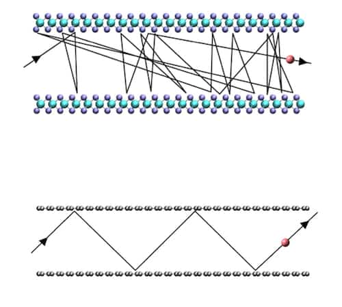
“Gas permeation through nanoscale pores is ubiquitous in nature but it also plays an important role in many technologies,” explains Boya Radha of the University of Manchester in the UK who led this research effort together with Nobel laureate Sir Andre Geim. “Since the size of the pores is usually smaller than the mean free diffusion path of gas molecules, we can describe this flow by conventional Knudsen theory. Here, the diffusing molecules randomly bounce back (or scatter) from confining channel walls, which reduces their flow.
“In channels with atomically-flat walls, however, such as those made from graphene (a 2D carbon sheet) and boron nitride (BN), which are flat at scales of 1 ångström (10-10 m) this theory breaks down. This is because the diffusing molecules only rarely scatter from the walls and so essentially permeate through the channel as if it weren’t there.”
Graphene is flattest
Geim, Radha and their colleagues obtained their results by measuring the rate at which helium gas diffuses through ångström-scale slit-like channels with walls made from cleaved graphite, hexagonal BN and molybdenum sulphide (MoS2). All these materials can be exfoliated, or thinned down, to monolayer thicknesses and have atomically flat surfaces. In the experiments, the researchers made channels using two thin (roughly 10–100 nm thick) crystals of each of the materials and used these as the bottom and top walls of a channel. They plasma etched a third thinner crystal so that it contained long narrow trenches. This crystal plays the role of a spacer between the top and bottom walls.
“We assembled the three crystals, which are held together by van der Waals forces, on top of each other,” explains Ashok Keerthi, who is first author of the study. “We could choose the spacing between them to be just one atomic layer thick or up to as many layers as we required.
“We found that the rate of helium transport is fastest through graphene and slowest in MoS2. Although all the 2D materials we studied are atomically flat, the minute differences in the atomic “corrugations” of graphene, hBN and MoS2 can be ‘felt’ by the scattered helium molecules. This can only be explained by quantum effects, that is the wave-like nature of matter (helium).”
In fact, when the (de Broglie) wavelength of helium is much larger than the atomic-scale roughness of the wall surface, as is the situation for a wall made of graphene, it is specularly scattered. Such scattering leads to frictionless gas flow and ballistic transport. In contrast, helium permeates through MoS2 at a rate predicted by Knudsen theory because it is rougher at atomic scales than graphite (and hBN). This roughness in fact comes from sulphur atoms protruding in between Mo atoms. These corrugations are almost an ångström tall, which is around the same diameter as the wavelength of helium molecules.
Confirming the matter-wave effect
The Manchester team backed up this matter-wave effect by measuring the rate at which deuterium (hydrogen’s heavier isotope) permeates through 2D channels made from graphene and hBN. “We found that hydrogen permeates faster than deuterium, even though it should be the opposite according to the classical Knudsen description,” says Geim. “While the size of both hydrogen and deuterium molecules are the same, their de Broglie wavelengths are not. The de Broglie wavelength of hydrogen is bigger than deuterium’s, and this leads to increased specular reflection of hydrogen from the channel walls and thus faster diffusion.”
These results back up previous findings from experiments on gas transport in various atomically smooth nanochannels, such as carbon nanotubes, nanoporous films made from graphene, graphene oxide and other 2D materials, adds Radha.
The researchers, reporting their work in Nature 558 420, say that they would now like to study size-selective separation of gases in even thinner channels. “While small molecules pass through these channels at high speed thanks to specular rather than diffusive scattering, the larger ones should be excluded due to steric effects,” she explains. “This could come in useful in filtration technologies since we would have fast flow and size exclusion at the same time in one system.”
Dentists are fascinating people. Multiskilled and multidisciplinary by nature, these renaissance women and men of our time need to be consummate and compassionate communicators, able and adaptable scientists, and skilled and artistic creators. Dentists need excellent communication skills to advocate smoking cessation, counsel people on maintaining good oral health, and treat anxious patients with compassion and care. Because dental technologies and treatments are in a state of continual evolution, dentists must also be scientifically literate, critical thinkers; it is essential for clinicians to stay up to date with developments throughout their careers. Since dentists restore teeth that have become unsatisfactory for many reasons – including pain, loss of function, or simply being unsightly – they must be artistically as well as technically skilled, and able to intervene in an environment that is cramped and full of nerve endings. And contrary to what you might expect, their involvement in healthcare is not limited to tooth and gum care: dentists are in fact gatekeepers of whole-body health, as oral problems and symptoms sometimes indicate systemic health conditions.
Tough materials
For a materials scientist like me, though, the most fascinating thing about dentists is that, whether they realize it or not, they are discerning and meticulous materials engineers. Materials underpin so much of a dentist’s daily life: whether they are filling cavities; fitting dentures, crowns and sports mouthguards; or applying tooth-whitening treatments, many of their standard processes require polymers, ceramics, alloys or other advanced materials. Rarely will a dentist pass a day in the clinic without using some sort of material that must function in intimate contact with the hard and soft tissues of the mouth.
The strictures that dentistry places on these materials are also unusually tough. Like any biomaterial, a dental material must be biocompatible – that is, it must not elicit any adverse reaction, and it must be stable and maintain its function for the required period (which could be anything from a few minutes to the lifetime of the patient, depending on the application). But over and above those standard requirements, the material must also be able to survive in a hostile environment: the mouth. As well as constant moisture, parts of the mouth experience pH values that range from near-neutral to less than 3.0 (cranberry juice, for instance, has a pH of ~2.8); temperatures that run from close to 0 to around 60 °C; and pressures of tens of MPa in normal function. Natural tooth tissues – the enamel and dentine – are well equipped to withstand damp conditions with huge variations in force and temperature, but they are less able to cope with the modern low-pH diet, which is a comparatively recent addition in evolutionary terms. Indeed, the tendency of hydroxyapatite – a calcium-containing mineral that makes up 70% of the dentine and more than 90% of enamel – to break down in the presence of acids is known to be the root cause of tooth decay: the acids produced by plaque bacteria cause a slow but steady dissolution of tooth mineral, ultimately leading to the formation of a cavity.
The biomaterials used in dentistry must, therefore, be carefully engineered to be strong in compression, tension and flexure. They must not corrode or otherwise deteriorate when wet, and they must be able to resist frequent and rapid changes in temperature and pH. In recent years the commercial aspects of dentistry have also driven more stringent requirements regarding the physical appearance of the material. In colour, translucency and fluorescence, patients are increasingly demanding materials that are indistinguishable from the adjacent natural teeth, to the naked eye at least.
Freedom to choose
Of course, all of these materials come with a price tag, and those with the best balance of physical, mechanical, chemical and biological properties can be very lucrative for manufacturers. This drives innovation, because dental materials are big business: the global market for dental restorative materials and adhesives was estimated at just over $1bn in 2016, with a compound annual growth rate of more than 5%. Equally important is the fact that dentists are able to innovate; even dentists who work in publicly funded health systems have a degree of choice when selecting materials, and in private dentistry that choice is even wider. This means that dentists can be, and often are, early adopters of new technologies. It is thus in the interest of dental materials manufacturers to continually innovate and release new materials onto the market in a bid to capture this enthusiasm for the “new and improved”; complacency leads to loss in market share.
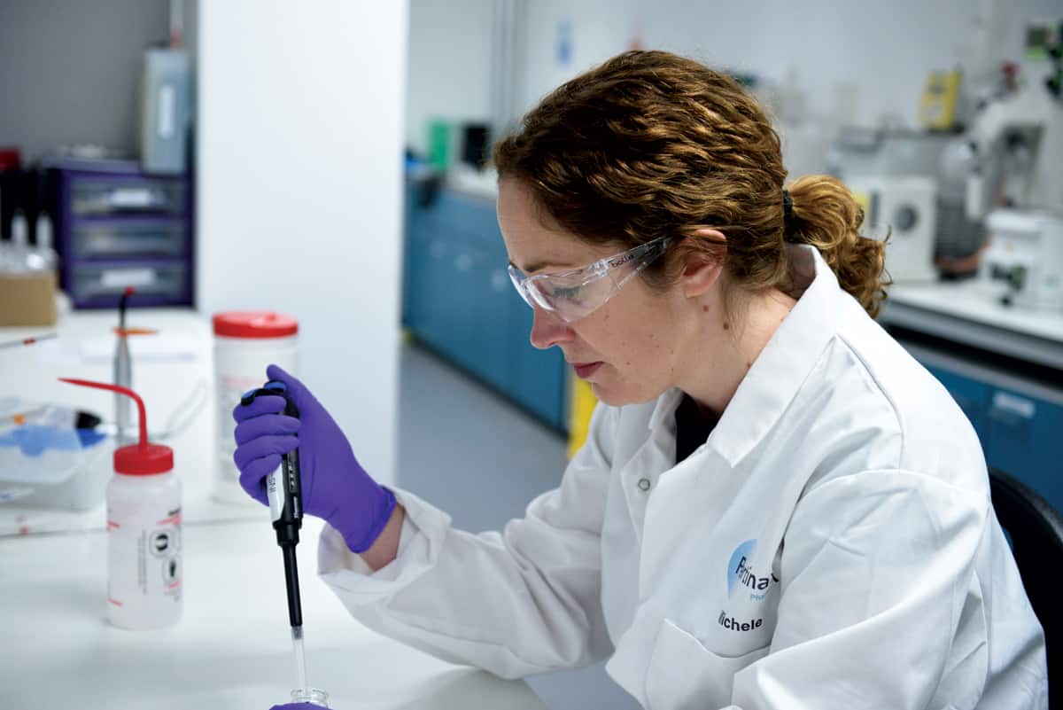
Developing new materials: The author in her lab. (Image courtesy: Pertinax Pharma)
This combination of factors creates a niche occupation: the dental materials scientist. This is the niche I found myself occupying at the outset of my academic career 12 years ago. The dental sector appealed to me because I wanted the materials I develop to have a direct and near-term impact on people’s day to day lives – both the clinicians who use the materials and the patients who benefit from the clinician’s skill. I also came to appreciate the culture of progress and innovation that has grown up around dental materials owing to dentists’ opportunity to choose. This opportunity is significantly greater for dentists than it is for, say, orthopaedic surgeons or urologists, who have a more limited portfolio of materials to choose from when installing a hip implant or a urinary catheter (to take just two examples).
Over the past decade or so, the field of dental materials has seen several interesting developments. The mechanical strength of light-cured polymer composites – which are used to restore teeth in cases where a good aesthetic outcome is essential, for example if the teeth are near the front of the mouth – has improved considerably, and innovations in polymerization methods have reduced the amount of shrinkage usually associated with similar polymers. The field has also been rocked by the Minamata Convention, a legally binding treaty that commits signatories to ceasing the use of mercury across almost all industries. The convention has been signed by 84 countries, and its adoption puts amalgam, an important tool in a dentist’s armoury, under threat.
Smarter infection control
In my opinion, though, the most interesting developments have been in the field of antimicrobial dental materials, where my own research lies. The starting point for our work is a common and widely used antiseptic called chlorhexidine. Dentists use chlorhexidine as a multipurpose antimicrobial for treating gum disease and oral infections; it is also applied preventively before dental surgery. Other uses in the wider medical world include skin disinfection (again before surgery), combatting MRSA outbreaks in hospitals, and as a component of wound dressings for chronic or surgical wounds. Chlorhexidine is used in veterinary practice as well, in wound care and to treat infections of the mouth and ear. Regardless of how they are applied, though, all current chlorhexidine products suffer from the same limitation: the chlorhexidine salts used are highly soluble, meaning that when they are placed in a damp environment, the local liquids rapidly remove the chlorhexidine. For this reason, its activity is limited to a short period after application and it needs to be reapplied regularly, at relatively high concentrations, to provide any lasting protection.
Our approach to this problem is a simple but very effective modification. By sequestering the chlorhexidine in salts of condensed phosphates, we create a material with much lower solubility, one that is stable when dry and that releases chlorhexidine at a steady (close to linear) rate in a wet environment. Naturally, given our background, our first thoughts were to exploit this new material in dentistry. The main cause of failure of modern dental fillings is secondary infection, where bacteria infiltrate the interface between the tooth and the filling material. By incorporating our sustained-efficacy chlorhexidine into the materials used at this interface, we can create a locally chlorhexidine-rich environment with a clinically optimal dose: sufficient to kill any microbes that penetrate, but not to adversely affect human cells. We can even incorporate a degree of “smart” behaviour, whereby the chlorhexidine release is accelerated in response to a fall in pH, which would indicate the very earliest stages of tooth decay in the area.
But of course, since chlorhexidine is also used widely in medicine and veterinary care, we have opportunities to exploit our technology in those areas as well. Although the material’s genesis was in a dental environment, our spin-out company Pertinax Pharma is now developing wound-dressing materials with a lasting antimicrobial effect for conditions such as burns and diabetic foot. The slow release of the antiseptic means that our prototypes do not elicit the adverse response from human cells associated with high doses of chlorhexidine and other antiseptics. We are also now developing a spray-delivery, resilient, water-resistant film of our material that could find applications in diverse fields. It could be used to prevent umbilical-cord infection in newborns, for example, especially in developing countries where the material’s low cost, long-term efficacy and benign storage requirements (with the consequent ease of transport) make it an appealing option. This material could also be used in an actual field, to treat infectious foot disease in cattle.
These are the applications I find the most exciting. There is a great deal of overuse and misuse of antibiotics, and although reducing this (and managing or combatting the antibiotic resistance that results from it) will require a multidisciplinary approach, the development of technologies such as ours – which can be used in place of antibiotics to give a clinically equal or superior outcome – can certainly play a part. I hope to see materials such as our novel chlorhexidine salts make a substantive difference to the global community not only via innovative dental materials that can protect against tooth decay, but in the wider fields of medicine and dentistry where sustained-efficacy antiseptics can serve a multitude of purposes.
A team of clinicians at the University of Wisconsin have used a ViewRay MRI-guided radiotherapy system to perform brachytherapy planning for cervical cancer. So, what motivated the team to use the ViewRay system to perform brachytherapy planning? And how does use of the system fit time-wise alongside its use for treatments?
Time saving
Writing in the journal Brachytherapy, the authors present the results of some 142 fractions of intracavitary brachytherapy, performed between April 2015 and January 2017 on 29 cervical cancer patients – the first ever clinical use of ViewRay-guided brachytherapy (Brachytherapy 10.1016/j.brachy.2018.04.005). They conclude that “time to treatment using this approach was shorter compared to diagnostic MRI” and that the ViewRay system also provided “significant advantage in visualizing the tumour and cervix compared to CT”.
As co-author Cindy Ko, a radiation oncology resident at the University of Wisconsin School of Medicine and Public Health, explains, the motivation to undertake this research came from necessity. This is because, although Ko and her team typically employ a diagnostic quality 1.5-3T MRI at diagnosis and for “at least the first fraction” of brachytherapy – a quality of imaging that they would ideally use for every fraction of brachytherapy planning – a combination of time constraints and limited resources means that they elect to use such MRI levels for only the first and third treatments.
“We used the imaging component of the ViewRay treatment system, which is in and of itself an independent treatment machine that can be used to directly visualize and treat cancers of the lung, gastrointestinal system [and others] with MRI guidance,” says Ko.
“The ViewRay system is within our department, unlike the diagnostic MRI machines, and thus it can save some travel time between the procedure suite, imaging, and back to the procedure suite for treatment,” she adds.
In addition to the benefits of proximity, Ko reveals that the ViewRay MR image acquisition is also faster, “on account of not requiring multiple sequences”. This is because there is only one proprietary sequence officially available for use with the system. This fast sequence, known as TRUFI, is a hybrid of T2 and T1 sequences that the team uses both with and without contrast for its treatments on the ViewRay system.
Scheduling
Since beginning to use the system in the department, Ko and her team found that scheduling a time to use the machine for brachytherapy planning between treatments worked well. Given the unpredictability of procedures, Ko reveals that they tend to “pink slip” individual brachytherapy cases into a block of time on the ViewRay system’s schedule – and continue to treat or simulate scheduled patients until the brachytherapy case is nearly ready. They will then “image [the case] briefly before going back to the other treatments or simulations on the schedule”.
According to Ko, this requires the therapists to develop a familiarity with how the logistics of a ViewRay imaging session with a brachytherapy patient works, as well as a working knowledge of how long the imaging session will take.
“We don’t find that one use of ViewRay system hinders the other – meaning that the schedule is mostly on time using this approach – and we are happy to find multiple uses for the same machine. The ViewRay schedule is busy, but no busier than our other treatment machines,” she says.
Ko also points out that the brachytherapy instrument placement itself “does not require much more than an ultrasound” for the first fraction, although for some patients, placement can take an hour or more.
In view of the fact that performing instrument placement within the ViewRay suite would probably interfere with the normal ViewRay treatment schedule – during which time the imaging function would not be used – the team found that transferring to the ViewRay system after instrument placement, and then adjusting if needed based on the MR image, was sufficient.
“Very seldom do we have to adjust instrument placement after viewing the ViewRay, MRI [or] CT images, but it can happen and, if so, we image again after adjusting to ensure good geometry,” adds Ko.
US-based researchers have confirmed that they can detect the same glow – invisible to the human eye – from trees, grasslands, crops, mangroves, marches and desert plants as the green things put chlorophyll to work and photosynthesise leaves, flowers, fruits and roots from atmospheric carbon.
The pay-off is simple: an easier and potentially more accurate way of calculating the global carbon budget and assessing the climate cost of human exploitation of fossil fuels.
But the same information will help biologists and geoscientists advance what is sometimes called earth system science: how carbon-based lifeforms make their living from sunlight, water and carbon dioxide in a continuous trafficking that has fuelled three billion years of evolution.
And at the heart of the study is a new realization that images from an orbiting satellite deliver better information in a reliable fashion.
Researchers from the University of New Hampshire report in the journal Global Change Biology that years of observation of solar-induced fluorescence – a glow from plants that no human could expect to see, but an instrument can detect – have confirmed that there is a direct relationship between gross primary productivity and the amount of fluorescence registered by the eye in the sky.
No exceptions
It means that what is true for the canopy of tropical forests in the Congo would also be true for a landscape of maize in the American mid-West, or the grasses and wildflowers of the savannah, the dusty maquis of the Mediterranean, or the swamps of the Louisiana bayous.
Up till now, researchers have tried to make accurate and reliable estimates on the ground, playing with air temperature, sunlight, rainfall and other factors to arrive at their conclusions about what they like to call carbon “sinks.” The message from OCO-2, the NASA orbiting carbon observatory, is that the gleam from the foliage below provides an answer more swiftly, and perhaps more surely.
“The importance of these results is that rather than look at several different types of data and computer-based models from information collected on the ground to monitor plant photosynthesis across the globe, using the satellite observations will provide a near real-time option that is simple, reliable and fast,” said Jingfeng Xiao, of the University of New Hampshire, the chief investigator.
“This is a big step towards being able to solely rely on satellite measurements.”
Researchers long suspected that the dielectric constant of water is lower at interfaces with other materials – what no-one knew was how much. “This is a huge issue,” says University of Manchester condensed matter lecturer and National Graphene Institute researcher Laura Fumagalli. “The value of the dielectric constant at the nanoscale was not clear at all and it has a lot of impact on a lot of phenomena.” These range from the study of proteins and DNA to electrochemistry and batteries. Now Fumagalli has teamed up with 2010 Nobel Laureate for the discovery of graphene Andre Geim, as well as colleagues in the UK, Iran, Spain and Japan, to report experimental evidence that the effect of interfaces on the dielectric constant of water is far greater than previously suspected.
The dielectric constant gives a measure of how well electric dipoles of molecules orient in an electric field. Water is a highly polar substance, so although the molecules can readily reorient in an electric field in the bulk, their alignment at surfaces can be inhibited, potentially diminishing the dielectric constant in interfacial water near surfaces compared with values found in bulk water. Establishing definite values for these effects has flummoxed researchers for decades.
Dielectric measurements get ultrasensitive
Fumagalli has long specialized in investigating the dielectric properties of structures at the nanoscale. In 2012 during her time at Institut de Bioenginyeria de Catalunya and Universitat de Barcelona, Fumagalli and colleagues in Barcelona and Madrid reported on a technique using electrostatic force microscopy with piconewton sensitivity that could identify nanoparticles with identical shape but different chemical composition by ultrasensitive measurements of the dielectric constant.
These experiments did not focus on water, but as Fumagalli points out, “Water is everywhere, even where you don’t want it there is a layer of water from the humidity of the environment.” Yet while her interest was piqued, applying the technique to water proved far from trivial. The success of the latest experiment hinged on Geim’s expertise in 2D materials.
Confinement device yields success
One of the challenges was producing a system to confine water at the nanoscale. Happily one of the many things 2D materials are good at is trapping water, so when Fumagalli joined the National Graphene Institute at Manchester, and spoke to Geim about the problem, a solution proved to be in sight.
“We started with something simpler but the results were not so convincing,” says Fumagalli. “We clearly needed the most advanced devices, and Andre Geim was able to produce them.” She describes how in 2016 and 2017 Geim introduced a new technology that allows the assembly of two-dimensional materials into devices with the smallest possible man-made channels. “Among many other possible applications, these devices allow us to study the transport and properties of water inside such tiny channels.”
The final system comprised slit-like channels fabricated from atomically flat crystals of graphite and hexagonal boron nitride. The researchers could set the heights of the channels to be as low as one nanometre in size so that they only accommodated a few layers of water.
“When you reduce the quantity of water you have, and the water is confined near surfaces so that you have just a few molecular layers there, the molecules are not free to move like in bulk water, and the dielectric constant goes down to two,” says Fumagalli. “This is the minimum value imaginable – so low people had not expected it.”
She highlights that this anomalously low value of the dielectric constant in confined water is in stark contrast to the anomalously high dielectric constant of bulk water, which is around 80. “Water is full of anomalies,” she adds.
Result’s impact reaches far
Water, described as the universal solvent because so many other substances are soluble in it, has been dubbed “the solvent of life”. These solvation properties are directly linked to the dielectric constant, which highlights the impact of these results. No small wonder then that the researchers in Manchester remain very interested in water.
“It would be interesting to see if it behaves similarly with other surfaces and how it behaves near bio surfaces,” says Fumagalli. “How water is polarized near biomolecules makes a difference to the forces they experience and has a huge impact on their structuring and functions.”
Geim also emphasized the significance of the results in a press statement: “This anomaly in the dielectric constant of interfacial water is not just an academic curiosity but has clear implications for many fields and for life sciences, in particular. Our results can help to improve the understanding of the role of water in technological processes, and why it is so crucial for life. Electric interactions with water molecules play an important role in shaping biological molecules such as proteins. One can probably claim that interfacial water shapes life as we know it, both literally and figuratively.”
Other substances that may be subject to the same effect include any polar liquids. Studies of those used in batteries for energy storage may have particular relevance for industry.