Flexible nanotubes pack a punch

Since they are flexible, high-aspect-ratio nanostructures store elastic energy. When released, this energy could be used to destroy bacteria by physically stretching and rupturing their cell membranes. This technique was inspired by the bactericidal nature of insect wings and destroys both Gram-positive and Gram-negative bacteria at extremely high rates. The nanostructures could make for a new type of highly efficient mechano-responsive antibacterial surface.

Antibiotic resistance is an increasing problem worldwide. In Europe, for example, around 25,000 people die every year from resistant bacterial infections. Without new antibacterial agents, routine medical procedures and operations could soon become impossible.

Antibacterial surfaces are used to prevent the formation of bacterial biofilms on a variety of surfaces, including medical tools and implants. Until now, these generally worked thanks to a chemical coating that slowly releases a biocidal agent and kills bacteria that come into contact with it. This approach does have some drawbacks, however, with the main one being that bacteria may develop resistance over time.

Cicada wings are natural bactericides

A team of researchers led by Elena Ivanova of RMIT University in Melbourne, Australia, recently discovered that cicada wings, which are covered with arrays of pillars roughly 200 nm high and 60 nm in diameter, are highly bactericidal. This property is purely physico-mechanical – the bacteria cell membranes stretch and rupture between the pillars, which is the point subjected to the highest mechanical stress.

The researchers found that the bactericidal efficiency of the nanostructured surfaces depends on the spacing and size of the nanofeatures. This suggests that the nanopatterns can be modified to increase the surfaces’ antibacterial properties depending on which type of bacteria need to be treated. Gram-negative bacterial cells like Pseudomonas aeruginosa, for example, are destroyed relatively quickly on cicada wings, while Gram-positive bacteria such as Staphylococcus aureus are more tenacious since they resist the stretching process better. The differences in the two types of bacteria might come from the mechanical properties of the bacterial cell membranes themselves.

Inspired by these natural nanostructures, Ivanova and colleagues decided to design similar surfaces using high-aspect-ratio vertically aligned carbon nanotubes (VACNTs) grown by chemical vapour deposition as the pillars. Carbon nanotubes are very attractive for biotechnology applications thanks to their high strength and stiffness and high electrical and thermal conductivity. VACNTs, which share many of the same properties as CNTs, are also highly flexible and resilient, and can be easily functionalized using a plasma treatment. They can be made in different lengths by adjusting growth times, which allows their bending stiffness, and thus the amount of elastic energy they store, to be controlled.

Shorter nanotubes are best

When the VACNTs come into contact with a bacterial cell, they bend and release their elastic energy. This bending stretches the bacterial cell membrane and destroys it, so killing the bacteria.

The researchers observed the bactericidal effect of the nanotubes using a variety of techniques, including focused ion beam-scanning electron microscopy (FIB-SEM) and scanning transmission electron microscopy (STEM).

“We found that shorter nanotubes around 1 micron in length are more efficient at inactivating bacteria thanks to their ability to store and release more elastic energy compared to longer tubes,” says Ivanova. “We achieved bactericidal rates of as high as 99.3% for P. aeruginosa and 84.9% for S. aureus by modifying the lengths of the VACNTs, which allowed us to determine the optimal length for efficiently killing different types of bacteria.”

A new platform to combat antibiotic resistance

“Modulating the CNT characteristics in this way will further improve our understanding of mechano-bactericidal mechanisms. It also shows that the antibacterial activity of high-aspect ratio nanofeatures can outperform both natural bactericidal surfaces and other synthetic nanostructured multifunctional surfaces reported previously.

“Indeed, we believe that such high-aspect ratio features are promising as a new platform to combat antibiotic resistance in pathogenic bacteria,” she tells Physics World.

The team, reporting its work in ACS Nano 10.1021/acsnano.8b01665, says that it is now busy optimizing the height of its high-aspect ratio nanopatterns to make them more efficient against a wider range of bacteria.

Implanted antennas track fracture healing

Monitoring the healing of severe bone fractures is important because patients with such injuries are particularly susceptible to complications. Delayed bone healing that is not treated in a timely manner may cause severe pain and, ultimately, a loss in muscle strength and mobility.

The first few weeks following trauma influence whether the fracture will heal properly. The current solution is to wait until some evidence of successful healing is seen, such as the ability to bear weight. Despite over 40 years of scientific studies, monitoring non-union fractures is still a challenge. In fact, the remaining bottleneck in fracture care is the lack of clinical tools that enable direct, quantitative assessment of fracture healing and early diagnosis of non-union conditions.

Several approaches have been proposed to quantify fracture healing in long bones, ranging from mechanical vibrations to electrical impedance. Both in vitro and in vivo experiments have demonstrated the capabilities of different techniques to monitor the properties of callus (bony tissue that forms around the ends of broken bone) during healing, and differentiate fractures that heal from those that fail to heal. None of these, however, translated into clinical practice, mostly because of the complexity of the musculoskeletal system surrounding the fracture site.

Study authors

To address this shortfall, researchers at Loughborough University are developing a novel technique for monitoring bone fractures healing by harnessing electromagnetic-related properties of bone tissue. They hypothesized that if the fixator screws implanted to align and support bone fragments could double up as monopole antennas, they could transmit a radiofrequency (RF) signal from one monopole to the other through the fracture, and thus monitor the unification process over time (Biomed. Phys. Eng. Express 4 045006).

Phantom studies

To determine the potential of an RF antenna system for monitoring bone fracture healing, lead author Symeon Symeonidis and colleagues created a proof-of-concept monopole antenna system and tested it on three heterogeneous bone phantoms.

Prior research showed that the permittivity and conductivity of blood are significantly higher than that of bone across a major part of the RF spectrum. The authors hypothesized that the reduction of blood inside and around the fracture, and the formation of callus during the healing process, would reduce losses in an RF signal propagating from one monopole to the other. Thus it should be possible to track healing by measuring the power transmitted from one monopole antenna to the other (S21).

The prototype fracture monitoring system consisted of two 4 mm-diameter monopole antennas, the size of an average fixator screw, containing a 2.5 mm diameter conducting part covered by biocompatible polymer insulation. The researchers created varying lengths and groundplane sizes, but each had a 5 mm gap of free space between the muscle tissue and groundplanes to prevent contact with the highly conductive muscle layer.

Bone phantom

The researchers developed tissue-mimicking materials for blood, bone cortical and muscle, and used these to create phantoms representing radius, phalange and tibia bones. They measured S21 as blood emulating liquid was injected inside the phantom emulating the conditions of a bone fracture.

For comparison, they simulated bone fractures in a voxel model of a 26-year-old female, with a fracture represented by two coaxial cylinders. The inner cylinder was injected with blood-simulating tissue to represent the dielectric properties of various stages of healing. The outer cylinder surrounding the “bone” was given the dielectric properties of blood. They simulated the maximum amount of haematoma at the initial fracture state, and gradually reduced it until it was removed entirely.

Finally, the team conducted an ex-vivo measurement using a lamb femur bone. Extensive testing confirmed that, in all cases, the power transmitted from one monopole antenna to the other through the fracture decreased significantly as the volume of blood increased.

The authors note that the pattern of change of S21 could be observed in much of the frequency range of 1 to 4 GHz. Monopole lengths could easily be adapted to fit the diameter and depth of any human bone likely to have external fixators. Because no two fractures are identical, considerable variation in S21 magnitude is anticipated. However, if a healing time could be estimated, the rate of change of S21 would expect to slow over this period. This information could indicate that bone healing had stalled.

While initial results are highly promising, there is still a long way to go before this technique can be used in clinical trials. Further activities will include comparison with other, more traditional techniques, moving slowly away from the workbench into clinical assessment.

Innovation: patent applications review

A round-up of the latest international patent applications in medical imaging.

Breast scanner combines microwave and ultrasound imaging

UK diagnostic imaging company Micrima has developed a combined microwave and ultrasound imaging system (WO/2018/078315). The device comprises a microwave antenna array and an ultrasonic array, with the ultrasonic transducers interspersed between the microwave antennae. The arrays may be formed on a hemispherical substrate contoured to conform to the body part. A processor receives the microwave and ultrasonic signals and generates an image of the internal structure of a body part, such as a breast. The microwave antennae and ultrasonic transducers may be wavelength-matched to allow similar data capture, processing and rendering for the two modalities. An actuator can rotate the arrays to position the antennae and transducers at previously unoccupied positions, thus creating a virtual array with a higher density of transceivers.

Electrical impedance tomography keeps track of neonatal lung function

Swisstom has published details of an electrical impedance tomography (ΕIT) system that non-invasively monitors lung function in neonates, accurately and in real time (WO/2018/085951). The system comprises an electrode array that’s positioned on the patient to measure impedance distribution. To simplify positioning of the electrode belt, the array contains at least one visual aid that visually indicates the position of at least one electrode or electrode pair. The system also includes a data entry unit that accepts data describing the position of the visual aid, as well as a unit that calculates the position of the individual electrodes relative to the patient’ s body and provides correction for the image creation algorithm, thereby enabling computation of reliable ΕIT difference images.

GRE metric creates accurate synthetic CT images

Researchers at Memorial Sloan Kettering Cancer Center have devised schemes for creating synthetic CT images from MR images, enabling MR-only planning of radiotherapy for cancer patients (WO/2018/089753). The approach involves application of a generalized registration error (GRE) metric that determines the goodness of local registration between MR or CT image pairs. The methods also feature image processing techniques that improve the similarity between CT and MR images prior to CT-MR image registration, as well as standardization of the MR intensity histograms prior to MR-MR registration. Application of these techniques results in more accurate assignment of the Hounsfield unit to each point in the synthetic CT compared with other atlas-based methods, providing more accurate dosing in MR-only radiotherapy simulation and planning.

PET images model the human heart

Aarhus University researchers have described a method for modelling a human heart and/or its chambers and cavities (such as the left and right atrium) based on a series of PET or SPECT images (WO/2018/086667). Each image represents concentrations of a tracer that was injected at a specific time. The method involves extracting tracer time-activity curves for a number of pixels/voxels, and identifying first-pass peaks of these curves, each peak corresponding to an arrival time of the tracer at the corresponding pixel/voxel. The system then defines a model comprising at least two portions of the heart, isolated by selecting pixels/voxels based on comparisons of the first-pass peaks against a threshold. Finally, the portions are arranged in relation to each other by comparing the arrival times of the first-pass peaks of the pixels/voxels therein. The result is a segmented model of the human heart, which can be used to estimate the volume of the left or right atrium.

Multi-energy CT quantifies luminal stenosis

A team at the Mayo Foundation has designed a system for determining the severity of stenosis (narrowing) in a subject’s vasculature using multi-energy CT (WO/2018/098118). The method includes acquiring multi-energy CT data using a CT system, performing a material decomposition process on the acquired data to generate one or more material density maps, and selecting – based on these maps – one or more regions-of-interest (ROIs) encompassing at least one vessel cross-section. The method also includes measuring iodine content in the ROIs, and determining a stenosis severity based on the measured iodine content.

Bioresin helps print biological tissue

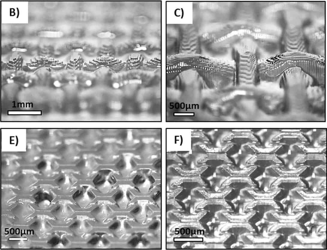
Microscope images of DLP-printed 3D constructs

Most biofabrication techniques make use of bioinks – hydrogel structures laden with cells and extracellular matrix components – that are extruded layer-by-layer to engineer functional biological tissues. But researchers are also developing cell-laden bioresins for lithography-based bioprinting technologies, such as stereolithography (SLA) or digital-light processing (DLP). These techniques could offer an attractive alternative, since they can create more intricate printed patterns that better mimic the complex tissue architecture.

A team led by Tim Woodfield at the University of Otago Christchurch in New Zealand and collaborators at University Medical Center Utrecht in the Netherlands has now developed a new bioresin that allows them to use DLP to bioprint sophisticated cell-laden hydrogel structures with high-resolution features ranging in size from 25 to 50 microns. “Such structures cannot be fabricated using extrusion bioprinting,” says Woodfield.

The resin is made from two different types of hydrogel (PVA-MA and Gel-MA) and a photoreactive compound that initiates a chemical reaction when illuminated with visible light.  When the resin is biofunctionalized with 1 wt% Gel-MA, more than 90% of the bioprinted cells survive for up to three weeks. The seeded cells also attach to the gel and successfully spread across it.

Bioresin opens up lithography

The DLP fabrication technique exploited by the team uses a digital micro-mirror device to project a patterned mask of light (usually in the UV or visible wavelength range) onto the bottom surface of a polymer resin bath. Specific regions of the resin are polymerized when exposed to light, and the platform then moves upwards as resin flows to produce a fresh layer before the process it repeated.

“Lithography-based fabrication technologies have long been used in jewellery making and in the automotive industry, for example, using a range of commercially-available resins,” explains study lead author Khoon Lim. “These resins often contain organic solvents or toxic chemicals and require photo-initiators that are only soluble in toxic organic solvents. To make our resins ‘bio’, we employed a combination of macromers (photo-responsive PVA-MA and Gel-MA) and the photo-initiator ruthenium. All these components are water soluble and not cytotoxic to cells.”

Best of both worlds

Lim explains that the macro-component in the bioresin is a mixture of synthetic and biological polymers, so it boasts the best of both worlds. “The PVA-MA has versatile physical and mechanical properties that we can tailor with no batch-to-batch variation,” he says. “And we know that PVA hydrogels are good candidates here because they have previously been used for multiple tissue engineering applications, including neuronal, cartilage and bone.”

The ruthenium-based photo-initiator is also highly efficient at the custom wavelength of 400–450 nm of the DLP machine, which allows the researchers to accurately produce hydrogel structures with superior spatial resolution. What’s more, the structures can be created with a commercial printing machine.

Controlling cellular behaviour

“It is the combination of all these components that allows us to fabricate biofunctional hydrogels with physico-mechanical properties that can be tuned to different tissue engineering applications,” Woodfield told Physics World.

According to Woodfield, the high resolution offered by the technique allows topological features such as gratings or pillars to be created on the surface of DLP 3D-printed constructs. It also allows cells to be embedded within these complex bioresin constructs with high cell viability and without cell settling.

“These features provide the physical as well as spatial cues needed to control cellular behaviour – something, again, that we cannot easily achieve with extrusion bioprinting,” explains Woodfield. “We can also fabricate convoluted structures such as intrinsic vascular networks or cell-laden macro- and microfluidic devices.”

The researchers say they are able to successfully synthesize bone and cartilage tissue using their bioresin. “We now plan to try it out in different applications, such as making liver models and cancer models for high-throughput drug testing and vascular engineering,” says Woodfield.

The research is detailed in the IOP Publishing journal Biofabrication.

  • Read our special collection “Frontiers in biofabrication” to learn more about the latest advances in tissue engineering. This article is one of a series of reports highlighting high-impact research published in Biofabrication.

Photonic crystals follow a straight path to absolute darkness

Just as there is no such thing as a complete vacuum, there is no such thing as complete darkness. This is because there are always continuous fluctuations of light in space, also known as light noise. Theory predicts that this light noise might be completely eliminated in photonic crystals, however, so allowing them to become absolutely dark.

Photonic crystals are nanostructured materials in which a periodic variation of the refractive index on the length scale of visible light produces a photonic “band gap”. This gap affects how photons propagate through the material and is similar to the way in which a periodic potential in semiconductors affects the flow of electrons by defining allowed and forbidden energy bands. In the case of photonic crystals, light of certain wavelength ranges can pass through the photonic band gap while light in other ranges is reflected.

Finite not infinite

For a photonic crystal to possess the extraordinary property of absolute darkness, theory has it, however, that its periodic structure must repeat indefinitely throughout the entire universe, explains team leader Willem Vos of the MESA+ Institute for Nanotechnology in Twente. But, real nanostructures are of course finite so we need to ask ourselves the question: how fast does the density of states in the band gap of a finite-sized crystal approach the infinite-crystal limit – that is, how fast can complete darkness be reached by making nanostructures larger and larger?

To answer this question, the researchers developed a new theory that describes how light noise is suppressed as a function of the crystal size. They began with a simplistic model that describes a photonic bandgap crystal. To introduce the effect of a finite support, they then considered a sphere made up of such a material and surrounded by vacuum. This vacuum interface serves to introduce optical states, they explain.

“We can analytically solve the local density of states in such a geometry and we use it to calculate the density of states inside the sphere,” they say.

Linear not exponential

“Until now, it was thought that light noise at one position inside a real finite crystal approaches the infinite size limit in an exponential way,” says Vos. “With our new calculations, we found that the density of light noise is in fact inversely proportional to the length of the crystal (as the number of unit cells in the crystal increases).” This process is much slower than the theoretically predicted exponential decrease in light noise.

This “straight path to absolute darkness”, as the researchers have dubbed it, applies not only to 3D nanostructures but also to periodic arrays of nanorods or nanoholes in a 2D slab and even periodic stacks of layers in 1D. “This discovery could help photonic engineers to design nanostructures of the right size as a function of the end-functionality required,” says co-team leader Ad Lagendijk who presented the work this week in a keynote talk at the EOS Meeting on Waves in Complex Photonic Media in Capri, Italy. Such nanostructures include on-chip miniature lasers, light emitting diodes or photovoltaic cells.

“Photonic crystals can be used to manipulate photonic density of states, which is potentially important for many applications,” comments Marin Soljacic of the Massachusetts Institute of Technology, who was not involved in this work. “This study calculates this effect explicitly and quantitatively, however, in realistic (finite size) systems like those that would actually be used in applications. The new results are thus useful to people trying to use photonic crystals in these applications.”

Vos and colleagues say they are collaborating with scientists from ASML, the world’s leading lithography company, and Philips Lighting and Lumileds. Their research is detailed in Phys. Rev. Lett. 120 237402. A preprint is available here.

Pulling water out of thin air

Getting clean drinking water is a challenge in many parts of the world, most typically in the desert. Yet while these regions are defined by their arid conditions, even in the desert there is some humidity in the air that, if retrieved, can provide a valuable source of drinking water.

To collect water from the air requires a material on which droplets will condense and then be released. In 2017 Omar Yaghi and Evelyn Wang in the US and Saudi Arabia and colleagues showed that metallorganic frameworks (MOFs) could fit the bill. These materials – metal ions or clusters coordinated to organic ligands – typically form porous structures in one, two or three dimensions. Their high surface area had already attracted studies into applications in catalysis, gas separation and storage and even dehumidifying. However, despite the low capture rate and high energy cost of releasing captured water from alternative materials such as zeolites, use of MOFs in water harvesting is a relatively new idea.

Yaghi and Wang used the porous metal-organic framework MOF-801 – [Zr6O4(OH)4(fumarate)6]. While they chose this MOF to minimize energy consumption, some power was still needed, which their device soaked up from the Sun, an abundant resource in the sun-scorched desert. In addition the nightly temperature plummet in the desert would aid water absorption that can be released during the hot days.

This week Yaghi and colleagues report in Science Advances results from testing their device in genuine desert conditions in Arizona, as well as improvements on the prototype that eliminate the need for any external cooling source. The device collects around 200 ml of water per kilogram of MOF per day–night cycle, and with a cheaper aluminium-based alternative MOF (MOF-303) the water captured could double.

Of course, water shortages are not just an issue in arid areas – two-thirds of the world’s population are affected. Where humidity levels are high, fog capturing meshes can help. There have been material developments to encourage greater drop-off rates, but the real limiting factor with these devices seems to be aerodynamic deviations that prevent the droplets being captured in the first place. This week Maher Damak and Kripa K Varanasi at MIT in the US show that they can resolve aerodynamic deviation issues by charging the droplets. The device can retrieve more than 60 g of water from fog per hour at an energy consumption of 2 kWh/m3 – a lower energy cost than current reverse-osmosis desalination procedures.

The fog catcher could also be used to catch steam from the chimneys of power plants where, as Varanasi points out, for a typical 600-megawatt power plant it could capture 150 million gallons of water a year, representing a value of millions of dollars. Varanasi and Damak have already co-founded a company with the aim of commercializing the device.

This week was World Environment Day, and around the world people celebrated proactive habit changes that might help save the planet. Besides concerns over plastic pollution, probably the most cataclysmic effect of current human activity on the planet is projected climate change, and the resulting large-scale desertification – while the homes of vast swathes of the world’s population are plunged under the sea. With this gloomy prospect in mind, it’s worth thinking what can be done to ensure the demand for fog and water catchers like these doesn’t sky rocket.

LaTeX emojis and salty sounds from the world’s oceans

Today is World Oceans Day, and to celebrate you can listen to “Music by the Oceans” by the conceptual artist and composer Stef Veldhuis and oceanographer Erik van Sebille – who are both in the Netherlands. According to the ERC=Science² website, “The project revives the romantic theme of music in a bottle, by transforming the data from submersible Argo robots across the oceans into melodies”. In case you are wondering, Argo is a global array of nearly 4000 buoys (see figure) that measure the temperature and salinity of the sea from surface level to a depth of 2 km.

Physicists have been in a love-hate relationship with LaTeX for 35 years. While the typesetting software excels at handling complicated equations, it resembles a programming language and can seem daunting to people who normally use WYSIWYG systems like Word. Now, computer programmer Mike Bostock has come up with a way of improving LaTeX – by incorporating emojis into equations.


Organic molecules found in ancient Martian rocks

Organic molecules have been found in ancient rocks under the surface of Mars. The discovery was made by NASA’s Curiosity Rover by drilling into mudstone that was laid down 3.5 bn years ago at the bottom of a Martian lake. The molecules found include sulphur-rich thiophenes, aromatic hydrocarbons, such as benzene, and aliphatic hydrocarbons such as propane.

While the presence of these molecules does not prove that life once existed on the red planet, the discovery suggests that conditions on Mars could have been like those here on Earth when life first emerged more than 3 bn years ago.

The discovery is reported in the journal Science by NASA’s Jennifer Eigenbrode and an international team of scientists. They used Discovery’s Sample Analysis at Mars (SAM) instrument to examine samples that had been gathered from Mars’ Gale crater using a drill that can probe 5 cm below the surface.

SAM works by heating rock samples to release any organic compounds that may be present. The emitted gases are then analysed using a gas chromatograph mass spectrometer and a laser spectrometer.

This is not the first time that Curiosity has detected organic molecules, but previous measurements were considered unreliable because of possible sample contamination and unwanted chemical reactions.

Curioser and curioser

While such organic compounds could have been produced by ancient life – or could have provided a food source for ancient organisms – it is also possible that the molecules were created in the complete absence of life. “Curiosity has not determined the source of the organic molecules,” explains Eigenbrode.

Apparently barren and devoid of life today, scientists believe that Mars may have once been a more hospitable environment. Data gathered by Curiosity in 2015 suggests that the Gale Crater was once home to streams and lakes of liquid water. Now, scientists know that some of this water contained molecules that could be associated with life.

NASA associate administrator Thomas Zurbuchen says the agency wants to keep searching for signs of life on Mars. “With these new findings, Mars is telling us to stay the course and keep searching for evidence of life”.

In a second paper in Science, NASA’s Christopher Webster and an international team describe how they have used instruments on-board Curiosity to measure a seasonal variation in methane levels in the Martian atmosphere. The study, which ran for three Martian years (about five Earth years), found that methane concentration in the summer was nearly three times higher than in the winter.  Webster and colleagues say that the variation cannot currently be explained by processes known to occur on Mars.

Sounding out lasing in silicon

Combining photonics and electronics would bring the advantages of speedy optical data transfer to the increasingly miniaturized world of electronics. Ideally photonics and electronics elements would be seamlessly integrated but many traditional photonics components are tricky to replicate in silicon, a fundamental challenge being a source of light on-chip. Now researchers at Yale University, Arizona University and the University of Texas at Austin show how this hurdle may be tackled using a silicon Brillouin laser.

When a material is strained, its refractive index changes due to “Brillouin scattering” from strain-induced deformations. In a crystal this strain may be due to acoustic vibrations in response to the electric field from an intense beam of light – stimulated Brillouin scattering. Put this scenario in a ring structure where the optical gain from stimulated scattering overcomes roundtrip loss and you have a Brillouin laser.

While the power and flexibility of Brillouin lasers has already attracted notice, as Peter Rakich, and Nils Otterstrom at Yale University and their co-authors point out in a report of their work, Brillouin interactions are markedly weak in conventional silicon photonic waveguides. The trick with this latest work was devising a silicon system with “unusually large Brillouin coupling”.

Narrowing in on Brillouin lasing in silicon

For the Brillouin lasing silicon system the researchers fabricated a racetrack structure from single crystal silicon on insulator. They then removed the insulator under the two long edges and it is these two suspended waveguide sections that produce large intermodal Brillouin gain.

Investigations of the system by injecting continuous wave pump light into an asymmetric cavity mode and analysing the “Stokes” scattered light emitted into a symmetric cavity mode revealed a 3% slope efficiency of input versus output power.

Another notable feature of the system’s response was the spectral compression – the curve describing the spectra of the Stokes emission narrowed by a factor of around a thousand to a line width with a resolution-limited value of just 20. This kind of monochromatic single-frequency output is a typical feature of lasers. Despite a high degree of acoustic spatial damping and lack of phonon feedback spectral compression occurred as long as the temporal acoustic dissipation rate was lower than that of the optical field.

Full details are reported in Science

Rio de Janeiro unveils huge science graffiti

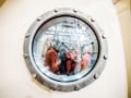
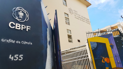
The Brazilian Center for Physical Research (CBPF) in Rio de Janeiro has today inaugurated what it calls the largest urban graffiti dedicated to science.

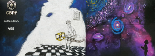
Spanning 240 m2, the graffiti occupies the walls that enclose the institute. It is divided into eight segments that each touch on a specific theme including “the particle that changed Brazil”, “from nano to the macro” and “in search of more”. The “highlight”, according to the CBPF, is “constructors of science”, which features the faces of 100 scientists and is inspired by the famous painting “Operários” in 1933 by the Brazilian modernist painter Tarsila do Amaral.

This work allowed me to see the world differently

Gabi Tores

The graffiti artwork was led by Gabi Tores who is an arts student at the University of the State of Rio de Janeiro. The mural took 600 hours to complete using 110 litres of paint and 324 spray cans. “I fell in love with the project when I realized that this interaction with the public would be unique and we were bringing an innovative concept to this type of urban art,” says Tores. “This work allowed me to see the world differently. In particular, I realize the value of science and how it is so close to us, but sometimes we do not even realize it.”

Hidden puzzles

The CBPF carries out research into a range of topics from nanotechnology to high-energy physics, is one of the top physics institutes in the country. It also serves as the main hub for the academic Internet infrastructure on which a variety of other scientific institutions rely, such as the Brazilian National Cancer Institute. During the 2016 Olympic Games in the city, the institute’s facilities were even used as the headquarters for the event’s digital security.

The graffiti also apparently holds various hidden puzzles that are associated with science and technology, the answers to which you can submit on the project’s website. Those that solve what the CBPF dub the “hardest” puzzles will even receive a prize, which may include a visit to the CBPF, or – if the budget allows – a trip to the CERN particle-physics lab near Geneva.

See more images of the artwork below.

Wall art
Wall art
Grafite da Ciência-Vista parcial da entrada lateral do CBPF com elementos do mural-Crédito NCS-CBPF
Grafite da Ciência-Vista parcial das áreas Abstracao e Misterio-crédito NCS-CBPF
wall art
Copyright © 2026 by IOP Publishing Ltd and individual contributors