
Rooted in physics

When I cycle through Hyde Park in London on my way to work, pausing as I cross the Serpentine to admire the view of the Houses of Parliament, I rarely think about what is going on beneath my tyres. There are just too many above-ground distractions to spend much time wondering about the subterranean network of plant roots weaving their way through the dark and mysterious soil below. And anyway, what have roots got to do with physics?

Roots are complex branched systems whose topology and structure determine the entire physiology of the plants above them. The tip of each branched root has distinct regions where different cell activities take place. At the very end there is the “meristem” where cell divisions occur. Moving up the root, an “elongation zone” comes next where the cells stop dividing and instead elongate. It is followed by a “differentiation zone” where the cells stop elongating and start differentiating.

The direction and rate of root tip growth are influenced by the signals they perceive from the environment, and the organization of the internal tissue within each root tip plays a big role in this root-soil interaction. As root tips explore, the signals help them seek out the most efficient paths to discover water and nutrients. The composition of the soil will influence the architecture of the root system, which ultimately determines the efficacy of the exploration. It seems that evolution has placed enormous pressure on root tips to understand their environments – and it has taken until now for us to start asking why.

The physics of the soil–root interactions that optimize subterranean structures is crucial for plant survival. The roots sense what is going on around them with remarkable detail at the appropriate length scale. “We are not talking about the scale of quantum mechanics, but interactions described by classical physics,” says Giovanni Sena, a development biologist at Imperial College London in the UK, who has a strong background in theoretical physics.

Roots are constantly exploring their space, steering through natural force fields – gravitational and electromagnetic fields as well as magnetostatic and electrostatic – and experiencing the mechanical and chemical properties of the soil they live in. Gradients in light, chemicals, temperature and oxygen provide positional information that allows the root to navigate and forage efficiently. These kinds of root responses are known as a “tropisms”.

Charged insides

The impact of mechanical forces on roots is relatively intuitive – stress and strain can change the shape, number and organization of growing tissue – and the alignment of root tips with the gravitational field is also relatively well understood. However, there is still no model that can effectively describe the much more subtle interaction between an external electric field and the internal electric field of biological tissue.

Keeping a plant alive without damaging it during measurements is no mean feat, and near-on impossible without understanding instrumentation and optics

We know that living cells actively maintain a negatively charged cytoplasm – the material inside a cell excluding the nucleus – and this establishes a non-zero membrane potential. Oscillations in the membrane potential create a temporal pattern, while a spatial pattern is produced by the differences in potential between cells. Yet there are still many unanswered questions. Could these patterns, for example, be crucial to tissue organization and morphogenesis, the process that causes an organism to develop its shape? Is spatial variability just stochastic noise? Or is it an actual pattern that is used as “positional information” during morphogenesis? Until now, how bioelectricity in plant tissue impacts their shape and function has never been defined.

One of Sena’s collaborators, Michael Levin at Tufts University in the US, is studying the bioelectrical cues of membrane potentials and using them to describe cell behaviour. Levin argues that cracking “the bioelectrical code” – understanding how patterns are encoded into biological networks – would allow us to programme cell activity and transform synthetic bioengineering and cancer treatment (Mol. Biol. Cell 25 3835). Sena’s group is collaborating with Levin and partially funded by the Allen Discovery Center at Tufts University to develop models of bioelectric circuits within roots that store patterning information during morphogenesis.

Cut to regenerate

For a physicist, Arabidopsis – a small flowering plant also known as thale cress – is an ideal subject to observe and model because it has transparent, slow-growing cylindrical roots. It is now well established that if you cut the end off its root tip, it will regenerate and reform with no external influences, as shown in figure 1 (Nature 457 1150). However, like a lot of developmental biology, a comprehensive understanding is still missing.

Electrotropism – the alignment of a root with an electric field – has also been observed for decades, but not explained, and Sena’s lab further identified that if you place the cut root tip in a weak electric field, it is more likely to regenerate (Regeneration 3 156). The field impacts some hormones more than others but cause no obvious changes in tissue patterning. These are fascinating results that science still can’t explain: why does an external electric field make tissue regenerate faster? And why does a plant root align with an electric field in the first place?

Understanding the regeneration of damaged tissues would be a game changer in controlling morphogenesis. For roots it can be modelled as a self-organizing 3D system of interacting and coupled cells, which divide with a certain probability – a complicated mathematical model. Instead of just being a cylinder, the root becomes a 3D complex system of interacting units, where the units respond to an electric field. Despite advances in the computational models for developmental biology, there is still no effective way to model the increased regeneration.

Unlike animal cells that can move and change shape because they do not have cell walls, plant cells have a rigid extracellular cellulose coat that restricts their ability to migrate when embedded in a tissue. The driving force to shape plant organs is instead cell division – exactly where and when cells divide will strongly influence the overall tissue shape. The dynamics of cell division events are entirely non-trivial during morphogenesis, and at present very little is known about their spatial and temporal correlations.

Physics tells us that if you have a complex system undergoing a transition there is a sudden change of the correlation length. Sena wants to identify how the spatial and temporal correlation of cell divisions change during regeneration, and how regeneration is influenced by the physical forces that the root tip is growing in.

Experiments with tiny plants

To investigate dynamic developmental processes, scientists need to image living tissues at cellular resolution regularly over an extended period of time. Keeping a plant alive without damaging it during measurements is no mean feat, and near-on impossible without an understanding of instrumentation and optics. To overcome these challenges, building on his previous experience as postdoc at Rockefeller University in New York (PLOS ONE 6 e21303), Sena’s team created its own automated light sheet fluorescence microscope (LSFM), combining optical sectioning with a miniaturized plant growth chamber.

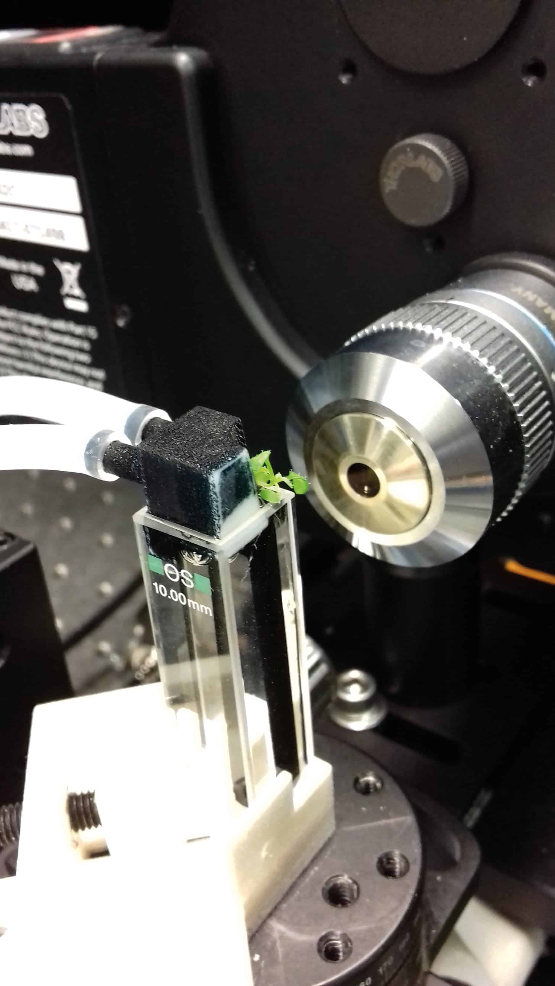
At any given time, the root is illuminated by a thin laser sheet (about 4 µm across), reducing the incident photo-energy and acquisition time by avoiding the need for lateral scans. The plant is held within a cuvette that is continuously illuminated and held at a constant temperature, with oxygen and an actively circulated liquid nutrient solution to keep it growing. Cell divisions are identified by tagging a protein – that appears just before and is destroyed just after cell division – with a fluorescent marker.

Because plants are so sensitive to gravitational fields, Sena had to play with the geometry of the microscope, with the fluorescent signal collected by a long-working distance lens normal to the excitation light-sheet (generated by a laser beam through a cylindrical lens) and the cuvette positioned vertical in the microscope. The stage is automatically repositioned to keep the root tip within the focus of the objective. As even slow-growing Arabidopsis can extend by a few millimetres per day, Sena and his group wrote an algorithm to compare the most recent image with the one before and automatically reposition the stage in 3D.

Before Sena’s dynamic LSFM, confocal microscopy only let researchers take daily images of regenerating root tips. If you wanted to know whether the root reforms or not, confocal microscopy was fine, but if you wanted to investigate regeneration at high temporal resolution, it was not enough. The LSFM allows Sena to grow a root and observe in 3D every cell division event every 10 minutes for at least one week at a time – and by measuring both cut and uncut roots, his team can compare the dynamics of regeneration against normal growth.

A glowing worm

On a computer, the process looks like a glowing worm wiggling across the screen, but it’s really the meristem of a root tagged by fluorescent proteins being tracked by Sena’s automated microscope stage. Whenever and wherever the root blinks, a cell division event has occurred. The video represents a 2D projection of a week of LSFM data collected every 10 minutes. For a non-expert, watching cell division is breathtakingly beautiful, but for Sena, it is just more intriguing physics and biology. Alongside the sped-up video, he can track the number of cell divisions, analyse their temporal and spatial distributions, characterize the bursts of proliferation activity and the more quiet periods, and in general compare normal and regenerating growth. The data are messy – well, we are dealing with biology, not electrons – but the implications are profound.

There is no doubt that physics contributes significantly to Sena’s team’s understanding of plant morphogenesis. In his lab almost all the researchers are experimental physicists – one even used to build particle detectors at CERN – while the others are biologists who have seen the light. If you gave their data to pure biologists, they’d jump immediately to genetics, and start a quest for “mutants” – genetic defects sufficient to modify the observed cell behaviour. Conventional plant science is familiar with visualizing root tips as static slices, but Sena argues that the dynamics are much more important – abnormal processes might look perfectly normal when viewed as static images. Plant morphogenesis, influenced by cell division, is an outcome of evolution – so how and why natural selection has resulted in these division dynamics is an underpinning question for biosciences. And the answers could provide a whole new perspective of those subterranean networks that I cycle above every day.

Optical tweezers create a single molecule from two atoms

A single molecule has been created by combining individual atoms of sodium and caesium, using optical tweezers to guide them into place. The technique, devised by Lee Liu and colleagues at Harvard University and Harvard-MIT Health Sciences and Technology, could help chemists to study chemical reactions far more precisely by giving them control over the individual atomic and molecular collisions. The team hopes that their method will be used in a variety of fields to create diverse, complex molecules, allowing for discoveries of previously unforeseen molecular properties.

Conventional studies of chemical reactions involve observing the macroscopic results of large numbers of collisions of atoms and molecules – rather than studying individual collisions. Currently, chemists need to compare experimental reaction rates with theoretical models to calculate the probabilities of individual collisions taking place – a process that is fundamental to the understanding of chemistry. An alternative, and more precise, technique is to study interactions between individual atoms and molecules – something that requires great experimental dexterity.

To begin their interaction process, Liu and colleagues use magneto-optical traps to prepare reservoirs of stationary atoms of sodium and caesium at just a few hundred microkelvin.  “Cooling and controlling atoms and molecules to temperatures where they are standing still allows for easier manipulations of their properties, interactions and reactions,” explains team leader Kang-Kuen Ni.

Polarizing forces

The chilly temperature means that individual atoms can be easily loaded onto optical tweezers – specialized, highly-focused laser beams that can trap dielectric particles including atoms. To confirm that the tweezers had been loaded successfully, the researchers use spectroscopic techniques to check for the presence of each atom.

One optical tweezer – based on 700 nm-wavelength light – holds the sodium atom, while another tweezer at 976 nm holds the caesium atom. To bring the atoms close together, the team move the tweezer containing the sodium atom so that it overlaps with the tweezer containing the caesium atom. Then, the 700 nm light is switched off. The 976 nm laser is capable of holding both atoms, so the sodium and caesium are trapped together.

The team found that simply bringing the atoms together is not enough to form a molecule – a pulse of light is required. “The atoms couldn’t turn into a molecule just through an isolated collision, as they had to simultaneously conserve energy and momentum – something very difficult to achieve in experimental conditions,” Ni explains. “To solve this, we shone another laser tuned to the resonance wavelength of the molecule, driving them to form the molecule.” The excited molecule which formed quickly decayed into a stable ground state, allowing for observations of its properties. “This was the first time that a molecule has been assembled starting from exactly two atoms, and establishing individual control over each of them,” said Ni.

New and unexpected

Even for such a simple molecule, the team observed new and unexpected properties in their creation. “We saw new molecular spectroscopic lines, and that contributed to a deeper understanding of the overall picture,” explains Ni. The researchers believe their technique could soon be expanded to study complex chemical processes.

“We are now thinking of working with many optical tweezers and many molecules simultaneously, allowing for studies of their interactions or reactions,” Ni says. With this technology available, it could soon be possible for chemists to synthesize their own designer molecules, with applications ranging from new medicines to qubits in quantum computers.

The research is described in Science.

Erosion may be carbon source, not sink

Researchers have challenged one of the few certainties of earth and climate history: the link between erosion and the drawdown of carbon dioxide from the atmosphere.

For decades, it has been a given that heavy rainfall on steep mountain slopes is likely to chemically weather the exposed rock and precipitate a chemical reaction that ends with carbonate minerals on the ground and with less of the greenhouse gas carbon dioxide in the atmosphere.

So geochemistry and the weather between them help moderate the planet’s climate.

But geologists and oceanographers who took another and closer look at the process in action – in the central mountains of Taiwan, hammered by three or more major typhoons each year – say they are not so sure.

They report in the journal Science that the same erosion process could be a source of carbon dioxide, releasing it into the atmosphere far faster that it can be absorbed by the newly exposed rock.

And the agency at work in this unexpected process could be biology: the researchers found evidence that tiny microbes in the mountain soils were consuming sources of organic carbon trapped in the rock, and releasing CO2 into the atmosphere.

On the face of it, the process may not be severe enough to upset the global calculations that add up to what climate scientists call the carbon budget – the annual traffic of carbon in the form of greenhouse gases from atmosphere to living things and then into the rocks and oceans – but it is yet another reminder that the climate machinery is still incompletely understood.

“This goes against a long-standing hypothesis that more mountains mean more erosion and weathering which means an added reduction of CO2. It turns out it’s much more complicated than that,” said Jordon Hemingway, of Harvard University, who led the study.

The Earth’s crust, powered by heat from the mantle below, is permanently in a state of levelling and reconstruction: powerful subterranean forces build mountains and the steady attrition of wind and rain immediately begin the process of wearing them down.

Complicated picture

But mountains are part of the climate machine. There is a theory that the rising of the Himalayas and the Tibetan plateau provided the barrier that made the South Asian monsoons possible, and a secondary theory that the increased rainfall on the freshly raised mountain slopes weathered so much rock that the planet’s levels of atmospheric carbon dioxide took a dive, to precipitate 30 million years of Ice Ages.

A closer study of the soil, bedrock and river sediments in Taiwan revealed a more complicated picture. The scientists found that almost 70% of the organic carbon initially present in the weathered bedrock had been oxidised by soil microbes, to put, for every square kilometre they measured, somewhere between six and 18 tonnes of carbon back into the atmosphere.

This is not enough to set alarm bells ringing. But it does suggest that the intricate details of the carbon budget depend not just on what happens on the planet’s surface, but also in the teeming life beneath – and sometimes far beneath – the surface.

This is basic research at a down-to-earth level: climate science can’t make sense of what is happening now without a better understanding of what has always happened, and of the swings in planetary temperatures over the past 4.5 billion years.

Clear understanding

Researchers are confident that they understand the cycle of Ice Ages, and they also have a clear idea that the biosphere plays a hand in keeping the planet at liveable temperatures, but they also know that the high altitudes are more than usually affected by climate change driven by ever-higher ratios of greenhouse gases released by the combustion of fossil fuels by seven billion humans.

Their fears extend to Alpine economies and the plants and animals that live in the mountains. Now it seems clear that some long-term questions require explanation at the microscopic level.

“Looking backwards, we’re most interested in how these processes managed to keep the levels of CO2 in the atmosphere more or less stable over millions of years. It allowed Earth to have the climate and conditions it’s had – one that has promoted the development of complex life forms,” Dr Hemingway said.

“Throughout our Earth’s history, CO2 has wobbled over time, but has remained in that stable zone. This is just an update of the mechanism of geological processes that allows that to happen.” – Climate News Network

• This report was first published in Climate News Network

If I’m stressed then you’re stressed

Stress is a major modulator of neuronal networks and its consequences can be transmitted to others. With this in mind, a team from University of Calgary embarked on a journey to decipher whether transmitted stress has the same consequences on synapses as authentic stress (Nature Neuroscience 21 393). Their findings in mice suggest that the lasting effects on synapses are the same for both authentic and transmitted stress, with implications for other stressful events.

Stress induces metaplasticity
In the experiments, the authors subjected mice to footshock stress for 5 min and, upon returning them to their homecage, to high-frequency electrical stimulation for 30 min. They discovered that acute stress resulted in persistent changes at glutamate synapses (glutamate is the major excitatory neurotransmitter in the nervous system) on corticotropin-releasing hormone (CRH) neurons. This enables the induction of metaplasticity, activity-dependent changes in neural functions that modulate subsequent synaptic plasticity. The CRH neurons are found in the paraventricular nucleus of the hypothalamus, one of the most important autonomic control centres in the brain, and their activation is necessary for the rise of the endocrine response to stress.

Control and footshock mice

Pheromones help transmit stress
To study the effect of social interactions on metaplasticity, mice were subjected to footshock or a novel environment for 5 min and returned to a same-sex control partner in the homecage for the next 30 min. Interestingly, stress is transmitted by the release of alarm pheromones, predominantly from glands in the anogenital area, which are sensed by the partner during investigative behaviour. The triggering of this type of behaviour and synaptic priming require the activity of CRH neurons (which the authors determined using whole-cell recordings from CRH neurons in hypothalamic slices).

This experiment suggested that transmitted stress primes glutamate synapses in male and female subjects with different sensitivity and, importantly, that the effects of authentic stress in females are reduced by the presence of a partner.

Transmitted stress

In another experiment, the team showed that the synaptic priming induced by transmitted stress can also be propagated by a partner mouse to a tertiary group member, with similar effects (the same synaptic load).

The findings of this study indicate the importance of mitigating the effects of stress and how the information extracted from the experience of a distressed subject can have adaptive benefits. Moreover, the impacted neuronal circuits can this way prepare the individuals for subsequent challenges.

Beyond maths to meaning

Everybody knows that quantum physics is weird, right? Indeed, quantum physicist Richard Feynman once said in a lecture: “The theory of quantum electrodynamics describes Nature as absurd from the point of view of common sense. And it agrees fully with experiment. So I hope you can accept Nature as she is – absurd.” Beyond Weird: Why Everything You Thought You Knew About Quantum Physics is Different presents a refreshing challenge to this viewpoint. In the book, science writer Philip Ball dares to take on Feynman, the closest thing physicists have to a patron saint, and suggests that we need to rethink that “weird” label.

It’s not that the author denies the existence of the strange phenomena of quantum theory. He simply points out that there’s nothing truly weird about it – this is just how nature is. Instead, Ball suggests that we perceive quantum physics as strange and mysterious because we are misled by our everyday experience. The weirdness is in our understanding, not in nature.

If this distinction seems worryingly philosophical, be prepared for a bumpy ride. Where most popular quantum physics books concentrate on the science and its applications, the core of Beyond Weird is the interpretation(s) of quantum mechanics, and that inevitably involves philosophy. It’s not that Ball ignores the science – his description of the Schrödinger equation and the wave function is one of the best I’ve seen in popular writing – but we are provided with this as background to interpretation.

Philosophy is not always popular in physics circles. Infamously, Stephen Hawking and Leonard Mlodinow in The Grand Design proclaimed that philosophy was dead, and that the serious questions that used to be the remit of philosophy are now in the hands of science. However, when it comes to getting a feel for quantum physics, Ball suggests that we can’t ignore the philosophers. The description of the difference between quantum theory and reality covered in Beyond Weird parallels Kant’s idea of the Ding an sich – an inaccessible underlying reality that we can only model through the results of our experience.

So, we are told, familiar concepts of the quantum world such as wave–particle duality and superposition – where a quantum object appears to be in more than one place at a time – do not describe the true nature of quantum objects. In fact, we can’t know what that really is. It’s not that a quantum object is sometimes a wave and sometimes a particle, but never both simultaneously. Instead, the way we perceive the object in our experiments will be one or the other. Quantum physics, it seems, is all about perception and information, not reality.

This approach is suggestive of the “shut up and calculate” school where, instead of worrying about what’s “really’” happening, you simply apply the maths. If we can never know what really lies beneath, what is the point of constructing interpretative models? Ball shows us that there is a lot we probably need to agree with in the traditional Copenhagen interpretation of quantum mechanics formulated by Niels Bohr and Werner Heisenberg in the 1920s. This probabilistic interpretation of quantum mechanics suggests that the observable properties of a particle do not have definite values until they are measured. But Ball also shows that there are multiple interpretations of this itself, as there was never a formal definition of the Copenhagen interpretation and the two scientists themselves had rather different ideas, meaning that it too is incomplete and ultimately, unsatisfying.

Why, Ball asks, does quantum physics, uniquely, need interpretations? None of these attempts to provide a bridge between the highly-successful mathematical models such as Schrödinger’s equation, and what is observed, is ideal. They all fall down, irrespective of whether someone supports the science fiction-like “many worlds interpretation” – the idea that every outcome of a quantum decision takes place in a parallel world system, which Ball argues is counter-scientific – or David Bohm’s universal interconnectedness; or more modern attempts.

The difficulty of interpretation is also not helped by the degree to which the fundamentals of quantum physics were plucked out of the air. As Ball tells us, “It was a hugely abstract and intellectual exercise, relying on informed guesswork to an extent we might consider both impressive and alarming.” The mathematical models match what was later observed impressively well, but it’s alarming considering we have no good reason for expecting these mathematical models to work.

The key to understanding the confusion caused by quantum theory, may be to consider information

The key to understanding the confusion caused by quantum theory, may be to consider information, suggests Ball. It seems that as quantum objects interact with their surroundings, they lose their quantum uniqueness. In this decoherence process, they go from a collection of probabilities to having measured values. In this picture, quantum phenomena are largely about transfers of information.

Through the book, it becomes clear that, while the mathematics of quantum theory is highly effective, we may need to approach it differently if we are ever to get beyond the numbers to an understanding. Towards the end of the book we are presented with attempts, that are still not fully developed, to reformulate quantum physics as a series of axioms that make sense in conventional terms, while still allowing most quantum phenomena to be deduced. If this all sounds diffuse and woolly, it’s not realistic to expect Ball to be able to make everything that is described as “quantum weirdness” go away. Rather, what he successfully does is to enable the reader to look at quantum physics in a different light, where these oddities become less challenging.

The only real negative about Beyond Weird, past a tendency to make the brain hurt, is the way that the book is set up as a series of challenges to “everything you thought you knew about quantum physics”. Ball is an urbane writer and the aggressive challenging adopted here sits uncomfortably with his usual style. This comes across particularly strongly in the early parts of the book, where page after page is taken up telling us that quantum physics isn’t really weird like we thought it was, but that it is merely surprising. Thankfully, this early confrontational tendency fades later, and should not take away from the fact that this is the most original and interesting book on quantum physics for the general public in a long while.

  • 2017 Bodley Head 384pp £17.99hb

Paris targets ‘more readily achievable’, Canadians say

The targets for climate change outlined in the Paris Accord are easier to achieve than commonly thought. That’s the claim of researchers in Canada, who have defined climate outlooks in a way that is both simple and easier to analyse.

The new definition relies on just two parameters – total energy use this century, and the carbon intensity of that energy – that can be mapped onto a phase-space diagram. By data-mining that phase space, the researchers found that current “business as usual” scenarios given by the Intergovernmental Panel on Climate Change (IPCC) assume increasing carbon intensity, or so-called re-carbonization – contrary to recent trends.

“By thinking that we’re definitely heading in a direction of re-carbonization before we institute climate policies, it makes climate policy that aims for de-carbonization look overly difficult,” said Justin Ritchie of the University of British Columbia. “If we update our outlook for business-as-usual, the necessary climate policies to guide a low-carbon transition appear far more achievable than previously thought.”

The creation of future climate scenarios is a complicated business. For many years, they have been based on integrated assessment models (IAMs) – computer models that explore the projected interplay of population, economics and energy use up to the year 2100.

Since the IPCC’s Fifth Assessment report in 2014, two more layers have been added. Above IAMs are shared socio-economic pathways (SSPs), which describe possible socio-economic developments; while the layer above SSPs consists of representative concentration pathways (RCPs), which define the levels of greenhouse gases that could ensue from the different underlying scenarios. RCP8.5 is the oft-called business-as-usual scenario, in which no strong policy action on climate change is taken.

The problem with this approach, according to Ritchie, is that the underlying IAMs are very time-consuming to run. “If they are used to map all the possible future developments based on different scenarios of population, GDP and energy use, it can get quite unwieldy,” he explained. “For example, there were 1,184 scenarios produced by about 20 different IAMs for the IPCC’s Fifth Assessment, and managing all those model outputs is a growing challenge… Further, most of those scenarios were based on a common set of population and socioeconomic projections, so while their number is quite large, they are actually surprisingly narrow in scope.”

Instead of this top-down approach, Ritchie and colleague Hadi Dowlatabadi, also from the University of British Columbia, generated climate outlooks the other way around, mapping out all the combinations of carbon intensity and energy use that could generate a certain level of carbon emissions as given by the RCPs.

Doing it this way reveals the full range of possibilities without resorting to complex simulations, says Ritchie. It “allows us to see that integrated assessment models may be too narrowly focused”.

Indeed, Ritchie and colleagues’ method revealed that RCP8.5 is mostly depicting a world in which coal remains the cheapest form of energy and outcompetes other types of energy generation.

“Since that looks increasingly unlikely, it means they are likely overestimating how difficult it will be to develop along technology pathways that lead to 2˚ of warming or less, in line with the Paris Agreement,” Ritchie said. “Renewables, energy-efficiency and oil and gas technologies have proved far more dynamic than anticipated. Thus, coal has been losing global market share for a long time.”

The team published the study in Environmental Research Letters (ERL).

PET/CT lines up for breast imaging

A dedicated breast PET/CT scanner that produces combined dual-modality images could improve the detection of breast diseases in challenging cases. A team at West Virginia University School of Medicine has been developing dedicated breast-PET imaging systems since 2000. Performance tests of their newest breast PET/CT prototype demonstrated that its performance was similar to that of current dedicated breast-CT and breast-PET systems (Med Phys. doi.org/10.1002/mp12780).

Women with dense, cystic, post-surgical and augmented breasts can be difficult to evaluate using current imaging techniques. PET is a good candidate for supplemental breast imaging of women with indeterminate mammograms, because tissue contrast is based on physiologic differences (characterized by radiotracer uptake) rather than tissue density.

Integrating CT with PET adds anatomical information regarding the size and shape of lesions identified with PET. CT data could also be used in corrections for physical processes that degrade PET images, such as Compton scattering and photon attenuation. Accurate quantitation of radiotracer uptake could also be improved by using CT images to calculate partial volume corrections for small structures identified in PET scans.

The PET/CT scanner
The PET/CT scanner developed by Raymond Raylman and co-authors consists of a PET scanner with two detector heads coupled to a 4×3 array of flat-panel positron-sensitive photomultipliers (PSPMTs). Specialized electronics reduce output signals from 64 to four channels for each PSPMT and produce an output signal whose amplitude represents the total amount of light detected by the PSPMT. The system design maximizes collection of scintillation light and light produced by elements at the edges of the scintillator array and reduces the processing load.

The dedicated breast-PET/CT scanner

PET data are acquired by rotating the detectors in step-and-shoot mode, with the dwell time at each position selected by the user. The estimated imaging time is approximately six minutes per breast. The nominal field-of-view (FOV) is 20 cm (transaxial) and 15 cm (axial), and the nominal reconstructed voxel size is 1 mm3. The cone-beam CT scanner comprises a tungsten filament pulsed X-ray source and a flat-panel X-ray detector operating in portrait orientation. Its nominal FOV is 16 cm (transaxial) x 20 cm (axial).

Both components are mounted on a computer-controlled rotating gantry, along with a three-axis rotating arm to hold a biopsy gun – enabling the new system to perform image-guided biopsies of suspicious lesions. The scanner elements are mounted on linear slides so that their distance from the centre-of-rotation can be adjusted according to the desired mode of operation (PET or CT) and breast size. Specialized software registers the CT and PET images, harmonizing image voxel sizes and aligning the images. CT images are segmented for use with the Compton scatter and attenuation corrections utilized in PET image reconstruction.

Performance testing
The team reported that the PET/CT scanner performed as expected during testing with the NEMA NU2-2009 protocol. The system exhibited a spatial resolution of 2.2 mm (using filtered-backprojection reconstruction) 5 mm from the centre of the scanner. Imaging of a micro-hot-rod phantom illustrated the potential utility of the dual-modality images.

Raylman told medicalphysicsweb that the tests demonstrated the systems’ high performance, although some improvements are planned. The team is now working to reduce CT detector binning and implement an enhanced iterative reconstruction algorithm. These modifications should improve CT resolution and permit reduction of radiation dose to the breast by reducing X-ray flux, without degrading image quality. They have also developed a patient bed that incorporates shielding to block the PET detectors from annihilation photons produced by radiotracer in the patient’s organs and minimize scatter of X-rays into the patient’s torso.

When these improvements are made, the researchers will perform a phantom study to estimate lesion detectability with the PET and CT components, and measure radiation dose to the breast and torso. Raylman said that the team hopes to start pre-clinical testing within six months.

PET/CT advantages
“The addition of CT to dedicated breast-PET scanners makes it possible to supplement PET images by detecting lesions that may not preferentially uptake the PET radiotracer used in the study,” said Raylman. “Future CT scanners may be able to detect some micro-calcifications, which are not visible with PET. Finally, the combination of CT with PET enables accurate attenuation and scatter correction methods to be applied to PET images; potentially enhancing lesion detectability and facilitating accurate quantification of radiotracer uptake in the breast and breast lesions.”

Raylman predicts that radiation dose to the breast from the PET/CT scanner may be lower than that from a 3D tomosynthesis digital mammogram. Meanwhile, the anticipated exam time of 12 minutes should be much faster than the time required to perform an equivalent breast-MRI exam. The breast PET/CT exam should also be less expensive than a comparable MRI scan. Finally, breast PET/CT scanners could also be more accessible to patients than breast-MRI scanners, because they will be less expensive to purchase and far less challenging to install in smaller imaging centres and radiology departments.

“Being able to perform a PET-guided biopsy will also be advantageous to confirm diagnoses of suspicious lesions. Our system is currently the only dedicated breast-PET/CT scanner capable of performing image-guided biopsy,” Raylman added.

This system is one of two under development in the USA. Another initiative to develop a dedicated breast PET/CT scanner has been underway at UC Davis Medical Center since the mid-2000s.

Atomic force microscope makes single-electron current meter

An atomic force microscope can be used as a single-electron current meter, according to new experiments by researchers at IBM. The technique, which measures the energy levels of single molecules on insulators for the first time, provides precious information on single-electron intermolecular transport. The work might ultimately help to make improved electronics devices in the future, by characterizing defects in chips, for example.

Electronic devices contain printed circuit boards, in which all of the components making up a device can clearly be seen. The conducting tracks, which carry electric current through the entire board, are visible too. The boards also include insulating layers that shield the tracks from current leakage.

In molecular electronics, we would see a similar set-up with single molecules as the conducting tracks and single electrons being transferred from the molecules, explain the researchers led by Gerhard Meyer at IBM Research-Zurich. However, the difference on this scale is that the underlying substrate produces supplementary effects that need to be analysed. Unfortunately, this is no easy task since the molecules being electrically characterized are on top of an insulator.

The reorganization energy

“While charging a molecule on an insulator, the atoms in the molecule will relax towards accommodating this additional charge, as will the nuclei in the insulator,” says Shadi Fatayer, who is the lead author of this study. “This change in the atoms’ position impacts their energy levels and is known as the ‘Marcus reorganization energy’. It drastically affects the rate at which single electrons are transferred between molecules.”

In their experiments, the researchers grew multilayers of sodium chloride, NaCl, which is an insulating material, on top of a metal substrate. Such a system allows the molecules that are then absorbed on top (single naphthalocyanine molecules in this case) to have stable charge states, since they are decoupled from the metal surface.

Reorganization energies are usually measured by analysing molecules in solution or with molecules on top of a metal, but until now, there was no way of doing this for individual molecules on top of an insulator.

Enter atomic force microscopy

Atomic force microscopy is a widely-used ultrahigh-resolution technique that allows researchers to observe extremely small objects, even down to single atoms. It works by sensing the topography of a sample as it scans across it thanks to a very fine probe (the cantilever), which has an extremely sharp tip at its end. The AFM measures the tiny forces between the tip and the sample, such as a molecule on a support as in this case.

Thanks to previous work in their lab, the researchers had already succeeded in using an AFM to measure different charge states on top of an ultrathin insulator – with single-electron sensitivity. They also managed to image stably-charged molecules and transfer single electrons between molecules on top of a thicker insulator. Being able to measure reorganization energies proved to be more difficult still, however, and meant that they had to measure the energy levels that corresponded to particular charge-state transitions.

“Before this work, we were able to measure the electric current through a single naphthalocyanine (NPc) molecule atop an ultrathin insulating NaCl,” says Leo Gross, who is a physicist at IBM. “However, this only works in one direction for a given electron orbital. When we could measure the energy needed to attach an electron to a certain orbital, we could never measure the energy to remove one electron from that orbital, for example. With our AFM technique, we can now measure the energy levels in both charge-state directions on a thin film substrate.

Very weak signals

“The signals we need to detect are very weak, however, because they come from weak forces associated with currents that are just zepto-amperes in magnitude. This means that we must perform many careful measurements for a proper statistical analysis.”

The team in fact employed the tip and the force exerted on the tip to count single electrons; “We adjust the tip height and voltage and then count how long it takes for one electron to go to (or from) the tip and from this you can obtain the energy levels,” adds Gross.

The technique is detailed in Nature Nanotechnology doi:10.1038/s41565-018-0087-1.

Nuclear clock could be one tick closer

The internal structure of the thorium-229m nuclear state has been studied in detail for the first time by physicists in Germany. Thorium-229m is a metastable (or isomer) excited state of thorium-229 that decays via the emission of an ultraviolet (UV) photon. This photon has much lower energy than most nuclear emissions and could form the basis of a “nuclear clock” that would be much more precise than existing atomic clocks.

Atomic clocks work by keeping a laser in resonance with electronic transitions between energy levels in atoms or ions – with the “ticks” of the clock being the frequency of the laser light. Although the best atomic clocks available today could keep time to within one second if they were left running for 13 billion years, physicists are still keen on boosting this performance. Clock performance is limited by the effects of stray electromagnetic fields on atomic energy levels – and this is where nuclei can help. Nuclei are hundreds of thousands of times smaller than atoms and bound together much more tightly – and this makes nuclear transitions less sensitive to external electromagnetic fields.

The problem is that nuclear transitions tend to occur at energies that are thousands or even millions of times greater than the photons produced by today’s lasers. However, the transition between the ground state of the thorium-229 nucleus and an excited state (thorium-229m) is expected to have only around 7.8 eV energy. This corresponds to the energy of ultraviolet photons, which can be laser-generated.

Narrow transition

The spectral width of this transition is extremely narrow, which is good for clock performance. However, this narrowness has made it very difficult to determine the actual energy of the transition. A breakthrough came in 2016, when  Lars von der Wense of Ludwig Maximilian University of Munich and colleagues throughout Germany compared the decays of thorium-229 atoms and ions, which allowed them to conclude that the energy of the ultraviolet photons is in the 6.3–18.3 eV range. Subsequently, the researchers were also able to measure the lifetime of the thorium-229m state – and important piece of information for those aiming to build a nuclear clock.

In this latest research, von der Wense and colleagues – including Christoph Düllmann of Johannes Gutenberg University Mainz – have taken a much closer look at the thorium-229 nucleus to further characterize its potential as a nuclear clock. They began their experiments by storing thorium-229 ions in an ion trap. Some of these are in the thorium-229m state, and the team used laser spectroscopy to measure the hyperfine structure of the ions. Hyperfine structure arises from interactions between atomic electrons and the nucleus and can provide important information about the structure of a nucleus.

From the spectroscopic studies, the team worked out the charge radius of the thorium-229m state as well as its magnetic dipole and electric quadrupole moments. These quantities are important because they define how the state interacts with external electric and magnetic fields. The value of the electric quadrupole moment, for example, suggests that a clock based on a crystalline solid doped with thorium-229 would have to deal with a substantial shift of the nuclear transition frequency caused by electric-field gradients in the crystal.

Testing a constant

Writing in Nature, the physicists say that if such a nuclear clock could be built, its timekeeping would be extremely sensitive to the value of the fine structure constant, which measures the strength of the electromagnetic interaction. This could allow physicists to test whether this constant is indeed constant, or if its value changes under certain circumstances.  If discovered, variations in the fine structure constant could point to physics beyond the Standard Model of particle physics.

Tiny positioning differences can impact survival

Corinne Johnson

Tiny differences in a patient’s position during radiotherapy for lung or oesophageal cancer can impact how likely they are to survive, according to research presented at the ESTRO 37 conference in Barcelona. The study found that differences of just a few millimetres can shift radiation targeted to the tumour fractionally closer to the heart, where it can cause unintentional damage and reduce survival chances. This finding suggests that survival could be improved by tightening up treatment guidelines to ensure patients are positioned more accurately.

Corinne Johnson, a medical physics PhD student at the Manchester Cancer Research Centre, and colleagues studied 780 patients with non-small cell lung cancer who were treated with radiotherapy. For each treatment, patients were positioned on the treatment system and an image taken to confirm that they lay within 5 mm of their original position.

The researchers used the data from these images to gauge how accurately radiotherapy dose was delivered over the course of treatment, and whether it was shifted slightly closer or slightly further away from the patient’s heart. Comparing these data with how likely patients were to survive showed that patients with slight residual shifts towards their hearts were around 30% more likely to die than those with similar sized shifts away from their hearts. Repeating the research in a group of 177 oesophageal cancer patients revealed an even greater difference, of around 50%.

“We already know that using imaging can help us to target cancers much more precisely and make radiotherapy treatment more effective,” Johnson explained. “This study examines how small differences in how a patient is lying can affect survival, even when an imaging protocol is used. It tells us that even very small remaining errors can have a major impact on patients’ survival chances, particularly when tumours are close to a vital organ like the heart.”

She suggests that imaging patients more frequently, and reducing the threshold on the accuracy of their position, could help lower the radiation dose to the heart and avoid unnecessary damage. The researchers are now examining the data in more detail to see whether particular regions of the heart are more sensitive to radiation than others. They also hope to investigate the effect of differences in patient position in other types of cancer.

Copyright © 2026 by IOP Publishing Ltd and individual contributors