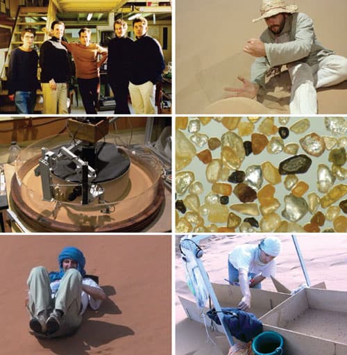
If you go to a sandy desert or beach on a blustery day, you might see a range of topographic features created by the wind. Looking down at your feet, you may notice centimetre-scale periodic ripples made by hopping grains of sand. You could also explore much larger sand dunes that form on length scales of tens of metres.
Ripples and dunes are thought to form in very different ways and in most sandy environments you will not see periodic structures at intermediate length scales – often referred to as the “forbidden region”. There are exceptions to this rule, and unusually-large “megaripples” are found in some places. Now, researchers at the University of Leipzig in Germany and Ben-Gurion University of the Negev in Israel have worked out that these structures form in much the same way as larger sand dunes.
Sand ripples are thought to be caused by a synchronization of the hopping of grains of windblown sand with emerging waves in the sand bed. The ripples are rarely larger than a few centimetres wide, and tend to be regularly spaced, with about 10 cm between peaks.
A mighty wind
Dunes result from the asymmetric way the wind blows over obstacles: “If you stand on top of a hill, the strongest wind is not exactly at the top: it’s slightly upwind of this position,” explains Leipzig’s Klaus Kroy. The maximum mass of a particle that can become airborne increases with the airflow rate, so, as the wind slows down slightly at the top of a dune, it deposits more sand, making the dune bigger.
Normally sand does not fly continuously in the wind but travels in discrete hops several centimetres long. For a dune smaller than about 10 m in length, this hop length is longer than the distance the point of strongest wind is displaced from the dune crest. Therefore, sand picked up from the windward side of a dune is not deposited on the crest and the dune is eroded by the wind.
These rare intermediate structures are known as megaripples and have, until now, lacked any theoretical explanation. As the name implies, they have generally been studied as unusually large ripples, but this has only deepened the mystery. For example, ordinary ripples tend to be periodic, and are neatly characterized by the distance between successive peaks. However, says Kroy, megaripples “don’t care where the nearest neighbour is”. Furthermore, megaripples tend to be covered with coarse sand: “People were saying to me things like ‘It’s really hard to understand why the coarse grains always accumulate on them,'” says Kroy.
Bimodal grains
Treating megaripples as dunes explains why they are not periodic, but raises other questions. They are often not much more than 30 cm in length, so how can they be stable? The researchers present a mathematical model of how, under specific geological conditions, the distribution of sand grain masses can become bimodal: fine, light grains perform large jumps in the wind and heavier, coarser grains creep along the sand bed in small jumps, pushed along by repeated impacts from the lighter grains.
These heavier grains respond to changes in wind speed on much shorter length scales than the lighter grains, so they can build much smaller dunes. Unlike large dunes, however, megaripples are transient features prone to destruction by storms. This, say the researchers, is because the coarser grains of sand will make large jumps in very high winds, making small dunes unstable.
The team confirmed its hypothesis by studying cross sections of megaripples in the southern Negev desert in Israel. They also used previously published data: “We show people how to look at the available data and reinterpret it in a different way,” explains Kroy.
Kroy suggests that the analysis technique could be further developed for use in other fields. “[Researchers have] found small ripples on the surface of the comet that passed near Earth recently, even though there’s no atmosphere,” he says, “Under such conditions, you can immediately see that there must – quite unexpectedly – be some wind.” He also says that studies of petrified megaripples could tell us something about conditions long ago on Earth when the features formed.
Geoscientist Nathalie Vriend of the University of Cambridge is impressed with the research: “There have been observations of megaripples before, but this is one of the first papers that really puts some physical modelling behind them,” she says. She cautions, however, that the researchers’ dynamical ideas about the effects of various wind speeds on sand grain size need field testing.
Over the past 40 years significant strides have been made towards tackling cancer. During this relatively brief period, survival has doubled in the UK and 50% of people will now survive the disease for a decade or more. At the core of this progress is research, bringing advances in screening techniques, diagnostic tests and treatments that together are helping to transform the outlook for this disease.
Despite these developments, now is not the time for complacency. Progress has not been uniform and for several cancers, such as oesophageal and pancreatic, survival has lagged far behind and seen little improvement.And for most patients whose disease has spread, achieving a cure remains a distant prospect. To ensure advances continue rather than stagnate, there is a clear case for innovation in the way we approach and carry out cancer research.
That is why in 2015 Cancer Research UK launched the Grand Challenge – an ambitious £20m programme to support scientists to solve some of the greatest problems facing the field. The grants not only seek to overcome major obstacles that stand in the way of progress, but also challenge more traditional ways of working. Where the increasing competitiveness of research funding may have sparked a trend towards safety as opposed to innovation, Grand Challenge seeks to encourage bold thinking and reward novel ideas and approaches to research.
A worldwide approach
With the complexity and scale of the issues at hand, from unravelling disease biology to overcoming treatment toxicity, the programme is not exclusive to the UK but open to researchers worldwide – encouraging collaboration across an exceptional pool of international talent. But collaboration requires more than the sum of many minds.
To tackle such significant problems we need a breadth of knowledge and expertise. This is why Grand Challenge is designed for the physical and life sciences to come together, forging truly multidisciplinary teams that can bring forward and capitalise on the range of unique skills that each discipline offers. By breaking down both geographical and disciplinary barriers, Grand Challenge ultimately seeks to transform cancer research and to beat cancer sooner.
While cancer research may traditionally be placed among the life sciences, physics has been pivotal to some of the greatest advances in treatment and diagnosis to date. It was physicists who pioneered the field of radiology as far back as the 1800s. The serendipitous discovery of the X-ray, by German physicist Wilhelm Röntgen in 1895, triggered a global change in diagnostics virtually overnight, turning previously invisible maladies into diagrams of ill health.
And it was the subsequent surge of interest worldwide that soon identified another purpose for radiation in medicine: radiotherapy. Now a cornerstone treatment that has helped save millions of lives, radiotherapy has had a century of research and refinement to turn it into the incredibly sophisticated and precise technique that it is today. Yet the work is not over. As new technology allows the limits of what can be achieved to be pushed further and further – not just in these fields but across the board – opportunities arise to explore previously uncharted territories in cancer science, making now a more exciting time than ever to be involved.
The inaugural round of Grand Challenges resulted in four groups being funded. One winner was Josephine Bunch from the National Physical Laboratory, who was awarded a £16m grant to map tumours and get down to their metabolic nuts and bolts at the molecular and cellular level. Bunch and colleagues will use an array of new mass-spectrometry techniques, and other methodologies they have pioneered, to detail everything from whole tumours down to individual molecules within the cells that comprise them. This ambitious project will involve physicists, chemists, biologists and technology innovators who all bring their own unique skills. Hopefully it can spur new developments in treatment and diagnostics that could ultimately lead to more lives saved.
Seed funding
For the programme’s second round, eight new challenges are looking to be solved. These range from developing artificial intelligence to help detect cancer earlier to defining mechanistic rules for treatment combinations. Ten teams have been shortlisted, which have each been given £30,000 of seed funding to get their ideas off the ground. The winners will be announced later this year.
Though applications for the current round are now closed, the 14 challenges that have so far been set are compelling evidence that applicants for round three will be no less audacious. The stakes are high, but the rewards are even greater. We hope that more physicists will take this opportunity to lead groundbreaking science that will revolutionize the field of cancer science.
Physics World published a special issue on the physics of cancer in July 2013
Decellularization of tissues for regenerative medicine applications, such as cartilage repair, is the subject of much research effort. The principle of naturally derived extracellular matrix (ECM)-based tissue engineering is to remove cells from the tissues without affecting their ECM, leaving an intact niche for cell repopulation and, hence, enabling enhanced healing (see: Decellularized and recellularized grafts repair injured cartilage).
Taking this a step further, the decellularization of whole organs offers the possibility of achieving the same goals in a complete structure such as a heart. This would revolutionize the fields of transplantation and regenerative medicine.
Whole organ decellularization is achieved through perfusion, which delivers the decellularizing agents (detergents and enzymes) using the tissue’s own vasculature. Obviously, this is a complex process and there are many variables that can influence the decellularization and affect the microstructure of the organ. In addition, research in this field has mainly focused on small animals like rats (which have much smaller organs), and is now more and more striving towards human-sized organs. Thus, the field of tissue engineering needs further research to understand the processing of whole organs by perfusion, particularly in large animal models.
Standardizing and optimizing
To shed some light upon the decellularization of whole hearts, Jörn Hülsmann, Hug Aubin and colleagues from the working group of Payam Akhyari and Artur Lichtenberg at Heinrich Heine University Düsseldorf have published a study in which they assess the processing of small and human-scale heart models. They performed decellularization of rat and ovine hearts and analysed the process by-products (perfusates or the solution that runs through the organ) and the effects on both organs. As result, they observed how the process presented varying characteristics that may help further its understanding and optimization (Biomed. Mater. 13 035014).
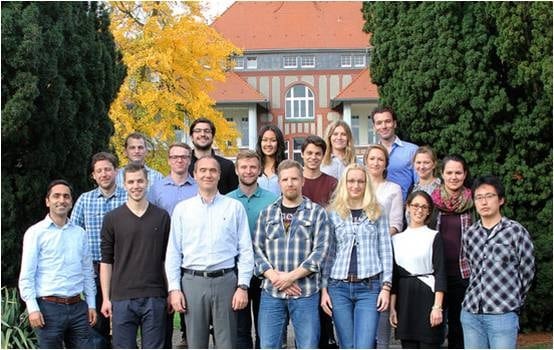
The research group of Payam Akhyari and Artur Lichtenberg at Heinrich Heine University Düsseldorf.
First, they confirmed the efficacy of the decellularization using macroscopic observation, histology and quantification of DNA and cell markers, which demonstrated a high removal of cellular material, while maintaining the tissue ultrastructure and extracellular matrix architecture. Later, the researchers investigated the dynamics of this process via analysis of the by-products or perfusates.
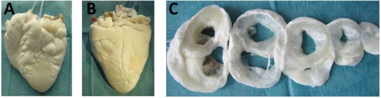
Ovine hearts decellularized by perfusion.
What are we losing?
Ideally, decellularization should only remove the cellular material, which comprises around 19% of the organ’s wet tissue weight. This can be observed after phase separation, drying and weighing of both hearts. Additional exposure to detergents harms the scaffold and, finally, may lead to its de-functionalization.
Surprisingly, after analysing the perfusates, the researchers observed that the detectable protein content was much higher, particularly in rodent hearts, where it represented 71% of depleted mass versus 37% in the larger models.
These differences between small and large organs may be attributed to varying physico-chemical interactions, potentially due to a size effect or to physiological differences (the lipid fraction in ovine hearts is higher, which can affect the action of the decellularizing agents). Furthermore, within the ovine hearts, important differences appeared between individual’s hearts. The researchers related such differences to different discharge dynamics (the speed at which a component is eliminated) produced by effects such protein interactions, debris formation or inherent organ characteristics that complicate the distribution of the solutions. These discordances are evidence for the need to further illuminate and optimize the decellularization of human-like organs.
A more detailed approach
From another point of view, this study presents a more detailed follow-up on the perfusion decellularization of organs. The combination of the analysis of discharged proteins and the viscosity of the decellularizing solutions offers added value that could be implemented into current protocols and improve the control, reproducibility and outcomes.
In future, such a protocol could improve the assessment of the efficacy of the decellularization process and the effects on the organ structure, and help optimize the process during its different phases. Furthermore, it could be a first step towards an automated set-up for production screening or industry production of decellularized whole organs. However, the authors point out that some limitations must still be solved with, for example, the use of more sophisticated and accurate detection systems.
How did you get involved in commercializing science?
When I moved to New Zealand in 2007, I had only ever done fundamental science – looking at molecules and how they absorb light. But in New Zealand, the funding system is such that if you want to buy a million-dollar piece of equipment, you have to find a way to cover the depreciation, which means your budget starts at $100,000 a year before you even get to do anything. That meant I had to get funding from applied research, so we started a lab called the Photon Factory to expose New Zealand’s scientists to the exotic laser pulses that we use. But of course, you’re not going to commercialize femtosecond spectroscopy directly. Nobody’s going to open a spectrum store. So I learned to go out and listen to what people need, rather than talking about what I can do.
How did Engender get started?
A dairy investor said, “Hey, you want to go grab a coffee? I have some ideas of things that needs to happen.” He told me there were five problems facing the dairy industry, and out of those five I picked sperm sorting because it seemed like it might have a physical solution. Then I went back to my lab and gave the first four students I encountered 24 hours to come up with six ways of sorting bull sperm into male and female. Four of their ideas were really stupid – I know there are no stupid ideas, but these were close – and of the other two, we chose the one that would damage the sperm cells the least. Then my postdoc and I did the background work, and when we’d finished I told the investor and our university tech transfer office. They put in a little bit of money and we started a company from a drawing on a piece of paper – and some due diligence, of course.
Why is sperm sorting important?
It enables dairy farmers to accelerate the genetic gain in the top half of their herd. Let’s suppose that one of your cows, Maisy, makes a tremendous amount of protein and fat in her milk. Naturally, you want to breed Maisy against a top dairy bull, and you would love it if she had a girl calf every time. Without sperm sorting, though, it’s a 50/50 chance. The other thing is that although dairy cows have to have babies to give milk, not all of those babies are equally valuable. In fact, it can be cheaper to kill the bobby calves, the males, for meat than it is to raise them. What Engender does is to allow dairy farmers to breed the bottom half of their herd for males, but against a beef bull, and that adds tremendous value to those bobby calves, boosting animal welfare. Finally, if you look at the developing world, right now it takes three Indian cows to make the same amount of milk as one New Zealand cow because they haven’t been using artificial insemination for as long. With sperm sorting, Indian dairy farmers might be able to produce the same amount of milk, but with a third of the cows, which would reduce the impact on the environment.
How does Engender’s technology work?
The incumbent technology uses flow cytometry, which basically takes the cells, squirts them through a nozzle, puts them in droplets, charges the droplets and uses an electric field to sort them. You tell them apart by staining the DNA content with a fluorescent dye: because the X chromosome is a little bit bigger than the Y chromosome, the females are a little bit brighter than the males (which I love; it’s only about 3%, but it makes a big difference). Anyway, squirting the cells through a nozzle damages them because it puts shear stress on the membranes, and they don’t like the electric field either. So instead, we use a microfluidic chip, where the laminar flow means there’s not as much shear stress. We also use a laser instead of an electric field to do the sorting. One complication is that the sperm cells are shaped like flat discs, 5 × 10 × 1 µm. That means the biggest difference in fluorescence is actually an indicator of cell orientation, not gender. However, because the cells are asymmetric, you can generate a torque by shining light on them, so we have a three-step process where we use one laser to orient the cells, a second to do the fluorescence measurement and a third to nudge the unwanted cells into different flow streams on the chip.
Where are you now in developing the company?
Engender was founded in 2012, and towards the end of 2017 we proved that we could sort and enrich sperm collections by either X chromosome or Y chromosome. We are now in the middle of raising series B funding of about $18m and that will get us to a full commercial product. At the moment, the device is still a prototype in our lab, but the business model is to get all the pumps and lasers into a box about the size of a large desktop printer, and also to provide consumable chips, one per bull – or, rather, one per ejaculate. You start using some unusual words for a physicist when you talk about this project, by the way.
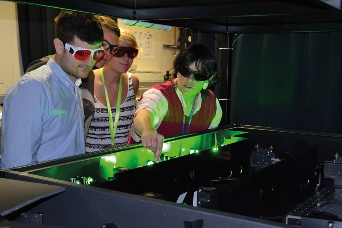
Not following the herd: For Cather Simpson (right) and colleagues, transforming a chip in a lab into a truly portable “lab on a chip” is the next challenge. (Courtesy: University of Auckland)
You sound enthusiastic about it, though. Are you sure it was just New Zealand’s funding system that pushed you into applied research?
I used to be in the “fundamental science is everything” camp, and when I gave lab tours I would say things like: “We do all this applied stuff to fund our basic science habit.” What changed my mind was a project I did with a US company called Intuitive Surgical, which makes a robot called Da Vinci. By chance, their chief research officer heard me say that femtosecond lasers can cut anything and her response was, “Can they cut bone?” So we travelled to Intuitive Surgical and all the people they funded were there talking about what they were doing. I just thought, “Holy crap! This isn’t about figuring something out that might be in a textbook in 15 years; I might actually be able to help real people in three years.” It was a transformative moment for me. Of course, we can’t do without fundamental science: you can’t apply something unless you understand it. But at the same time, I look at the world around us as a challenge-rich environment. Our job, as scientists, is to help meet those challenges.
What do you know now that you wish you’d known when you were getting started?
I wish I’d known more about how the commercial world works. Early on, I couldn’t have told you the difference between series-A and series-B funding, and I had no idea that there were so many ways for the people on the team – mostly students, postdocs and engineers – to reap financial benefits. Engender started with an idea and a tiny amount of “okay, prove it might work!” funding. The university was a major shareholder, a venture capital group put together most of the rest, and as the chief scientist, I have some founding shares as well.
I had no idea that a founding shareholding was different from any other shareholding, but it turns out that the university’s policy is to give a third of what the university owns back to the inventors. That means that our first-phase team will get a third of the revenues from whatever the university eventually realizes on Engender. In contrast, the second phase, where the team showed that each of the individual steps could all happen on the same chip – a really important job – was done as a research contract. Members of that second-phase team got paid for their work, of course, but it wasn’t until afterwards that I realized that their “skin in the game” was different from the first crew’s. Now that Engender’s more established, there are option-related and milestone-linked incentives for the R&D team, and I had to learn how those worked. Luckily, it all worked out so that the R&D team – including me – is properly rewarded and motivated to make sure Engender succeeds, and the intellectual property is linked to the inventors’ properly all the way along. But I still wish I’d had a business class, or that I’d done the reading and picking the brains of experts before starting a company.
Once you step past the money stuff, I think the hardest lesson to learn is about failure. In the real world, you can fail for reasons that have nothing to do with your science. For example, not understanding timelines and deliverables can lead you to fail because you’re not connected with what the real world is anticipating for your business. And failure is not always met with the same equanimity as it is in science. You can’t go to the board of your company and say, “Oh, I tried to sort sperm, but it didn’t work, so I went down this other interesting pathway instead,” like you can with, say, a National Science Foundation grant. That happened to me on a grant earlier in my career and I called up my programme director and said, “Look, you know how I had this whole elaborate five-year plan? There’s no point because my first hypothesis was wrong.” And he said, “Oh, well, that’s all right. Come up with another one.” That would not happen in the business world. I’d be fired. It wouldn’t be my project anymore.
That brings me to a second hard lesson. Right now, Engender is going from a laboratory prototype to a commercial effort. Sometime over the next year, I might not be in charge of the D part of the R&D anymore. As we grow, the company might hire someone who has more experience with that stage. And I’ve been surprised by how much I care about that. You’ve taken this thing, and you’ve put your heart and soul into making it work, and suddenly you realize that actually you don’t get to make the decisions anymore. So, success means that I’m not needed in some ways. That is a little bit hard.
Do you have any advice for anybody thinking of starting up a company in photonics and optics?
Find someone in the business world who you trust and ask lots of questions. The assumptions that people go in with are amazing. I have colleagues who are trying to start companies, or who are trying to do company-facing research, who assume that because it’s their idea, they should own 100% of the intellectual property. But that’s not how business works. As academics we tend to think, well, I wrote the paper on that, therefore I should get the credit. But it’s not about getting credit: it’s about risk and reward, and as soon as you wrap your head around that, you understand the language and the conversations a lot better. You also understand whether it’s for you or not because, for a lot of people, it’s not.
Applying a magnetic field to graphene superlattices produces room temperature quantum oscillations created by delocalized quasiparticles known as magnetic Bloch states. These states should be generic to any superlattice system, not just graphene, and the new finding will be important for fundamental electron transport studies. It will also be crucial for characterizing and understanding novel superlattice devices based on 2D materials like graphene.
“Our study proves that it is possible to create new metallic systems within the same material by applying a magnetic field,” explains team member Roshan Krishna Kumar of the University of Manchester, UK. “These new metals have unique properties compared to those to which no magnetic field is applied, thus providing a new playground for studying condensed matter physics.”
New class of metallic system
The new result comes hot on the heels of work done last year, in which the researchers, led by Sir Andre Geim, observed robust (Brown-Zak) quantum oscillations in the magneto-conductivity of graphene (a sheet of carbon just one atom thick) at certain values of magnetic field. This study, published in Science DOI: 10.1126/science.aal3357, showed that these magnetic Bloch states, could be observed well above room temperature. The new work, detailed in PNAS this time, reveals high-order magnetic Bloch states that represent a new class of metallic system.
“Usually such experiments are done at liquid helium temperatures (around 4 K) to reduce lattice vibrations and study quantized phenomena,” says Krishna Kumar. “The data obtained can be rather difficult to interpret, however, because there can be multiple effects happening all at once. In graphene superlattices, for example, Brown-Zak and Shubnikov-de Haas oscillations co-exist at low temperatures. Our new approach involves heating samples to 100-200 K. This ‘smears’ most of the quantized effects so that our measurements only capture the behaviour of magnetic Bloch states.”
Fractal pattern in the magneto-conductivity
As reported in the PNAS paper, these states are present in second-, third-, and fourth-orders, and this full hierarchy creates a fascinating fractal pattern in the magneto-conductivity, Krishna Kumar tells nanotechweb.org. The pattern is intimately related to the way Hofstadter butterflies (striking fractal patterns that describe the behaviour of electrons in a magnetic field) originate. This complete fractal structure can indeed only appear due to high-order magnetic Bloch states and had never been seen in a graphene superlattice experiment until now.
A Bloch state is the quantum mechanical description of an electron in a solid-state crystal. It explains one of the puzzling questions in condensed matter physics: how can a negatively charged electron move through the crystal without bumping into the positively-charged cores?
Electrons carry on moving as if there were no field
“If we apply a strong magnetic field perpendicular to the crystal, we destroy this Bloch state because electrons experience the Lorentz force and become ‘stuck’ on closed orbits,” explains Krishna Kumar. “In our experiments, we show that electrons can move freely as a Bloch state under a certain applied magnetic field (one whose ‘magnetic length’ is commensurable with the periodicity of the superlattice). In other words, the electrons carry on moving as if there were no field applied.”
Mathematically, this effect occurs each time an integer number of magnetic flux quanta (φ) pierce an integer number of crystal unit cells, according to φ = SB = φ0p/q, where S is the area of a crystal’s unit cell and p and q are integer numbers. “It is impossible to find such magnetic Bloch states in typical crystals because we would require unfeasibly high magnetic fields (of 10 000 Tesla) to see them, says Krishna Kumar. However, the beauty of graphene superlattices is that they are significantly larger than naturally occurring crystals (50 times larger in our case). In these systems, we only have to apply field of around 4 Tesla to observe the magnetic Bloch states, which is relatively easy with the magnets available today.
Any superlattice
We should be able to observe these Bloch states in any superlattice, not just graphene, he adds. “Our findings will be important for electron transport studies, since the physics involved is one of the most fundamental aspects of magneto-transport in solid state-crystals. Our work will thus help us better characterize and understand novel devices based on 2D superlattice materials.
“These states are essentially a new class of material with different properties compared to ordinary graphene. For example, many of the first-order magnetic Bloch states host pronounced band gaps, in contrast to graphene, which is a zero-gapped semi-metal. Now that we have identified higher-order states, our next step will be to understand exactly how they behave.
Like many 2D materials, gallium sports some fascinating properties when in the form of atom thick layers. It has a superconducting transition temperature at 5.4 K and interesting superconductor-metal transitions with increasing magnetic field. In addition, like bulk gallium, which is widely used in electronics in III-V semiconductor compounds, 2D gallenene may also be useful in optoelectronic devices. In a recent 2D Materials report, Jun-Zhong Wang and colleagues at the School of Physical Science and Technology Southwest University in China show how to grow gallenene epitaxially on the (1 1 1) plane of silicon, helping towards the possible incorporation of gallenene in future electronic and optoelectronic devices.
Despite the success of epitaxial growth to produce graphene and other 2D materials, with its low melting temperature growing 2D gallium has presented difficulties. As Wang and colleagues point out the 7 × 7 (1 1 1) plane of silicon – the crystal orientation of the wafers commonly used for silicon electronics – has dangling bonds that interact strongly with gallium atoms and inhibit their migration along the surface.
Previous work by researchers at Rice University and Hysitron Inc in the US reported gallenene production by exfoliating from molten gallium but Wang and colleagues have shown that it is possible to epitaxially grow gallenene on silicon.
Layered success
The researchers deposit a third of a monolayer of gallium on the Si(1 1 1) − 7 × 7 surface at room temperature and anneal it at 550 °C for 30 min to produce a layer of √3 × √3 gallium. They then grow a buffer layer at 50 °C using a beam of gallium at a flux of 0.2 monolayers per minute. On this buffer layer they could readily grow honeycomb 2D gallanene.
They use scanning tunnelling microscopy alongside first principal calculations to derive the structure of the layers, identifying a 4 × √13 structure for the buffer layer. They also confirm the metallic behaviour of the gallenene top layer.
“As we all know, silicon is the backbone of the traditional semiconductor industry,” explain Wang and colleagues in their report. “If gallenene can be synthesized on the surface of silicon, its potential applications in optoelectronic and microelectronic devices will be greatly improved.”
Bone is certainly an impressive natural material because it is both stiff (it endures load without deforming much) and tough (it endures load and deformation without breaking). Stiffness and toughness are rarely found together in materials. The strength of bone comes from two main components: the mineral calcium phosphate (of the apatite variety), and the protein collagen.
The way in which bone collagen and mineral organize themselves has proved difficult to understand, however, because the way they assemble is complex at every length scale. Scanning transmission electron microscopy tomography experiments by researchers led by Roland Kröger of the University of York and Molly Stevens of Imperial College London, both in the UK,have now revealed, among other things, that bone mineral is hierarchically organized from the nanoscale upwards. It is composed of curved needle-like crystallites that merge into twisted plates that form larger aggregates, thus creating a bridge between adjacent collagen fibrils.
The findings could be important for a variety of fields, including osteology and archeology. They will also provide valuable insight into skeletal diseases, growth and development, and might even help in the design of novel biomimetic materials.
Evaluating the mineral phase of bone in 3D
Kröger and Stevens set out to evaluate the mineral phase of bone in 3D. They used electron tomography – that means collecting images at many sequential angles or “tilts” – to reconstruct 3D images of their samples.
One of their observations, for example, was the slight curvature of crystallites across their lengths. Another was stacks of parallel twisted crystallites around irregular voids in the oblique direction. Finally, they saw tight packing of hexagonal crystals into rosette shapes in perpendicular projections. “We developed a model that puts all these observations together that shows that crystals are curved, hierarchical and splaying to form a continuous cross-fibrillar network,” explain Kröger and Stevens.
Fractal-like helical motifs
“The twisting crystals in bone assemble with twisted collagen chains and form fibrils that are twisted themselves”, they add. “As known from earlier studies, the fibrils twist into concentric layers that gently twist themselves around tiny capillaries in bone, which in turn twist around the bone’s long axis, which is often curved into a helix (like a rib, or a collar bone).”
The researchers say that the helical twist is self-similar, or fractal-like. Fractal-like helical motifs are ubiquitous in nature (think of ferns or cauliflower). “This type of organization might have evolved over millions of years to optimize bone structure and function,” lead author of this study Natalie Reznikov tells nanotechweb.org.
Yong Mao of the University of Nottingham in the UK agrees: “This is certainly an interesting study, contributing to the understanding of bone structure,” he comments. “I think researchers are increasingly appreciating the fractal/hierarchical nature of the world, which generally offers an evolutionary advantage. These finer structures are becoming more accessible with the development of additive manufacturing (or 3D printing). I expect this to be an exciting field of development in the future.
Individual meal choices have a profound effect on our environmental footprint, but much of the analysis has been based on aggregated or stereotyped diets. Now researchers have examined the food choices of more than 16,000 US residents on a single day.
The study shows that an environmental saving equivalent to cutting 661 million passenger vehicle miles could occur if just the top 20% of most intensively emitting meals on a given day were replaced with a diet with the average impact – 4.7 kg of carbon dioxide equivalent.
What’s more, these savings soon add up. The group reported that such a hypothetical diet shift – if implemented every day – could bring the US 9.6% closer to achieving UN Framework Convention on Climate Change targets for 2025.
“One of the key outcomes is being able to represent the greenhouse gas emissions and energy impacts associated with the US diet as a distribution across the population,” said Martin Heller of the University of Michigan.
To examine greenhouse gas emissions and energy use meal by meal, Heller and colleagues linked food life cycle assessment studies to the US National Health and Nutrition Examination Survey (NHANES). Featuring over 16,000 responses, the analysis offers a nationally representative snapshot based on self-selected diets across the country over one day.
The linked database reveals some striking differences in food choices and could help to shape future educational campaigns and policy efforts aimed at encouraging diet shift.
The analysis required pairing over 6000 as-consumed foods and dishes with more than 300 life cycle assessment studies. But the challenges didn’t stop there.
“We had to fill in gaps in the literature by making informed proxy assignments,” said Heller. “However, it turns out these proxies have very little effect on the overall results because they tend to be foods that are consumed in low quantities in the US diet and have relatively low impacts.”
So what advice does the team have so far? “The first recommendation is to pay attention to nutritional needs and avoid excess calories as more food equals more impact,” said Heller. “The second is to be mindful of the consumption of animal-based foods.”
The study highlights that even modest reductions in meat and dairy intake can make a big difference in terms of greenhouse gas emissions saved. Adding to this is the issue of food waste.
“NHANES doesn’t have direct data on food losses, but using other data from USDA that we connected to our databases, we estimated that food losses, at both the retail and consumer level, represent about a quarter of impacts from producing the US diet,” said Diego Rose of Tulane University.
The team’s approach opens the door to assessing how diets contribute jointly to both environmental and health outcomes.
“Establishing a framework for linking the environmental impact of foods to individual diets opens up a whole host of research questions, including further exploration into policy options, economic and social justice considerations,” said Heller.
Future plans include expanding the research to factor in other environmental impacts such as water use and land use.
The ESTRO 37 meeting in Barcelona saw Accuray unveil a range of product advancements, including the launch of CTrue IR, an iterative reconstruction algorithm currently available on the Radixact System, which provides image-guided, intensity-modulated radiotherapy.
The Radixact treatment device encompasses integrated 3D volumetric imaging, using low-dose fan-beam CT images for daily set-up and registration. The system also offers Delivery Analysis software that leverages the same integrated detector to perform pre-treatment dose calculations and post-treatment analysis of exit fluence through the patient for every fraction, and generates warnings if any parameters fall outside of clinically acceptable windows.
The new image-guidance software can reconstruct these CT fan-beam scans with enhanced image quality, while maintaining a low imaging dose and the ability to perform live reconstruction as the images are acquired. The main benefit of iterative reconstruction is the much improved signal-to-noise ratio, which in turn increases the soft-tissue contrast.
“Iterative reconstruction reduces the noise by over a factor of two and helps improve image uniformity,” explained Accuray’s Andrea Cox. “It particularly enhances images in the superior-inferior direction, so that the slice transitions are less visible, and improves soft-tissue visibility in all image planes.”
The CTrue IR software is now installed at three customer sites, one in Europe and two in the US, with the first clinical images recorded just a couple of weeks ago. Cox described one of the first clinical test cases that used the new software release – an obese patient with a hip prosthesis.
Such a case could result in a sub-optimal CT scan, due to photon starvation artefacts (which occur due to photon attenuation through obese patients) and metal artefacts arising from the prosthesis. Using the CTrue imaging system with the new iterative reconstruction capability, the image exhibited excellent uniformity, with no problem with artefacts.
“The first customer to use this software found much improved image uniformity,” said Cox. “The therapists are more confident now in the positioning of the patient, they’re really happy with it.”
Construction of a new Super Cryogenic Dark Matter Search (SuperCDMS) dark-matter detector can begin now that the next round of funding for the experiment has been has been approved by the US Department of Energy (DOE). The detector should start taking data in the early 2020s and will be located 2 km below the Earth’s surface at SNOLAB in Sudbury, Canada. The experiment will be at least 50 times more sensitive than the previous SuperCDMS experiment, which ran in 2012-13 in the Soudan Underground Laboratory in Minnesota.
The DOE will contribute $19m towards building the detector, while the US National Science Foundation will pay $12m and the Canadian Foundation for Innovation will fork out $3m. The SuperCDMS collaboration involves 111 physicists at 26 institutions in the US, Canada, France and the UK.
The detector will look for weakly interacting massive particles (WIMPs), which are hypothetical dark-matter particles that could account for 85% of the matter in the universe. While indirect evidence for dark matter abounds in astronomical observations, physicists have yet to make a direct detection of the mysterious stuff. Doing so would be a major leap forward in astrophysics and could also point to new physics beyond the Standard Model of particle physics.
Extreme sensitivity
WIMPs are a family of hypothetical dark-matter particles that interact very weakly with ordinary matter. This means that if physicists want to detect WIMPs, they must use an extremely sensitive detector – ideally located deep underground, where it is shielded from cosmic rays and other background radiation.
According to Richard Partridge, head of the SuperCDMS group at the Kavli Institute for Particle Astrophysics and Cosmology at Stanford University, the new experiment will be “the world’s most sensitive for relatively light WIMPs – in a mass range from a fraction of the proton mass to about 10 proton masses”. He calls this sensitivity “unparalleled” and says it will “create exciting opportunities to explore new territory in dark-matter research”.
The SNOLAB experiment will look for WIMPs using silicon and germanium crystals that will be cooled to about 10 mK and surrounded by superconducting aluminium. The hope is that WIMPs will interact with an atomic nucleus in a crystal, creating tiny vibrations (or phonons) that propagate to the edge of the crystal and enter the aluminium. Once in the aluminium, the phonons transfer their energy to superconducting Cooper pairs, causing pairs to break and leading to a large increase in electrical resistance, which can be detected. WIMP-nucleus interactions should also produces electrons and holes in the crystal, which will be detected electronically.
Oversized hockey pucks
Six crystals – each resembling an oversized hockey puck (see figure) – will be arranged in a “detector tower” and the new SuperCDMS will comprise four towers along with the associated electronics, cryogenics and shielding. A detector prototype has been tested successfully at SLAC National Accelereator Laboratory in California and the first detector tower should be delivered to SNOLAB by the end of 2018.
“The detector towers are the most technologically challenging part of the experiment, pushing the frontiers of our understanding of low-temperature devices and superconducting readout,” explains SuperCDMS team member Bernard Sadoulet of the University of California, Berkeley.