Hopes rise for some coral survival

Researchers have raised hopes that limited coral survival may be possible, allowing one of the world’s best-known reefs to survive a little longer.

Although corals are highly sensitive to ocean warming, and notoriously bleach when temperatures exceed a certain limit, a new study has shown that at least one coral can evolve tolerance to excessive temperatures.

The implication is that even though other teams have repeatedly warned that the world’s reefs are in peril as the world warms because of ever-greater ratios of greenhouse gases in the atmosphere, as a consequence of human combustion of fossil fuels at a profligate rate, the world’s great reefs may survive for perhaps another century, rather than perish within the next 50 years.

“It means these corals will still go extinct if we do nothing,” said Misha Matz, of the University of Texas at Austin, who led the study. “But it also means we have a chance to save them. It buys us time to actually do something about global warming, which is the main problem.”

The argument is based on Darwinian logic: coral colonies produce colossal numbers of larvae each year, set adrift on ocean currents to colonise new reefs. As conditions change, those corals that by an accident of genetic inheritance have the traits needed to cope with environmental challenge will get a foothold, and flourish. Those that don’t will fade out. Natural selection will respond.

And this is hopeful news, if only because the world’s reefs are under threat as never before. Bleaching – the response to heat in which coral rejects the algae with which it normally lives in symbiosis – has always happened: research earlier this year suggests it could become five times more frequent, and reefs such as Australia’s Great Barrier would have no time to recover.

Some reefs have already been pronounced too damaged ever to be restored. This is bad news not just for the coral animals: the tropical reefs are just about the richest habitats on the planet, and of profound economic importance to humans too.

A partnership of US and Australian scientists reports in the Public Library of Science journal PLOS Genetics that computer simulation models and genetic evidence of variation from one species of staghorn coral, called Acropora millepora, together show that the coral could in theory adapt over a stretch of 20 to 50 generations.

“This genetic variation is like fuel for natural selection,” Dr Matz said. “If there is enough of it, evolution can be remarkably fast, because all it needs to do is reshuffle the existing variants between the populations.

“It doesn’t have to wait for a new mutation to appear; it’s already there. The problem is, when the genetic variation is exhausted, it is over and the future is unclear.”

Tentative conclusions

There are problems with such studies. This one is based on genetic evidence from one species of coral. But the 2,300 km Great Barrier Reef of Australia is home to at least 411 species of hard coral. It is based on a mathematical model, not on observed change in the reefs.

And global warming is not the only challenge to coral reefs, which are also threatened by human exploitation, pollution and increasing acidification of the surrounding seas, again as a consequence of ever higher levels of carbon dioxide in the atmosphere.

“Corals live in a symbiotic relationship with zooxanthellae, which are plant-like cells hosted in surface tissues that provide up to 90% of the energy to the colony,” said Stephen Simpson, a marine biologist at the University of Exeter in the UK, commenting on the study.

“Whether there is also sufficient genotypic variation in the zooxanthellae to tolerate further warming remains to be seen. While the fact that one species may do well is good news, there are many other reef organisms that may fare far worse, so it is easy to envisage a future with a few winners but many losers, threatening the functional integrity of reef ecosystems.”– Climate News Network

Laser bioprints stem cells

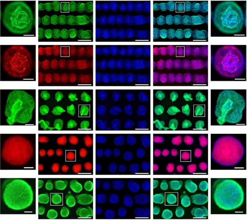
Researchers in Germany have succeeded in laser printing a special type of stem cell – so-called human-induced pluripotent stem cells (hiPSCs) – for the first time. These hiPSCs are prized for their ability to be differentiated into any type of human cell, a property called pluripotency, plus they can be generated from a patient’s own cells to avoid the risk of an immune response when they are implanted back into the body.

As a result, many scientists believe that printed hiPSCs offer the best option yet for making replacement organs or organ-on-chip systems for personalized drug testing. The problem is that these cells are notoriously fragile and difficult to handle, especially when dissociated into single cells.

Laser printing of stem cells

“Dissociation is required for printing these cells in high resolution patterns, but it induces programmed cell death (although this can be delayed by adding specific culture medium supplements),” explains Lothar Koch of Laser Zentrum Hannover, lead author of the study published in Biofabrication. “They are thus difficult to print with established techniques such as extrusion, ink-jet printing, acoustic droplet ejection techniques, laser-guided direct write, and laser bioprinting.”

What’s more, continues Koch, the cells’ pluripotency and directed differentiation are affected by environmental factors, such as the composition of the culture media in which they are grown and the bioinks used to print them. They are also sensitive to mechanical forces, such as the shear stresses that can occur during bioprinting processes.

The new laser printing technique developed by Koch and colleagues exploits laser pulses to expel tiny droplets of a bioink containing suspended hiPSCs from a thin layer of bioink deposited on a glass slide. “The main difference with previous approaches, such as extrusion or ink-jet printing, is the absence of a nozzle,” says Koch. “Although this makes preparation and application of the bioink more complex, it does mean that we avoid the high shear forces that usually occur in small nozzles.”

Any 2D pattern and 3D layer-by-layer patterns

The technique combines small droplet printing, down to a few picolitre volumes, with high-viscosity bioink printing and high cell densities of up to 108 cells per millilitre. “Each of these points can be achieved with other printing techniques too, but not in combination,” adds Koch.

The laser bioprinting set-up comprises the laser and two glass slides. “We coat the top slide with a thin layer of laser-absorbing materials,” says Koch. “This might be a biocompatible metal such as gold or titanium, or a polymer such as triazene or polyamide, or even a hydrogel like gelatine. Then, we deposit the biomaterial to be printed – usually a cell-containing sol – as a second layer on top of the absorption layer.”

The coated glass slide is mounted upside-down above a second slide, and 10 ns laser pulses are then focused through the upper slide into the absorption layer. “This causes a vapour bubble to expand and propel a small volume of the biomaterial towards the lower glass slide,” explains Koch. “By moving the laser and the glass slides, we can print any 2D pattern and also generate 3D layer-by-layer patterns. It is also possible to put substrates or scaffolds on the lower glass slide and print the biomaterial directly onto the substrate or scaffold.”

Survival rate of nearly 100%

Koch told Physics World that all the cell types tested in these experiments survived this printing procedure, with a survival rate of nearly 100%. The technique also retains the pluripotency of the cells, and allows them to be printed into highly controlled patterns to generate functional tissue substitutes.

The researchers tested a variety of hydrogels as bioinks and culture substrates for printing, and found that fibrinogen, blood plasma, Matrigel and hyaluronic acid were particularly suitable. “Hyaluronic acid is naturally found in the human body; it is generated in the early stage of embryogenesis and is abundantly found in stem cell niche environments,” explains Koch. “It enhances the proliferation of stem cells and supports pluripotency too. It is also good for the laser-printing process and allows us to fine tune the bioink’s viscosity.”

While Koch believes that hiPSCs are the most promising types of cells for printing tissue or organs, he highlights the need to investigate the process in more detail. “An important question we must ask ourselves is: which stage of differentiation is the optimal one for printing? To answer this, we need to investigate the printability of hiPSCs in all possible stages of differentiation.”

The team, says that it will now be developing a more advanced bioink to print more complex 3D structures. “Another interesting application for printed hiPSCs is to use them to generate cell constructs that mimic embryoid bodies for modelling human development or investigating diseases,” says Koch. “We will be looking to print such embryonal organoid models with our technique.”

  • Read our special collection “Frontiers in biofabrication” to learn more about the latest advances in tissue engineering. This article is one of a series of reports highlighting high-impact research published in the IOP Publishing journal Biofabrication.

Functional MRI scans could help identify brain disorders

AuntMinnie logoUsing functional connectivity MRI (fcMRI) scans to show how various brain networks interact and activate to perform basic daily tasks may someday help diagnose abnormalities in people with brain disorders, according to a study published April 18 in Neuron.

While the findings are preliminary, researchers from Washington University in St. Louis are hopeful that the approach could provide insight into variations in cognitive ability and personality traits, as well as brain dysfunction.

For the study, nine researchers took turns undergoing a variety of MRI scans late at night, dubbing their group the Midnight Scan Club, according to a release from the university. During the scans, each person performed tasks related to vision, memory, reading or motor skills, or the person rested quietly. The group then analysed data from more than 10 hours of fcMRI scans and 10 separate one-hour sessions performed on each person (Neuron 98 439-452.e5).

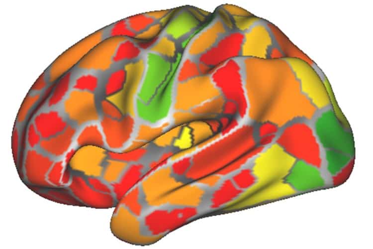
A heat map using data from nine brain networks

From the data, first author Caterina Gratton created a dynamic functional connectivity map of the brain’s outer surface and activity changes in 333 regions over time. The goal was to identify areas that became active and inactive in unison. Network maps were also created for each individual, showing patterns of correlation between parts of the brain.

The vast amount of data for each person allowed Gratton to determine how much an individual’s brain networks changed from day to day based on different mental tasks. The conclusion: There is very little change at all.

“Whether someone’s watching a movie or thinking about their breakfast or moving their hands makes only a small difference,” she said in the release.

That consistency actually makes fcMRI a promising diagnostic tool to identify brain disorders and diseases. While the technique’s potential was noted years ago, fcMRI-based diagnostic tests have not become part of the clinical routine because clinicians cannot tell which scans reflect fundamental, stable features of the brain or if the brain changes with every passing thought.

More data are needed before researchers can accurately determine the difference between normal variation in brain activity and a disorder or abnormal activity.

“The individual differences were really easy to pick up, even in a population that is really very similar,” Gratton said. “It is exciting to think that these individual differences may be related to personality, cognitive ability, or psychiatric or neurological disease. Thanks to this work, we know we have a reliable tool to study these possibilities.”

  • This article was originally published on AuntMinnie.com.
    © 2018 by AuntMinnie.com. Any copying, republication or redistribution of AuntMinnie.com content is expressly prohibited without the prior written consent of AuntMinnie.com.

Early universe simulated in a cloud of ultracold atoms

The early universe has been mimicked in the lab using a ring-shaped Bose-Einstein condensate (BEC) of ultracold atoms. Gretchen Campbell, Stephen Eckel and colleagues at the Joint Quantum Institute at the University of Maryland expanded the size of their ultracold atomic cloud at supersonic speed and observed several effects believed to be associated with the inflationary epoch of the early universe. This epoch is thought to have occurred less than 10–32 s second after the Big Bang when the universe expanded at an exponential rate.

Bose–Einstein condensates (BECs) are formed when identical atoms with integer spin are cooled until all the atoms are in the same low-energy quantum state. This means that a BEC comprising tens of thousands of atoms behaves as a single quantum entity. A BEC can be thought of as a vacuum state for phonons, which are quanta of vibrational mechanical energy. The team used this phonon vacuum as an analogy for the quantum field vacuum in the early universe.

In one experiment, the researchers introduced a sound wave onto their cloud to see how it evolved during expansion. The phonon wavelength increases (or redshifts) as the expansion occurs, thereby providing an analogy with how photons redshift in an expanding universe. They also saw that the amplitude of the wave decreases during expansion. The team has made a tentative connection between this effect and a strange cosmological damping phenomenon called “Hubble friction”.

End of an epoch

In another test, the team expanded the BEC with no sound waves, and watched it stabilize after it reached its maximum radius, a state analogous to the end of the universe’s inflationary epoch. Here, energy that had been powering inflation quickly translated into unstable BEC solitons and vortices, producing phonons.

“We see the creation of excitations in a way that is reminiscent of preheating and reheating in the early universe,” explain Campbell and Eckel. “After inflation, all of the energy in the universe was presumably contained within a quantum field that drove inflation called the ‘inflaton’. That field decayed, depositing its energy into lower mass particles causing the universe to heat up (hence, the term reheating).”

The team is now working to improve their expanding ring BEC model to better observe the damping they associate with Hubble friction. Looking future ahead, the physicists hope to generate correlated pairs of sound waves that mimic Hawking radiation from black holes.  They also hope to simulate cosmological horizons by creating casually-disconnected regions in a condensate.

“Beautifully executed”

Although unconvinced by the connection with preheating and reheating, Silke Weinfurtner of the University of Nottingham thinks the experiments are “beautifully executed” and “a significant step forward in carrying out table-top analogue gravity experiments”. “Overall, analogue gravity cosmology experiments allow us to test some of the fascinating physics in a controlled laboratory setting,” she adds.

Yet as with other analogue cosmology experiments, the key test will be if the researchers uncover new physics or confirm new cosmological theories: “We have already learned a lot from cosmology, but it’s not yet clear if we will in turn guide cosmology,” note Campbell and Eckel. “Our hope is that our system could provide a testbed where we could actually study new models and see what happens.”

The research is described in Physical Review X.


Funnelling charges to boost solar-cell efficiency

Funnels are efficient tools for channelling liquids into containers with narrow openings. Now, researchers in Exeter have demonstrated the first funnel for electrical charges on a chip. The discovery builds on the ability to oxidize the atomically thin semiconductor, hafnium disulphide (HfS2), with a high-intensity UV laser. The non-uniform strain between oxidized and non-oxidized regions, and the subsequent band-gap modulation, generates electric fields, which effectively funnel the charges in the semiconductor flakes to areas where they can be more easily collected. This concept could enable a new generation of solar cells with 60% efficiency (currently around 21%), thanks to the increased efficiency in collecting photo-excited charges and the potential for hot-carrier extraction.

Intense laser light means oxidation, oxidation means strain

In general, bulk semiconductors can only sustain strains up to 0.4% before breaking. However, a layer of semiconductor that is only a few atoms thick can support strains of up to 25%. This amount of strain changes the band gap in the energy dispersion by up to 1 eV.

In this work, Saverio Russo and his group at the University of Exeter, induce the strain in the HfS2 using a 375 nm laser to remove sulphur atoms, which are then replaced by oxygen atoms. According to calculations performed using density functional theory, the hafnium atoms have different separations in HfS2 and HfO2. This produces a 2.7% strain at the boundary between the oxidized and non-oxidized regions. Electrical contacts anchor the material to a substrate, so a strain gradient is present across the whole flake, shifting asymmetrically the conduction and valence bands to higher energies, and opening the band gap by 30 meV.

Collecting the funnelled charges

Another key to making a useful charge funnel is ensuring that the charges will move towards an area where they are more easily extracted. In most direct semiconductors (such as GaAs), the funnelling effect pushes charges towards the strained region. By using HfS2, which is an indirect semiconductor, the charges were pushed away from the strained region, towards the electrodes at the edge of the flake, as observed in inverse charge funnelling.

To test the inverse charge funnelling, a field effect transistor was made using a flake of patterned HfS2/HfO2. The researchers used a laser to excite charges in the flake and scanning photocurrent microscopy to measure the current from these charges. Comparing the same device before and after a small area was oxidized, they measured a 350% increase in response in a region near the interface between HfS2 and HfO2. Their theoretical calculations of the system supported the results, and also revealed that the charge carriers had an increased lifetime of around four orders of magnitude – another indicator of the charge funnelling effect.

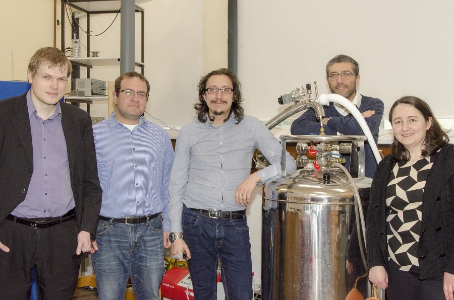
The team from the Quantum Systems and Nanomaterials group at the University of Exeter

Looking to the future

So how can this new phenomenon be implemented in future technologies, and what benefits will this lead to? Lead author, Adolfo De Sanctis, had this to say: “The electrical measurement of the charge funnelling effect could be revolutionary for photovoltaic devices. Two-dimensional semiconductors are truly unique when it comes to strain, and they offer the best platform for future ‘straintronic’ devices. The next steps will be to implement this technique on a larger scale, optimize the strain gradients, and study other materials in order to demonstrate the feasibility for photovoltaic solar energy conversion.”

Read the full article in Nature Communications.

Light is confined to an atom-wide gap

Light has been confined to a gap just one atom wide by an international team of researchers. While their work is very preliminary, it could find a range of applications including the development of tiny but powerful lasers and the extreme miniaturization of devices that use light to transmit and process information.

Photonic devices that use light pulses to transmit and process information offer important advantages over conventional electronics. Optical pulses travel faster through optical fibres and suffer much lower losses than electrical pulses sent along metal wires – and multiple signals can be sent down the same fibre without interacting with each other.

However, miniaturizing components to create nanophotonic devices remains an important challenge. Whereas electronic transistors less than 10 nm in size can be made, optical fibres are best suited for transmitting infrared light with a wavelength of about 1550 nm. Normally light cannot be confined in spaces smaller than its wavelength and so this puts a lower limit on how small photonic devices can be made.

Excited electrons

Particle-like collective excitations of electrons on the surface of a metal – called surface plasmons — could offer a way forward. Plasmons in a narrow gap between two metal surfaces can couple to a photon of light to create a surface plasmon polariton (SPP). This is a photon-like entity that can be confined in regions much smaller than the wavelength of the original photon. What is more, when the SPP reaches the end of the confining gap, the photon can be re-emitted. Another benefit of using plasmons is that they offer new ways for photons and electrons to interact, which could lead to new types of optoelectronic devices.

There are limits, however, on how narrow the gap can be. Below about 15 nm, the electric field of the confined light penetrates below the metal surface, exciting plasma oscillations that dissipate energy in a process called Landau damping. An alternative is to excite plasmons in graphene, which is only one atom thick. However, this has previously required patterning the graphene into nanoribbons: “When you pattern graphene, you have to etch it away where you don’t want it,” explains Frank Koppens of the Institute of Photonic Sciences in Barcelona. “You get rough, non-crystalline edges, and those affect the electronic and optical quality.”

Koppens and colleagues in Spain, France, Portugal and the US have now taken a different approach to using graphene. They begin with a single, crystalline sheet of graphene, that they cover with a layer of insulator. Then they add a periodic array of gold bars on top of the insulator. By shining an infrared laser on to the top of the structure, the researchers were able to excite plasmons in the graphene through the electric field in the tiny gap between the gold bars and the graphene layer. By patterning the external environment rather than the graphene, says Koppens, the researchers could neatly sidestep graphene edge effects.

Plasma frequency

The team experimented with different thicknesses of insulator and found that, down to about 3 nm thickness, the plasmons stayed well confined in the gap and did not penetrate significantly into the gold. Even when the gap was reduced to an atomic layer of boron nitride just 0.7 nm thick, the electric field leaking into the metal did not lead to Landau damping. The researchers believe this is because the resonant frequency of the graphene plasmons is much smaller than the plasma frequency in the metal, so even when the electric field spreads into the metal, it does not excite oscillations or dissipate energy.

In the near-term, says Koppens, the research could offer significant interest for enhancing nanoscale light-matter interactions, allowing much more powerful on-chip infrared emitters and possibly infrared lasers. The researchers also plan to investigate the possibility of plasmonic interconnects for electrical signals: “We have shown already that you can efficiently go from plasmon to electron, but we want to show also that you can go from electron to plasmon,” he says.

“This work is very exciting to me because, ideally, it provides a playground for how we can transmit and modulate light in an atomically thin waveguide structure,” says nanophotonics expert Nicholas Fang of Massachusetts Institute of Technology, who was not involved in the research. He cautions, however, that much work remains to be done before viable devices can be made.

“Massive paradigm shift”

Graphene expert Andrea Ferrari of the University of Cambridge agrees: “If we take this to its most extreme and futuristic implication, then we’re opening a new field where you could have light-driven optoelectronics at a scale similar to electronics: that would be a massive paradigm shift in technology,” he says. “I’m not saying that tomorrow or even in 20 years we’re going to have that, but even if it never goes to practical applications, the conceptual result is it. If you asked me or anybody else a few years ago, everybody would have said this was completely impossible.”

The research is described in Science.

Vertual brings the linac into the classroom

When pilots learn to fly a plane, they train on a flight simulator that artificially re-creates the aircraft environment – enabling them to hone their skills and practise expected and, importantly, unexpected flight scenarios with zero risk. So why not employ the same approach for radiotherapy? That’s the underlying premise of VERT (virtual environment for radiation therapy training) – a training simulator developed by UK company Vertual.

“Around 2000, I was teaching radiotherapy at Sheffield Hallam University,” explained Andy Beavis, Vertual’s CSO and radiotherapy director. “It would have been a lot easier to teach them in a linac bunker, but you can’t do that because it is being used to treat patients. So I had the idea of bringing the linac into the classroom instead.”

To do this, Beavis and colleagues established Vertual, a spin-off company from Hull University and Hull and East Yorkshire Hospital NHS Trust, to create tools for radiotherapy training and education. He notes that the development came about due to a collaboration between scientists from different fields, with his background in radiotherapy physics complementing that of the other founders, who were computer scientists. “It is truly one of those examples where the parts are greater than the whole,” he said.

Vertual’s product – VERT – creates a detailed simulation of a treatment room and linac, and uploads a patient model, using DICOM to transfer real patient data if required. The interactive 3D display shows the planned beam delivery, runs a virtual treatment and can even show the dose deposition inside the patient.

“VERT performs 3D linac simulations, using authentic hand controls from an actual linac, to give students hands-on experience of a treatment machine,” explained James Ward, managing director and a co-founder of the company. “It uses the same rationale as flight simulators, providing a completely safe environment for training. No-one is going to harm the patient. It’s the only simulator of this type in the world.”

When the company launched in 2007, VERT was rolled out to all radiotherapy training schools in England. “In the UK, we changed the way therapy radiography was taught,” said Beavis. “It was a great way to begin, as it also gave us a large bank of users who fed back to us and helped improve the software.”

Today, there are 138 VERT systems installed at 130 sites in 26 countries. While the main users are therapy radiographers, the system is also used to train medical physicists, dosimetrists and oncologists. It has also been used as a tool to explain the concept of radiation treatment to patients and their families.

Next step: protons
At the ESTRO 37 congress in Barcelona, Vertual was showcasing its newly launched Proton VERT, a simulator for proton therapy training. “Adoption of proton therapy has really accelerated, there are systems in operation in the UK now,” explained Ward. “This is a natural extension of our product and we anticipate it being useful for our customers in many countries.”

The initial release incorporates an interactive simulation of the Varian ProBeam proton gantry. It also includes a functional model of the ProBeam robotic treatment couch and integrates the ProBeam hand controls. The company notes that future developments will include modelling of other vendor’s proton systems.

As with its linac counterpart, Proton VERT can be used throughout all stages of the proton therapy process, providing 3D visualizations of treatment plans displayed on the machine, as well as simulated beam delivery. “Proton VERT provides tools to help people thoroughly understand the nuances of proton therapy, and to ensure that treatment is implemented in the right way,” said Beavis.

Beavis noted that one major advantage of both the linac and the proton version is that trainees can simulate what would happen if a treatment goes wrong. “They can see the effects of dose delivered to the wrong place, with no harm done,” he explained. “You’d struggle to find a pilot who hasn’t trained on a flight simulator first. We want to see the same here.”

Why aren’t China’s wind farms producing more electricity?

China has made large investments in wind power over the past decade, but estimates suggest that Chinese wind farms are producing less electricity than hoped for. Writing in Environmental Research Letters (ERL), a team advises that if the country is to achieve national goals and increase utilization up to US levels, in addition to addressing the much-discussed issues related to grid integration, China will need policy measures to address turbine siting and technology choices.

The study concludes that the gap between actual performance and technical potential is driven by delays in grid connection (14% of the gap) and curtailment due to constraints in grid management (10% of the gap). But the analysis doesn’t stop there.

“Our findings show that China’s underperformance is also driven by suboptimal turbine model selection (31% of the gap), wind farm siting (23% of the gap), and turbine hub heights (6% of the gap) – factors that have received less attention in the literature and, crucially, are locked-in for the lifetime of wind farms,” explain the authors, who were supported by Harvard University, US, and are now at the University of Minnesota, US, Florida State University, US, and Cambridge University, UK.

China aims to reach 210 GW of grid-connected wind capacity by 2020, as part of its 13th Energy Technology Innovation Five Year Plan . Today, it’s thought that the country has a capacity in the region of 169 GW, compared with 154 GW for the EU and 82 GW for the US.

However, as the team points out, it’s important to look beyond the installed capacity and examine how much electricity is fed into the electric grid to displace conventional, polluting power sources.

“In 2016, the European Union generated 36% more electricity per unit of installed wind capacity than China, and the United States generated 93% more,” calculate the researchers in their study.

The scientists note that China generated in the region of 241 TWh of wind power from its installed capacity of around 169 GW. These figures imply an approximate capacity factor of 16.5%, which compares with 32% for the US.

By digging deeper into the reasons, scientists hope to improve China’s prospects of maximizing the human health and environmental benefits that wind power has to offer.

A key feature of the work is the use of wind farm-level data to reveal the relative importance of each driver. These rankings highlight that design choices such as location, tower height and turbine model play a major role in whether a project meets its objectives. The overall picture, though, is one of being able to strike a balance.

Trade-offs include land availability and prices, wind resource quality, proximity of the site to the electric grid and service roads, and the level of available feed-in tariffs to guarantee revenue.

Another recommendation from the team is to increase access to multi-year wind measurements at specific locations, particularly where planners currently rely on short-term wind measurements or measurements from nearby weather stations.

Now the researchers are looking to apply these results to inform China’s national greenhouse gas emission reduction policies.

Plasmonic windows by design

A new composite material developed by physicists at Ohio University in the US, together with colleagues in China and Canada, can be used to create cost-efficient glasses and films based on plasmonic crystals that block all ultraviolet and infrared radiation while remaining transparent to visible light. The researchers investigated how nanoparticles of different shapes and materials interact with light, and their findings could help in the development of windows that would reduce the amount of heat entering a room or vehicle, thus ultimately reducing the need for energy-hungry air-conditioning or other cooling systems.

There are several ways to make spectrally selective materials, explain Alexander Govorov and Lucas Besteiro in Ohio, who led this research effort. One involves covering glass with a multi-layered film that reflects infrared light, while transmitting light in the visible part of the solar spectrum. Another is to use near-infrared interacting materials that can block certain solar light wavelengths.

‘Metaglasses’ containing plasmonic nanocrystals

“In our work, we describe an approach to create passive infrared-blocking ‘metaglasses’ containing plasmonic nanocrystals. We show that mixtures of specially shaped plasmonic nanocrystals made of noble metals (silver and gold) and alternative plasmonic materials (titanium nitride, aluminium and copper) can efficiently block infrared solar radiation.”

Plasmonics is a branch of photonics that exploits surface plasmons (SPs) for enhancing light–matter interactions. These SPs arise from the interaction of light with the electrons, which makes them oscillate within the metal. Plasmonic metamaterials are artificially engineered collections of metal nanostructures that can be fine-tuned to interact with light in very specific ways. With these materials, it is possible to adjust the shape, size and arrangement of the structures to support SPs at specific frequencies.

Classic electrodynamic simulations

“Our methodology relies, first of all, on classic electrodynamic simulations of the different nanoparticles that we are studying,” Govorov and Besteiro tell nanotechweb.org. “These calculations provide us with profiles of how the nanoparticles interact with light. Using that information, we can compute the expected light transmission of a glass containing a given ensemble of plasmonic nanoparticles. We can then look for the ensembles that better reproduce our ideal transmission profile target – that is, one that blocks all ultraviolet and infrared solar radiation while remaining transparent to visible light.”

The shape of the nanoparticle is important too. Indeed, the researchers found that glasses designed with plasmonic nanoshells were better at specifically blocking infrared radiation than nanorods and nanocups.

Nanoshell’s rotational symmetry is important

“One of the reasons why nanoshells are better is because they have rotational symmetry,” explains Besteiro. “This means that when they are dispersed in the supporting media (glass or a polymer, for instance), all the particles with a similar size resonate at the same (infrared) frequency.

“Let’s compare this to the behaviour of rods: although we have a good degree of control over their (infrared) resonant frequency (by changing their aspect ratio), only a fraction of the nanorods in a randomly oriented ensemble will be excited at the main resonant mode of the particle, which for a rod is along its longitudinal axis. This means that to achieve a certain level of opacity at that frequency, we must increase the number of rods. This, in turn, increases the overall volume of the material, which has the adverse effect of blocking light at frequencies other than that of the longitudinal mode’s.”

“This is not good for us,” add Govorov and Besteiro, “since we want to avoid increasing their extinction in the visible range, at which we want the glass to remain transparent. A similar argument holds for nanocups. These shapes also have the added disadvantage of having wider light extinction profiles that make it even harder to limit their interaction with visible light.”

Towards creating functional prototypes

That said, these three geometries (shells, rods and cups) are nevertheless better suited for infrared-blocking window applications than the comparatively simpler spherical-shaped nanoparticles. The spectral properties of these spheres cannot be tuned as easily by varying their size. More importantly, they interact strongly with visible light.

The team, which includes researchers from the University of Electronic Science and Technology of China and the Institut National de la Recherche Scientifique in Quebec, Canada, says that its approach could help in the development of cheaper energy-efficient windows. “The next logical step is to work on actually creating functional prototypes of these windows and compare them to those already on the market – not only in terms of optical performance but also regarding their cost. We are talking to applied research labs and companies that work on glasses and windows to this end.”

The research is detailed in Nano Letters 10.1021/acs.nanolett.8b00764.

Upconverting nanophosphors make good radiotracers

A new technique to synthesize radioactive upconverting nanocrystals that was previously used to extract uranium from ore could be used to make efficient radiotracers for biomedical imaging, say researchers at the University of Pennsylvania in the US. The nanophosphors, which emit beta-particles, could be ideal in theranostic applications and even targeted in vivo imaging when combined with CT scanning.

Upconverting materials emit light at a wavelength that is shorter than the wavelength of light they have been photoexcited with and are promising for applications in biomedical imaging. The so-called anti-Stokes shift in these materials limits the autofluorescence of nearby molecules within a sample. This significantly reduces background signals, allowing for better target detection.

Rare-earth compounds for upconversion

Although there are several materials and molecules capable of upconversion, rare-earth compounds are particularly good at converting near-infrared (NIR) light to visible light. These materials transfer the energy from the absorbed NIR photons in the form of excitons (excited electron-hole pairs) so that they are emitted with a higher energy (or shorter wavelength).

Another form of imaging commonly employed in nuclear medicine relies on unstable isotopes within radiolabelled molecules or nanoparticles (known as radiotracers) to produce a signal around the target. Radiotracers do not need to be externally excited, which allows the signals coming from them (beta-emission, for example) to penetrate deeper into tissue than optically-stimulated nanoparticles.

Beta-emission and upconversion in one system

A team led by Christopher Murray made the nanophosphor sodium yttrium fluoride (NaFY) doped with the rare-earths erbium (Er) and ytterbium (Yb), and radiolabelled with 90-Y. This nanocrystal upconverts near-infrared light to the visible thanks to the rare-earth dopants and emits beta-particles thanks to the 90-Y.

The presence of both beta-emission and upconversion in one system is very rare in nature, explains team member and lead author this study Stan Najmr, and allows for deep tissue imaging because the two modalities are involved.

Hydroxide metathesis method

The researchers made their nanophosphors using a hydroxide metathesis method that was previously used to extract uranium from ore. “This method allows us to exchange one anion (for example, chloride) with another (trifluoroacetic acid), which is crucial for homogenously incorporating the radioactive element into the host matrix of the nanophosphor,” says Najmr.

The nanophosphors are produced through the rapid thermal decomposition of the rare-earth trifluoroacetates. “Once they have been synthesised, we silica coat them using the ‘Stober technique’. This allows us to disperse them in water, which is required for biomedical applications. Since these nanocrystals are fluorides, they are fairly inert to their environment.”

Extending the technique to other radioactive rare-earths

“This particle system is an excellent candidate for biomedical trials, especially in theranostics,” Najmr tells nanotechweb.org. “Combined with CT scanning and surface functionalisation, these nanocrystals could be used for targeted in vivo imaging.”

The researchers hope that their work will encourage other groups to consider this synthesis route for developing rare-earth nanocrystals and silica architectures. “For our part, we are eager to extend this technique to other radioactive rare-earths, such as lutetium (Lu) and samarium (Sm),” says Najmr. “These elements will provide new signals like gamma radiation that might lead to additional pathways for multimodal imaging.”

The research is detailed in in Nano Futures 2 025002.

Copyright © 2026 by IOP Publishing Ltd and individual contributors