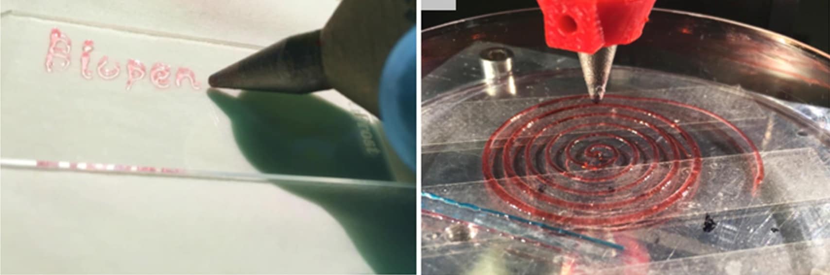
As 3D printing technology evolves and advances, we are getting ever closer to the goal of being able to implant 3D printed tissues inside the body. At present, 3D printed cells must first be cultivated and then allowed to grow into viable tissue – which normally takes a few days. But a new instrument, dubbed the biopen, could help to speed up this process. The biopen, developed by researchers in Australia, could allow surgeons to repair damaged bone and cartilage by “drawing” new cells directly onto bone during surgery and then filling in any damaged areas.
The researchers, led by Gordon Wallace of the University of Wollongong in close collaboration with Peter Choong, an orthopaedic surgeon at St Vincent’s Hospital in Melbourne, first reported their technique in the journal Biofabricationin 2016, and have now tested it out in experiments with sheep.
“The data obtained to date is highly promising,” says Wallace. “We are continuing to optimize printing protocols to demonstrate the versatility of the biopen.” The team is also now working with a manufacturer to produce a commercial device.
The prototype pen described in Biofabrication is made of medical grade ABS-like material and titanium 6Al4V alloy. It works by extruding a bioink, composed of stem cells inside a biopolymer, onto the surface of bone or cartilage. The ink is then solidified by photocuring it with light from an ultraviolet source fitted to the side of the pen. The device draws the stem-cell-containing structures directly onto a defect, and these structures subsequently differentiate into cartilage.
Specific clinical applications
In their paper, Wallace and colleagues report on using biopolymers such as those based on alginate, a seaweed extract, which was in turn protected by a hydrogel. Since then, they have been trying to identify other naturally occurring sources of biopolymers that may be suitable for bioinks, subject to some chemical modification.
“The biopen was our first foray into customized printing approaches for specific clinical applications,” Wallace told Physics World. “We now have an advanced programme for developing specially-designed printers and bioink formulations for use in a number of areas.”
“One of these is treating eye damage in the iFixpen project, which is a collaborative venture with the Sydney Eye Hospital,” Wallace continues. “Another is fabricating 3D-printed structures containing human islet cells for transplantation, which we are working on in association with the Royal Adelaide Hospital.”
A third project – which aims to create 3D printed ears for children with part of an ear missing – has also brought some interesting challenges, he adds. In this case multimaterial and stem-cell-containing structures must be printed in the appropriate shape, which has required the researchers to develop multiple ink formulations and a multihead printing system that will be provided by the Royal Price Alfred Hospital in Sydney.
“We also have a new fundamental research programme looking at printing and developing stem cells, such as induced pluripotent stem cells, and how they develop into neurons,” reveals Wallace. “This work will help us to obtain new insights into neural diseases.”
This article is one of a series of reports reviewing progress on high-impact research originally published in the IOP Publishing journal Biofabrication.
What was Stephen Hawking working on just before his death last week?
While I’m sure he had several irons in the fire, he had just put the finishing touches on a paper about inflation and the multiverse – which he co-authored with Thomas Hertog of the University of Leuven in Belgian.
“A smooth exit from eternal inflation?” was uploaded to the arXiv preprint server in July 2017 and was updated on 4 March, just 10 days before Hawking’s death. According to reports in several media outlets, the paper has been submitted to a journal for peer review.
The paper presents preliminary calculations that combine quantum and classical physics. The research explores whether an “infinite fractal-like multiverse” was created by the cosmic inflation that occurred just after the Big Bang. Hawking and Hertog’s calculations seem to say no.
Researchers in Beijing have engineered the protein SDF-1α (stromal cell-derived factor-1α) to tether to collagen, enabling controlled release of SDF-1α to promote cell migration towards the site of injury in tendon (Biomaterials 162 22).
Tendon injury is common, and not only in athletes. Unfortunately, current treatments do not provide satisfying results due to a high tearing recurrence. To address this matter, many studies are investigating how to associate cells to a scaffold to promote regeneration. However, the administration of exogenous cells can lead to ethical issues and also raises the question of which cell source is the most suitable.
Another strategy is to attract endogenous cells to the scaffold using chemo-attractant molecules. SDF-1α induces the migration of different cell types, including mesenchymal stem cells, dermal fibroblasts and Achilles tendon fibroblasts. A limitation in the use of SDF-1α – despite the fact that it has been shown to trigger regeneration of several organs, including tendon – is that it diffuses too fast in vivo, which reduces its local concentration and thus its efficacy.
Jianwu Dai and his team at the Chinese Academy of Sciences have engineered SDF-1α by adding a protein fragment that can bind to collagen (collagen binding domain, CBD) to control its release. The authors chose collagen type I as the scaffold due to its good biocompatibility and mechanical properties. Also, type I collagen is the main component of the extracellular matrix (ECM), which is the scaffold that cells secrete to support themselves.
The researchers attached this CBD-SDF-1α to a type I collagen sponge and studied its behaviour in vitro and in vivo (in a rat Achilles tendon defect model). SDF-1α acts on cells through a receptor named CXCR4 localized on the cell surface. Tracking cells that express this marker provides a way to determine whether SDF-1α triggers their migration.
CBD-SDF-1α attachment and release
The authors first checked that the addition of the CBD to SDF-1α didn’t alter its bioactivity, and that it can attach to the collagen scaffold. They found that release of CBD-SDF-1α from the scaffold was slow. After nine days, 61% of the molecules were released, with more time required for complete release. This report showing a slow release profile of the molecule-of-interest is one of the few in the field.
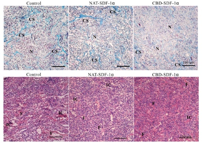
Later, the researchers observed in vivo that CBD-SDF-1α scaffold triggers the recruitment of more CXCR4-positive cells than the controls of collagen sponges without SDF-1α and with native SDF-1α (not modified). These cells deposit ECM and tenascin C, a marker of the healing tendon. They also saw no excessive amount of cells from the immune system migrating to the injury area, suggesting that CBD-SDF-1α didn’t induce immune cell reaction.
CBD-SDF-1α scaffold triggers cell migration
Formation of new tendon
The scaffold gave place to the formation of new tendon tissue after one week, although more time is required for it to resemble to a native tendon. The mechanical properties of the neo-tendon (the force that the tendon can withstand and the stiffness) were superior to those of the control systems. These superior properties are explained by the fact that, in vivo, tendon is made of collagen fibrils, which are highly organized and aligned. In this study, collagen fibrils in the CBD-SDF-1α group were larger and aligned, and the cells aligned with them.
Staining of regenerated tendons
One limitation of most investigations is the formation of bone instead of tendon at the site of injury, mainly due to the differentiation of stem cells to bone cells. In contrast, in this work, CBD-SDF-1α scaffold didn’t promote bone formation.
This study is highly encouraging for the development of collagen sponges tailored with specific features for tissue engineering. In future work, this CBD-SDF-1α sponge should be studied in larger animal models and for other applications, such as healing bone, cartilage and many more.
Coral reefs are under a barrage of threats. Ocean acidification alone could decrease the density of coral skeletons by up to 20%, with reefs close to the equator affected most.
Declines in coral skeletal density could have wide-reaching impacts on reef structure. Reefs are a natural coastal defence, shielding coastlines from storms by dissipating much of their energy. But “less dense skeletons will provide a weaker barrier,” said Nathan Mollica of Massachusetts Institute of Technology, US.
Such a decline could also render reefs more vulnerable to erosion. “There is some evidence that a lower density skeleton may be more easily bored into by bio-eroding organisms,” said Mollica, suggesting there may be wider implications for reef health.
The study, published in PNAS, showed that reefs in the coral triangle – a diversity hotspot in the western Pacific – are likely to be most at risk. Large increases in seawater acidity are predicted for this region. In areas that experience only small changes in acidity, coral skeletons could well stay strong.
Ocean acidification reduces the availability of the carbonate ions that corals need to grow. However, studies of coral growth in acidified environments in the lab and field have yielded mixed results, making it hard to predict how corals will respond to future change. The answer, it seems, lies in understanding the different ways that these organisms grow.
Corals develop in two ways: by extending outwards at the edges, and by thickening bundles of existing crystals to make the coral skeleton denser. This thickening is vulnerable to ocean acidification.
Scientists believe that corals have more control over their outwards growth than the thickening of their central skeleton. As these areas are under less control, they may be more susceptible to changes in seawater chemistry.
Coral polyps
By separating the two key components of coral growth, the team simulated how coral skeletons will change by the end of the century, validating their model with reef data from around the world.
Many studies have seen a relationship between seawater chemistry and skeletal growth. “Our model builds on that by allowing us not only to predict how corals will respond to [acidification], but put a number on it which is really valuable when making long term predictions,” said Mollica.
Currently, the model works best for Porites, a stony coral that dominates tropical reefs around the world. With some calibration it could apply to other taxa, allowing researchers to unpack the impact of ocean acidification on other corals.
“The future for coral reefs is certainly scary but all is not lost,” said Anne Cohen of Woods Hole Oceanographic Institute, US. “We have to continue to work hard to stabilize and even reduce carbon dioxide in the atmosphere and in the meantime take very good care of our reefs to optimize their ability to deal with the coming changes.”
Researchers have used genetic modifications to directly control structural colour appearing in nature. The work is an important step towards understanding how nature has evolved to effortlessly grow and control functional nanostructures, in the hope that we can harness these tools. The knowledge can be applied to create novel, biodegradable optical materials and sensors with myriad possibilities: for example, photonic materials designed so they are self-healing and can easily interface with living tissues. The findings by a collaboration of researchers from the University of Cambridge and Hoekmine BV are published in PNAS.
When a Hoekmine BV team unexpectedly discovered that they had isolated a previously unknown, brilliantly green coloured strain of bacteria, they contacted researchers at University of Cambridge to investigate this phenomenon further. According to Villads Egede Johansen, co-first author of this work, “from that point onwards, we were both driven to explore what can be done to alter and influence this system”.
The ensuing experimental work represents the first systematic study linking genetic markers to structural colour, with the hope that more studies will now be sparked in this direction. Striking examples of structural colour, which refers to colour that is not obtained through pigmentation, are found in natural phenomena such as butterfly wings and peacock feathers. Genetic control of macroscopic properties such as colour opens the door to an almost limitless number of material and device fabrication options.
From bacteria to sensors
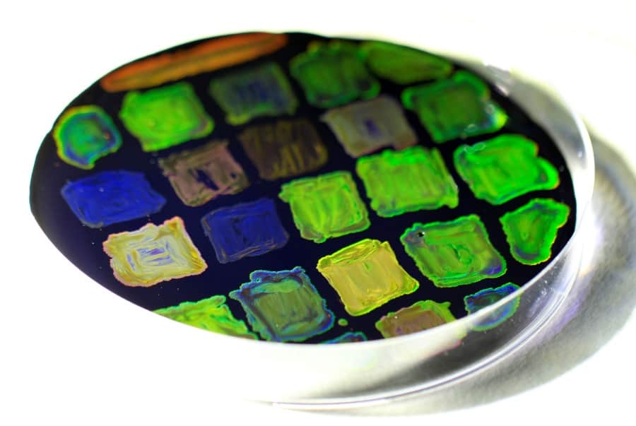
The bacteria used as a model system in this research is the Flavobacterium IR1 strain. These are rod-shaped bacteria that are able to pack together through gliding and growing mechanisms. Under different genetic and environmental conditions, Flavobacterium colonies form ordered nanostructures that interfere with light to give distinctive bright green, yellow, blue and red iridescent colorations, spanning the entire visible spectrum.
The iridescent colours are highly distinctive, meaning they have great potential for use as cheap and effective chemical sensors. For example, the bacteria could be genetically engineered to lose colour upon exposure to a specific chemical compound. This builds on existing concepts such as using sensors to “sniff out” drug production in sewers and using bacteria to detect explosives such as landmines. Other applications being considered are biodegradable paints and colorants, giving rise to the idea that we could even grow our own customized paints from different bacterial colonies.
Microscopic Petri palette of dried up genetically altered bacteria colonies.
A brightly irridescent future
To drive this research forwards, it is imperative to further understand the biological functions of the packing that leads to this distinctive expression of colour. The researchers were the first to link these packing mechanisms to both optical response, or manifestation of colour, and to certain genetic markers.
However, the eternal ‘nature versus nurture’ issue must also be considered, as it was observed that not only do genetic markers influence how the colonies exhibit colour, environmental changes have a distinctive effect. Fucoidan is a sulphated polymer derived from brown algae that the scientists found enhances the structural coloration. This discovery highlights the need for further research in this field to fully understand genetically modified colour.
Using genetics to alter and influence the material response of biological systems paves the way forward to a bright and shimmering future, filled with biodegradable, eco-friendly nanofunctional materials grown to fulfil our planet’s ever-increasing demands on resources.
My first thought as I stood in a narrow corridor filled with bright displays, music and booming, cheery announcements was that I had been transported to Walt Disney World, circa 1986. I was, in fact, at the headquarters of a $30bn, multinational materials science and adhesives corporation – 3M Company (3M), the inventors of the Post-it Note. I was here in St Paul, Minnesota, US, to learn about 3M’s contributions to the science and innovation of adhesives and tapes. I had been expecting scientists – not an amusement park for sticky stuff.
The visit began normally enough. In the vast entryway of the 3M Innovation Center, I was greeted by Stefanie Giese-Bogdan, a communications manager with 3M. Inviting me to follow her, she took me down the tunnel-like corridor, before closing the door to leave us standing in silent darkness. Then, the walls, which were covered in huge screens, flashed on to give a sleek multimedia presentation on the history and current work at 3M.
When the intro was over, double doors swung open at the other end of the tunnel. Ahead of me, in a cavernous void, I could just make out a few chairs set up like a planetarium, letting you lean back and gaze at the ceiling. Grinning mischievously, Giese-Bogdan suggested I take the middle chair on the second row for the best view. As I leaned back another presentation began, this time projected on the curved ceiling. And then suddenly, like a true Disney experience, spotlights flipped on all around me.
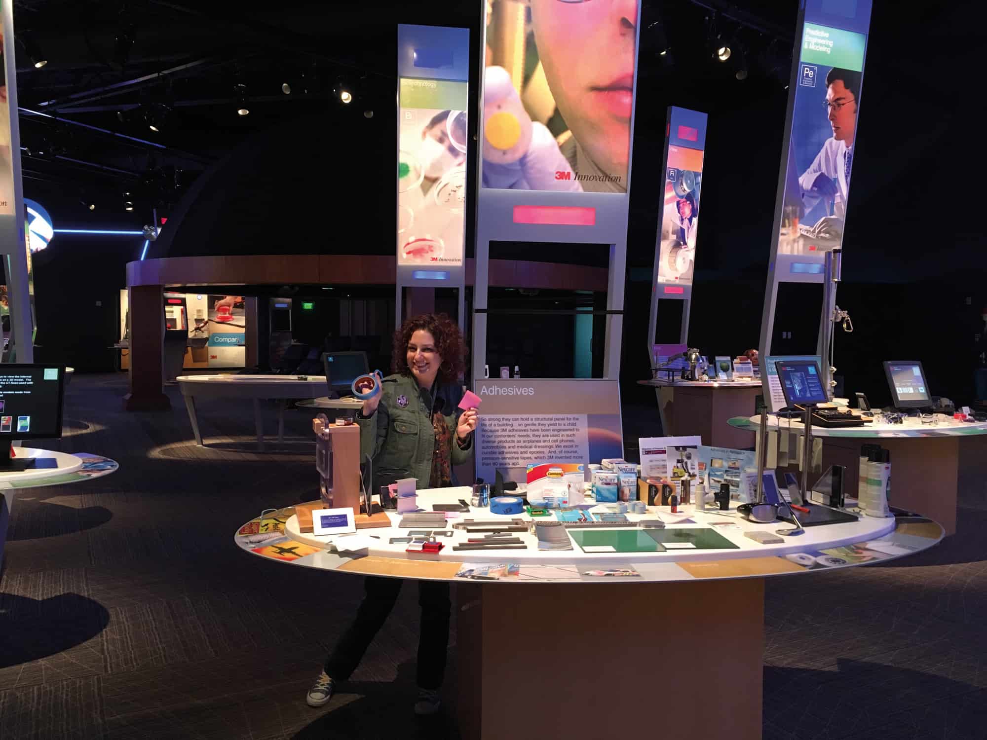
I was staring at the huge 3M World of Innovation – a massive room at the heart of the 3M Innovation Center, where 3M technologists meet customers to discuss how to incorporate 3M inventions into their products. “Innovation starts with imagination,” says Giese-Bogdan, who has a PhD in analytical chemistry, as we walked around the room’s 27 stations, each devoted to a specific 3M core technology. “At 3M we combine imagination with collaboration and communication. We solve problems by making uncommon connections of technologies and by applying science to life.”
The Industrial Adhesives and Tapes Division at 3M is the largest department in the 116-year-old firm, with more than 6100 employees worldwide, as well as research and development (R&D) centres in 10 countries and manufacturing locations in 26 nations. Adhesives are one of 46 core technologies that 3M puts into its products, but because adhesives are useful in so many sectors – from the aerospace and automotive industries to medical equipment and electronics – they are found in more than 30,000 of the company’s products. And then of course there is the retail and consumer side, with your friendly neighbourhood Post-it Notes and Scotch brand tapes.
The 3M World of Innovation, which is not open to the public, is a showroom and playground. It’s where 3M invites its customers to brainstorm with scientists and discuss how each technology might benefit them. The customers get to play, experiment and discover. They witness demos, learn about new innovations and, together, the 3M specialists and the customers try to solve problems. This is where my tale of tape truly unrolled, as I messed around at an adhesives station and Giese-Bogdan, as one of the hosts of the exhibition, explained to me how the innovation game is played. Like the time the Minnesota Zoo called and explained it had a whale with a wound and needed a special bandage to stick to its slimy lip. In fact, the solution the 3M researchers came up with proved so valuable and flexible that it became incorporated into an entire product line of human bandages.
The science of sticky
To find out more about the science of adhesives, I sat down with Cristina Thomas, senior technical leader in corporate R&D. Thomas has a PhD in chemical engineering but considers herself a polymer physicist because much of her technical career has been in the field of computational materials modelling. “The world around us uses many more adhesives than we realize, because we often need to bond things to surfaces,” she says. Even the room we’re in uses them, as Thomas points to the carpet, ceiling and walls. They are in the room’s electronics, inside the display and even holding the microchips in place. The building is kept energy efficient by film stuck to the windows and stretchable adhesives – 3M Command Strips –hold up the posters. These specialist products can bond to a surface and then once stretched again can be removed without any residue.
An adhesive is a substance capable of holding materials together by surface attachment. Generally made of polymers, modern adhesives allow things to be stuck together without needing to create discontinuities, such as holes, in the substrate materials. They work by creating the same strong molecular forces that hold materials together normally. But as polymer adhesive scientist Anthony Pagliuca from 3M points out on the company’s wesbite, to do so an adhesive must first “wet out” the substrate – it needs to flow over and cover the surface uniformly to maximize the contact area. The extent to which a liquid can wet out depends on surface energy and, for an adhesive to be effective, its surface energy must be equal to or lower than that of the substrate. Adhesion then occurs via interactions such as hydrogen bonding, mechanical interlocking and chemical bonds.
A brief history of 3M
Staple without staples 3M’s Post-it Note is a stationery must-have. (Courtesy: iStock/Rawpixel)
3M was founded in 1902 as a small-scale mining venture under the name Minnesota Mining and Manufacturing Company. But the founders’ original goal, of mining one type of mineral from one mine, turned out to be neither feasible nor sustainable. So they looked at other materials and products and quickly found that they could harness science and engineering to develop and improve products across multiple sectors. Early products from the 1920s included the first waterproof sandpaper and masking tape, which launched the company’s interests in adhesives and tapes. Over the years, 3M has grown into a firm employing 90,000 people, and with 60,000 products used in homes, businesses, schools, hospitals and more, touching most industries on Earth. One-third of 3M’s sales come from products that were invented within the past five years.
One product that 3M is especially well known for is the ubiquitous Post-it Note, which famously was discovered almost by accident. In 1968 Spencer Silver, a 3M scientist, was busily researching adhesives in the laboratory. In the process, he discovered something special: an adhesive that stuck lightly to surfaces but didn’t bond tightly to them. “It was part of my job as a researcher to develop new adhesives, and at that time we wanted to develop bigger, stronger, tougher adhesives,” said Silver. “This was none of those.”
What Silver discovered was something called microspheres, which retain their stickiness but with a “removability characteristic”, allowing attached surfaces to peel apart easily. He started sharing his adhesive with other 3M scientists, trying to discover a problem that the adhesive could solve. Almost six years later Art Fry, another 3M researcher, realized that Silver’s microspheres could serve as an adhesive for a bookmark-type product that can be placed on paper then removed and re-stuck somewhere else. Fry figured out how to manufacture it by 1977, got the green light from management in 1978 and the Post-it Note was officially launched in the US in 1980, and in Europe and Canada in 1981.
The rest is history – Post-it Notes hit the world by storm, and 3M continues to expand the product line, most recently shifting to plant-based adhesives for all its Post-it Notes. Other recent 3M milestones include the company earning its 100,000th patent in 2014, and unveiling a new, state-of-the-art, $150m R&D laboratory on its Minnesota campus in 2015. But Post-it Notes remain one of 3M’s most highly visible product lines and brands.
Adhesion scientists consider three factors when they design their products. First, there is the adhesion mechanism – how the adhesive sticks to and interacts with the various surfaces. Second, the scientists have to identify and examine the forces that will act on the adhesive-containing product while it is being used. These forces, such as shear forces or peel forces, impact the performance of the adhesive and can affect its integrity and function. Finally, there’s the durability, which includes examining how the environment will influence the effectiveness of the adhesive.
Given these factors, it is not surprising to learn that physics plays a huge part in thinking about and designing effective adhesives and tapes. After all, the basic mechanical properties of the adhesives have to be precisely calculated and tested and clarified. Indeed, there is an entire building of labs at 3M dedicated to just this task. It has every piece of equipment you’d expect in a corporate analytics and materials-processing facility.
Managing how polymers flow and understanding their stability during and after application are important aspects of developing both structural and pressure-sensitive adhesives. How will the adhesive be deployed onto a surface? What is the application mechanism? What is the curing mechanism? Some adhesives have to stay sticky while they hold two surfaces together, whereas others become solid or foamy upon curing. Knowing the adhesive’s function and the environment in which it will operate determines how to distribute it on a surface and how strong the chemical bond should be between the adhesive and that surface. Viscosity and elasticity are characteristics of adhesives that matter greatly too. Specialist engineers are needed to design, for example, adhesive storage cartridges and the nozzles from which they are ejected in large-scale industrial systems.
Materials science also plays a role in formulating adhesives and the substrates holding them, like tapes. Depending on the application, adhesives or tapes may need to provide other properties in addition to just good bonding performance. For example, 3M makes a pavement marking tape with a specific optical performance to help motorists. These tapes have a layered structure in which the adhesive is on the side contacting the road while reflective beads are embedded in the top layer so that drivers can see the markings under various environmental conditions. “The adhesive is a mechanism to deliver information to the driver via an additional aspect of physics, which is optics,” notes Thomas.
Sticky solutions Alaina G Levine plays at 3M’s World of Innovation. (Courtesy: Alaina G Levine)
Moreover, with adhesives and adhesion science embedded in many of 3M’s products, the sticky science is often combined with other technologies. For example, 3M can borrow from one technology, such as its tape formulations, combine it with cross-linking chemistry and create a strong water-based adhesive. For one project, 3M scientists infused viscoelastic foam with adhesives to create strong bonding tape ideal for use in the car industry. They also took ordinary elastomeric materials and developed a proprietary high-strength matrix so manufacturers can create more impact-resistant products. Adhesive experts, meanwhile, dipped into the science of microreplication – one of 3M’s biggest scientific assets. The microreplication process involves melting plastic pellets and squeezing them into rolls of plastic film to create microscopic sculptures on the surface. There can be thousands of features per square centimetre, arranged uniformly, that can change the physical, optical and chemical properties of the surface. One application where 3M scientists have used the technology is to make road signs. Here, the film is typically adhered to aluminium substrates, and thousands of tiny prisms reflect a car’s headlights back to the driver, resulting in signs and markers on roads appearing significantly brighter than normal.
Adhesives also have to withstand a spectrum of environmental stimuli – anything that could impact the function or durability needs to be addressed. Those pavement markings, for example, are exposed to water, oil, atmospheric compounds, such as pollutants, and many other chemicals and concoctions. They have to stay in place and be able to withstand constant shocks, vibrations, and movement from vehicles. Adhesives additionally have to cope with changes in pressure, temperature and a host of other variables.
In one of 3M’s many labs, I met Aaron Hedegaard, a chemical engineer who studies the viscosity and elasticity of different formulations of adhesives to quantify their strength and stickiness. He does this by smearing a sample of an adhesive on a rotational rheometer and measures its stiffness, dissipation factor, and its response to being heated and chilled, even to temperatures well below a typical Minnesota winter. “I develop new test methods, not just running the standard tests,” he says. “I am trying to push the boundaries to make the next standard test.”
3M's SEALS
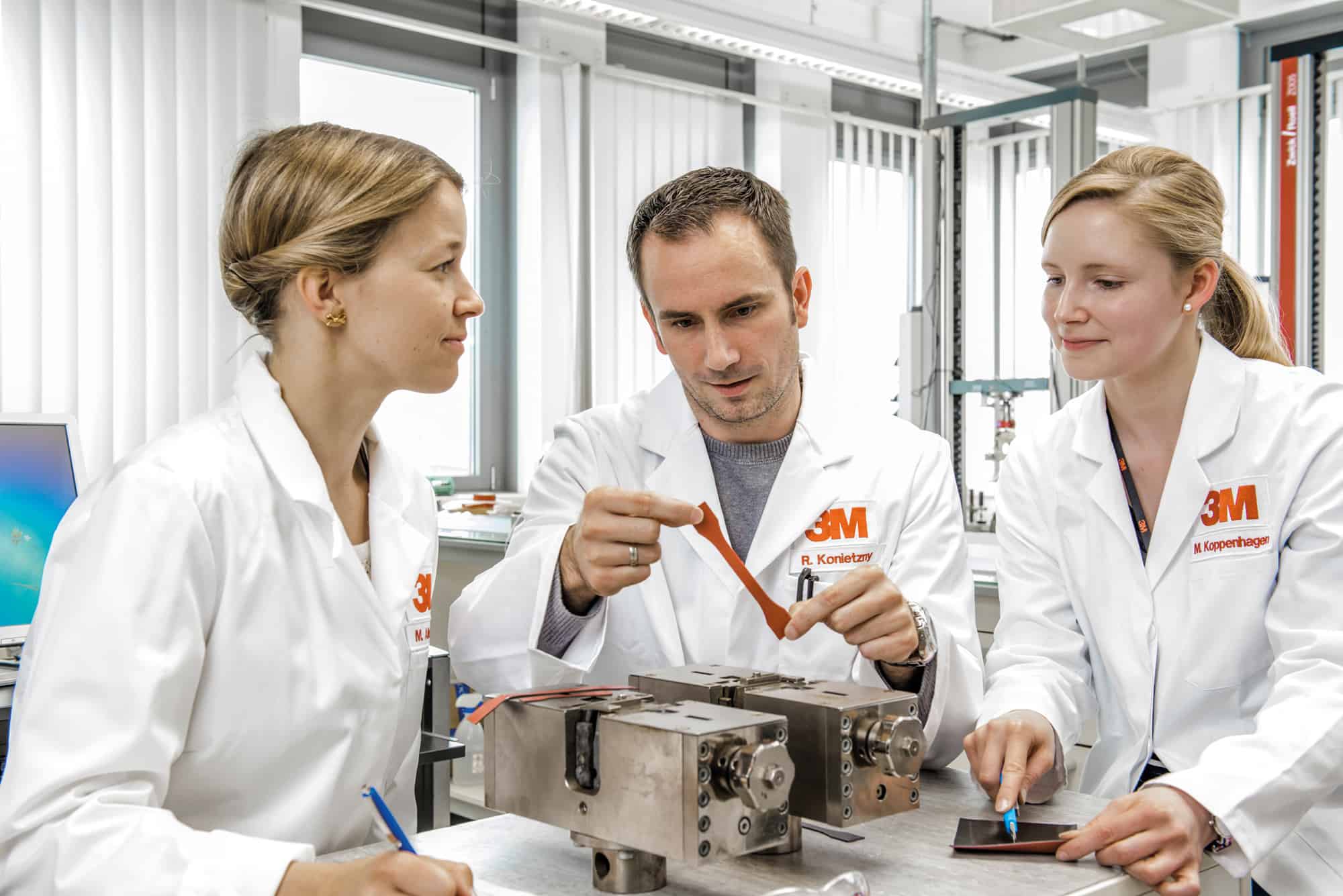
A scientist’s seal 3M’s experts work with customers to create their ideal adhesive. (Courtesy: 3M/J Rolfes)
When designing adhesives for customers, 3M experts ask questions based on the SEALS acronym:
S: Substrate – What is the nature of the substrate?
E: Environment – What is the bonding environment? What environment will the bond be subjected to (internal/external, high/low temperatures, chemicals, salt)?
A: Application –What are you doing with your component? What application characteristics do you need in terms of speed of cure, open time and rheology from the adhesive?
L: Load – What are the stresses on the joint in type, magnitude and direction?
S: Size – For industrial adhesives, how many units are you producing – per month, per quarter or per year?
Overcoming physics
Physics breakthroughs have impacted 3M adhesives in fascinating ways, according to Thomas. To a chemist, a polymer is a macro-molecule composed of repeating smaller molecules or units, but people now understand that these long molecules can be approximated as chains and so their behaviour can be explored and explained using statistical methods from physics.
“We can treat polymers using simplified models where the molecules are represented by individual units or beads that replace the group of atoms,” says Thomas. Macromolecules within the adhesion polymers can then be investigated using physics principles, enabling scientists to understand their behaviour under certain conditions. “If I’m doing a phase separation when it’s a polymeric system, it’s going to behave differently than if I am separating smaller molecules – this is something that really advanced the adhesion science field.”
Depending on temperature, some adhesives behave like glass (solid-like) and some like rubbers (fluid-like), so Thomas and her team draw ideas and knowledge from complex systems too. When you “chemically cure” an adhesive with the aim of converting it into a solid, the adhesive is going through a chemical reaction and, once cured, it is able to provide high strength and resistance to temperature, humidity or chemical exposure. Depending on whether you cure with light, heat or another mechanism, you are enhancing the performance of the adhesive due to the formation of an adhesive network that behaves like a glassy polymer.
But just because there is a lot of physics in adhesives doesn’t mean that 3M scientists are shackled to it. Back in the 3M World of Innovation, Giese-Bogdan showed me the Multi-layer Optical Film – a silvery, reflective piece of film that is used with a special tape. “If you are tilting a reflective surface there is an angle at which it is no longer reflective – that angle is called the Brewster’s angle,” she explains. “The Multi-layer Optical Film does not have a Brewster’s angle, it is reflective at any angle.” The film is used to make light shafts in buildings and, since Brewster’s angle is not a consideration, it can be placed around bends. It can also be used in, for example, electronics – from phones to TVs – to save energy, and as a parabolic mirror to direct light onto solar cells. It is one of the many product examples that Giese-Bogdan has ready to showcase in the World of Innovation. “How often can you say you beat the laws of physics?” she teases. “Well we did. We beat Brewster’s angle.”
The 3M Innovation Center gave me a glimpse into the amazing world of adhesives, and as I left, my take-home message was that adhesives are sticking around and expanding their reach, strength, and diversity of use. “Many of the things that we see every day have adhesives or result from the use of adhesion science. There is a lot of physics, of fundamental understanding behind that,” says Thomas. “Even things we take for granted every day, such as a marking on the road [are bonded by adhesives].” A huge diversity of good adhesives allows lorries to go over those tapes but also allows Post-it Notes to be reused, and allows medical tapes to cover fragile or sensitive skins, as well as countless other applications for tapes and adhesives across the globe.
An optical atomic clock that, if left running for the entire life of the universe, would neither gain nor lose more than 100 ms has been created by physicists in the US and Italy. The device has a relative precision of 2.5 x 10⁻¹⁹ – surpassing the 3.5 × 10⁻¹⁹ figure achieved by US members of the team in 2017. It is the most precise optical atomic clock ever made and it vastly outperforms the caesium fountain clocks that act as national time standards, which operate at about 1 × 10-16 relative precision. As well as providing a highly-reliable time standard, the technique used to create the clock could be used to study fundamental physics such as unconventional superconductivity.
The new clock is located at the National Institute of Standards and Technology (NIST) in Boulder, Colorado and uses thousands of strontium atoms confined in a 3D optical lattice. A laser is used to excite an extremely stable, high-frequency transition in the strontium atoms – thereby creating a frequency standard that provides an extremely precise measure of time.
To achieve their record-breaking feat, NIST’s Jun Ye and colleagues had to contend with a number of factors that tend to degrade clock performance. Interactions between atoms, which have a negative effect on the quantum coherence of the clock, were suppressed by cooling the atoms to a chilly 15 nK. This allowed the atomic ensemble remain coherent for up to 15 s – which contributed to the record-breaking precision.
Local effects
Errors can also arise because each atom in the lattice exists in a slightly different local environment than its neighbours, and this means that different atoms can have slightly different transition frequencies. The team addressed this problem by combining ultraprecise optical spectroscopy with high spatial resolution imaging to measure the clock frequency at different locations in the lattice. These data were used to create a “frequency map” of the clock that was used to identify and mitigate various sources of frequency irregularity.
The combined measurement technique also allowed the researchers to find the “magic wavelength” of the laser used to create the lattice. Operating the laser at this wavelength eliminates its perturbing effect on the frequency of the atoms, boosting the performance of the clock even further.
Fundamental physics
While the researchers were not able to image individual atoms in the lattice, this could be possible with further improvements. Indeed, single-atom resolution has already been achieved by others under different experimental conditions. The ability to make extremely sensitive measurements on individual atoms at lattice sites could be used to study a range of fundamental phenomena in physics. In the future, the team intends to us their technique to study few- and many-body physics, quantum magnetism and unconventional superconductivity.
Ye and colleagues also want to use their lattice of atoms as gravitational sensor to see the interplay between quantum mechanics and general relativity at the millimetre scale for the first time.
CT imaging is ubiquitous in lung cancer management – with scans employed for diagnosis, radiotherapy set-up and response assessment. But according to Iain Phillips from Royal Surrey County Hospital, the information contained within a standard CT scan could be used for a lot more.
Speaking at the recent MediSens conference in London, Phillips described how texture analysis of lung CT scans could be used to identify whether a lung cancer patient is fit enough to undergo radiotherapy. “Texture analysis is based on the idea that imaging provides a pool of unmined data, and that we can get more technical information from a standard image and greater value from fewer tests,” he explained.
Lung cancer patients often have multiple morbidities, such as chronic obstructive pulmonary disease, for example, which causes breathing difficulties. As a lower post-operative lung function increases mortality, patients scheduled for surgical lung cancer treatment are classified as fit or unfit for surgery beforehand, using simple tests such as spirometry.
Spirometry assesses lung function by measuring how much air a patient can breathe out in one forced breath (FEV1, the forced expiratory volume in one second). Physicians also assess the transfer factor TLCO, which measure’s the lung’s ability to transfer oxygen into tissue.
When considering surgery, a patient with an estimated post-operative TLCO or FEV1 of below 40% predicted is considered at high risk. But for radiotherapy, there are no standard thresholds in use and no model that describes the impact of lung function on radiotherapy side-effects. It is likely, however, that a lower lung function will lead to more adverse effects.
Texture mapping
To investigate the use of CT scans for assessing patient fitness, Phillips and colleagues devised an analysis methodology. Using a cylinder of lung tissue, they perform a voxel-by-voxel analysis to create a texture map of this region-of-interest (ROI). Each voxel is assigned a density value and an entropy score. The entropy score is based on the similarity of grey levels in adjacent voxels, with a high entropy describing a low uniformity, and vice versa.
Phillips described a texture analysis and lung function study of 30 fit and 30 unfit patients who had undergone radical radiotherapy (chemo-radiotherapy or stereotactic ablative radiotherapy). Patients were defined as fit if they had FEV1 and TLCO values of 50% predicted or higher; if either parameter was below 50% the patient was classified as unfit. The researchers used the patients’ 4DCT scans to perform a retrospective imaging review, calculating the mean, median and mode values of density and entropy.
Looking at the texture maps created from the CT scans, maps from fit patients included more white (heterogenous) regions, whilst maps from unfit patients had more black (homogeneous) regions. This homogeneity is likely due to emphysema, the presence of holes in the lung tissue, as seen in less well functioning lungs. Plots of density versus entropy for the two patient groups clearly revealed different regions of data cluster.
The study concluded that patients with good lung function exhibit texture maps with voxels of higher entropy and higher density, whilst poorer lung function corresponds to lower entropy and lower density. “It appears to be possible to differentiate between fit and unfit patients just by looking at the CT image, and getting functional data from the CT scan,” said Phillips.
To use this technique clinically, Phillips suggests a “simple ROI” scheme, in which the ROI is identified automatically following CT acquisition, then texture analysis is performed, and only at this stage is the clinician required to review the results. “This offers a simple way of including advanced image analysis into the normal radiology workflow,” he explained.
Phillips suggests that this CT-based approach could be used as a screening test for both surgery or radiotherapy, enabling faster decision-making at multidisciplinary team meetings. It could also play a role in resource-limited settings. “In terms of use in clinical practice, we can start applying this to patients, to assess whether they are fit for treatments,” Phillips concluded.
Smoke from biomass burning in south and central Africa brightens low-level clouds over the southeast Atlantic, cooling the climate. That’s according to researchers from the US and China who reported their work in PNAS.
“Our group is the first to quantify this brightening effect,” said Xiaohong Liu of the University of Wyoming, US. “This (smoke aerosols in clouds) reflects more solar radiation to space, which results in less solar radiation reaching the Earth’s surface. This creates a cooling effect.”
Previously scientists though that smoke diminishes the cooling from these clouds, by absorbing light that the clouds would otherwise reflect. But the team found that the smoke and cloud layers are closer together than expected so that aerosol particles from the smoke act as nuclei for cloud droplets to form around. These droplets are numerous but small and reflect more light than a collection of fewer, larger droplets.
In smoky conditions, the team found, there are almost twice as many cloud condensation nuclei per cubic centimetre. The result is a cooling effect that outcompetes the factors acting to reduce cooling by the clouds.
Since the Industrial Revolution, carbon dioxide from human activities has created a greenhouse effect of 1.66 W per square metre worldwide. During the fire season, smoke results in a cooling of 7 W per square metre over the southeast Atlantic.
The fire season in southern Africa runs from July to October. The fires, a mix of wildfires and fires set to clear farmland, create enough smoke to be visible on satellite images. The aerosols travel west over the southeast Atlantic Ocean, where they interact with the stratocumulus cloud beneath them, about 1 km above the sea.
Next the team would like to improve how global climate models account for clouds and interactions with aerosols from sources such as power plants, vehicles, deserts and oceans.
“Now that we know there are two competing mechanisms, and the seeding effect is winning, we can see whether climate models consider these processes properly when they predict the weather and climate in this area,” said Zhibo Zhang of UMBC, US.
In 2020 NASA is set to launch the PACE mission, which will be able to detect polarized light.
“With the new satellite you can look at things from different perspectives,” said Zhang. The plan is to develop three-dimensional models of the interactions between aerosols and clouds. “Hopefully we can look at this phenomenon even better.”
A new photo-active and rechargeable nanofibrous material that can efficiently destroy bacteria and viruses could one day be integrated into personal protective equipment (PPE) to prevent the outbreak of emerging infectious diseases. The material, which works by producing biocidal reactive oxygen species (ROS) in response to sunlight, is active even in dim or dark conditions, unlike previous such photo-antimicrobials that needed light irradiation to function.
Emerging infectious diseases (EIDs) are a serious global health problem. Such diseases include severe acute respiratory syndrome, bird flu and Ebola virus disease (EVD). The 2014 EVD outbreak in West Africa, for example, killed nearly 40% of the 28,646 infected civilians and more than 50% of the 852 diagnosed healthcare workers.
To prevent EID spread, healthcare workers are advised to wear PPE such as face masks, bioprotective suits and medical gloves. Although these minimize pathogen transmission, they do not completely eliminate the risk of catching an infection. Biocides, such as triclosan, nisaplin and solutions containing silver nanoparticles, can be used too, but they need to reapplied frequently.
ROS kill bacteria and inactivate viruses
A team led by Gang Sun of the University of California at Davis made its polymer-based nanofibrous membranes using an electrospinning technique. The membranes contain benzophenones and polyphenols, which are widely employed as photosensitizers in biochemistry and organic synthesis. These compounds rapidly generate ROS when exposed to sunlight in the presence of oxygen thanks to a photoreaction that involves hydrogen abstraction by the nanomembranes and subsequent oxidation.
“Once the pathogens have been intercepted and are in contact with the surface of the nanofibres, the photoactive biocides produce various ROS, including hydroxyl radicals, superoxide and hydrogen peroxide, explains team member Yang Si. “These ROS kill bacteria and inactivate viruses by damaging DNA, RNA, proteins and lipids.”
Rechargeable, so work even in dim or dark conditions
“The photoactive materials we used can store the biocidal activity under light irradiation thanks to their rechargeable function and readily release ROS even in dim or dark conditions,” he says. “In comparison, previous photo-antimicrobial materials could only work when irradiated with light and many of these even required high-energy UV light.”
The membranes quickly and effectively kill pathogenic bacteria and viruses when in contact with them. “For example, over 99% of bacteria (such as E. Coli and L. innocua) are killed in less than two hours and over 99% of viruses (such as T7 phage) in less than 30 minutes either under light exposure or dark conditions,” Si tells nanotechweb.org. “To compare, previous such membranes required 10 to 20 hours of contact with the bacteria or viruses.”
Towards commercialization?
The researchers have shown that the membranes can be used as a biocidal layer in many routinely employed PPE – for example 3M’s N100 respirator and DuPont’s Tyvek protective suit. In the respirator application, they can filter out and kill E. Coli, for example, in aerosol form. “They might also be used as a protective layer in face masks and medical gloves to defend again pathogens in either aerosol or liquid forms,” adds Si.
The team, reporting its work in Science Advances DOI: 10.1126/sciadv.aar5931, says that it is now busy developing PPE containing its nanofibrous materials. “We will try to produce these materials in large quantities and, in collaboration with industrial partners, integrate them into existing nonwoven production lines, which could allow us to commercialize the technology.”