Novel phantom simplifies MRI-Linac QA

Images recorded by any MR scanner, including MR simulators and MR systems integrating a linac, will suffer from some level of geometric distortion. Such distortions can arise due to inhomogeneity in the main magnetic field or from nonlinearity in the gradient coils. And while manufacturers supply distortion correction algorithms to apply to their systems, these are based on factory measurements and cannot account for the specific environment in which the scanner is installed.

Enter the QUASAR MRID3D, which can measure both the main field inhomogeneity and the gradient nonlinearity, reporting the results separately as geometric distortion vector fields. The system comprises the phantom itself – a lightweight hollow acrylic cylinder containing 1502 precisely machined fiducial markers filled with mineral oil (the control points) placed around the phantom boundary – plus the image analysis software.

“We are finding that the greatest demand for the QUASAR MRID3D is for use with MRI-Linacs for guided radiotherapy treatment,” said Enzo Barberi, director of MR product development at Modus QA. “Our device is being used on several MRI-Linacs worldwide at early adopter sites, and also selected by MRI-Linac manufacturers who have either purchased or are evaluating the phantom. Another key application is within MR simulation, where MR images are used for radiotherapy planning.”

At the Henry Ford Cancer Institute in the USA, for example, the team is evaluating the QUASAR MRID3D for distortion characterization in its ViewRay MRIdian MRI-Linac. “The phantom will be used to benchmark our systems against other MRI-Linacs; we are also exploring its use for routine distortion assessment,” explained Carri Glide-Hurst, director of translational research in the department of radiation oncology at the Henry Ford Cancer Institute.
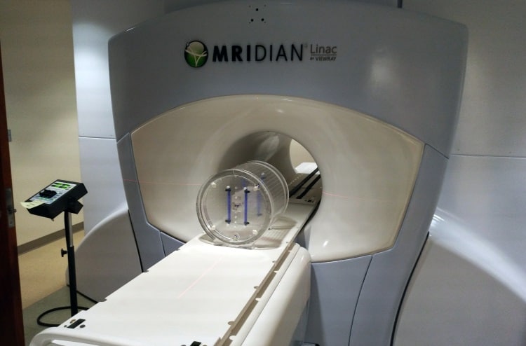
The QUASAR MRID3D phantom inside the MRIdian MRI-Linac

“The phantom has intelligent design features, such that it is lightweight, easy to setup and has excellent interactive software to analyse different sources of distortion,” Glide-Hurst added. “Previous work by our group showed that gradient non-linearity distortions, even after vendor corrections are applied, are non-negligible and may require corrections in an MR-only planning workflow. This emphasizes the need to have the proper phantoms and robust software to assess this distortion. The QUASAR MRID3D meets this need with ease.”

System development
Modus QA realized back in 2010 that MRI-guided linacs were being developed and that, once implemented in the clinic, would present specific QA requirements. The company also recognized that GRID phantoms – conventionally used to measure image distortion in MR scanners – would be either too small or too heavy for such applications, as well as highly expensive to manufacture.

As such, Modus QA teamed up with medical physicist Teo Stanescu from the Princess Margaret Cancer Centre in Canada to develop a completely new design of phantom, along with software to measure MR image distortion using a novel approach based on the harmonic analysis method.

Harmonic analysis is a well-established mathematical tool used to solve electromagnetism problems with well-defined boundary conditions, for example MRI gradient coil design, as well as B0 shimming. The QUASAR MRID3D extends this approach by measuring the boundary condition on the phantom and then using harmonic analysis to calculate the magnetic field distortion inside.

“We take an MR image of the phantom and if there is distortion on the boundary, the control points in this image will have shifted,” Barberi explained. “The software automatically locates the control points and compares them against the CAD files. The differences in their locations is the distortion vector field on the surface, which provides the measured boundary condition. The software then uses harmonic analysis to calculate the entire 3D distortion vector field within the phantom boundary.”

Lighter and faster
The QUASAR MRID3D uses far fewer control points than the 5000 or more required in an equivalent-sized GRID phantom. As well as reducing the weight, this design also enables faster QA. “Speed is a very important component of QA,” noted Barberi. “If we can achieve accurate analysis in a shorter time span, sites are more likely to perform regular QA.”

With scan times as low as 3-5 minutes at 3T, the QUASAR MRID3D process involves set-up, scanning, data transfer and data analysis in less than 10 minutes. As such, says Barberi, some sites are using the phantom every other week to monitor gradient coils and field inhomogeneity. “This was a suggestion from some of the early MRI-Linac adopters, who have implemented this in their QA programme. Our solution meets the demand for fast, accurate analysis with efficient workflow,” he explained.

Another team currently working with the QUASAR MRID3D is the MR Linac group at The Institute of Cancer Research, London, which is using it mainly for QA of their Elekta MRI-Linac installation. “Since the system has received several hardware upgrades, we tried to assess their impact on the geometrical distortions,” explained MR physicist Andreas Wetscherek. “We have used the phantom on clinical systems too, to assess whether the vendor-provided distortion correction is sufficient for radiotherapy applications.”

The QUASAR MRID3D phantom inside Elekta's MRI-Linac

Wetscherek says that the group was looking to replace an old in-house distortion phantom with one that covered a large field-of-view and, importantly, was supplied by an independent company rather than the scanner manufacturer. “We were attracted by the spherical harmonics concept of the MRID3D and convinced by the expert knowledge of the Modus QA representatives,” he told medicalphysicsweb.

Elsewhere, Rob Tijssen from UMC Utrecht in the Netherlands is using the QUASAR MRID3D as an independent check on vendor-supplied geometric QA solutions. “We use this clinically on our Philips Ingenia MR-RT systems,” he said. “We have also been using it in a multi-institutional MRI QA study, in which we are benchmarking the Elekta MRI-Linac systems that are currently installed at the different consortium sites.”

Tijssen also emphasized the importance of being able to perform measurements over a large field-of-view, as enabled by the MRID3D phantom. “We have a well-established working relationship with Modus QA and they have been very responsive to our feedback, which is also an important factor to us,” he added.

Future proofing
Following the launch of QUASAR MRID3D at the start of 2016, Modus QA has implemented software updates roughly every six months. The company has introduced several new features, such as 3D visualization tools with a region-of-interest selector for users to specify custom volumes in which to characterize distortion. “Another new feature in development will give users the ability to define distortion levels that they consider acceptable and the software will report the volume that conforms to that requirement,” said Barberi.

The image analysis softwa

Looking further ahead, as the emerging MRI-guided radiotherapy systems continue to evolve, the associated QA must develop alongside. Already, some manufacturers are looking to add functional imaging capabilities to their MRI-Linacs, such as diffusion weighted imaging and diffusion tensor imaging. In such applications, the diffusion coefficient is particularly sensitive to gradient nonlinearities.

“There is a need for a tool that can measure and output the spherical harmonic coefficients, then you can measure gradient nonlinearities specifically on your system,” Barberi explained. “This provides a much better diffusion coefficient correction technique. That’s something that we are also working on.”

Another future project involves using mathematical techniques to extend the harmonic analysis outside of the control point boundary. While a typical treatment volume will be well contained within the phantom volume, some specialists or device manufacturers are interested in measuring system performance outside the region of typical use. “This is something that OEMs have asked us about,” said Barberi.

“The Modus QA team is constantly trying to improve the software and adding new features that are requested by the users,” added Wetscherek. “I see the strengths of the QUASAR MRID3D in MRI for radiotherapy, where high spatial accuracy and confidence is relevant, or in centres with many different MRI scanners.”

  • Individuals quoted are sharing their own views, not necessarily those of their organizations.
Rising stars show their stripes

Reggie Bain is on the verge of completing a PhD in theoretical physics at Duke University and is about to begin a teaching position at the University of Houston

Most people come into graduate school with the mentality that they’re going to stay in academia. There is a core of people where that’s what they want to do, their mind is set and then if they don’t end up in academia it was because of forces beyond their control. But I would say that there seems to be a growing sentiment where people want to learn more about physics but aren’t necessarily married to the idea of pursing a research career for the rest of their lives.

Chani Nava is a PhD student in astrophysics at Harvard University

I think that there is this tendency to see scientists as an elite group of sorts. And I think there’s an inclination to see us as being hard to communicate with, maybe socially cold or awkward. I think that’s fair in certain aspects and unfair in others. I believe that the scientific community as a whole could certainly make more of an effort to communicate and have a relationship with the general public.

Will Chen recently completed a Master’s degree in quantitative ecology at the University of Washington

At the moment we’re sort of in this period of uncertainty where science and scientific thinking is being challenged. Given our political climate, we’re in an era where science is not necessarily taken as something that can be believed, or even something that can be trusted. You have people like Neil deGrasse Tyson who say that science is true whether or not you believe in it. I think there’s some thought that needs to go into how we actually portray science. It’s clashing with people’s personal beliefs and I don’t know if that’s the right message.

Grayson Doucette is a PhD student in materials science at Pennsylvania State University

I think that scientists have a responsibility to ensure that their science gets to the public. It’s all well and good making discoveries and making breakthroughs. But if we don’t take the next step and actually make sure we get it to those who can take it to applications, or to the general public to be aware of what’s going on behind our lab doors, then I think we’ve not done justice to our jobs.

Khady Sall is a PhD student in molecular biology at Oregon State University, originally from Senegal

I’ve learned a lot from people here. Before, I was learning in French, in France, and then I came to the US. It’s a more diverse country in terms of nationalities. I met a lot of people from different countries and I feel like that experience shaped me as well. It made me more open and also more aware of what’s going on in the world, and I gained a different perspective from people who are actually from that country.

Michael Graw is a PhD student studying oceanography at Oregon State University

The oceanography field as a whole is really enthusiastic about early-career researchers but there definitely are challenges. Once you transition away from being a student it gets increasingly hard and you’re sort of dropped into competition with researchers who have been in the field for 20, 30 years and are much bigger names. There’s that air of moving very quickly from a somewhat sheltered situation to really being on your own to sink or swim. Bad oceanography joke!

  • Each day this week, we will be publishing a couple of new video interviews from delegates at ComSciCon 2017, including all the above. You can watch the first two films today.

Hand movements preserved in the brains of amputees

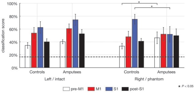
A new study has shown that the representation of hand movements in the brain continues to exist in above-elbow amputees, years after amputation. Researchers used machine learning (a linear classifier) to “decode” the brain activity related to attempted hand gestures performed by the phantom limb, compared with the amputees’ intact hand, as well as gestures executed by non-amputee controls. This work lends support to the use of decoded brain activity patterns in the development of brain-computer interfaces (Brain 10.1093/brain/awx274).

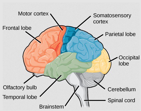
The cortical representation of touch and movement are encoded in somatosensory and motor brain regions, respectively; together, they are known as the sensorimotor cortex. Previous studies using functional MRI (fMRI) have shown activity in said cortex among amputees, indicating that, following loss of nerve supply (denervation) to the limb, the responsible brain regions are still operative.

Illustration of the areas of the brain

However, it is unclear how detailed and specific the activity is, and whether complex “attempted” movements by the phantom limb can be discriminated. Here, researchers from the Brain Center Rudolf Magnus at UMC Utrecht decode brain activity corresponding to six attempted hand gestures during a fMRI scan.

Decoding brain activity
A typical fMRI analysis comprises inputting voxel-wise data (each 3D “pixel” in the brain will have a time-series of activity) into a statistical model, then comparing the outputted model weights to every other voxel in the brain. If the test statistic for this weight is significantly above chance, then that voxel is said to be “active”. Unfortunately, this methodology is unable to look at patterns of activity derived from multiple voxels.

Multi-voxel pattern analysis (MVPA) is one way to overcome this. In the simplest case, a machine learning algorithm, known as a classifier, is trained on a subset of the data and is then used to classify the remaining data into provided categories, such as faces and houses, for example.

Phantom movements are preserved
In this study, the researchers decoded the fMRI BOLD signal changes (an indirect measure of neural activity) resulting from six different attempted gestures, in four sub-regions within the sensorimotor cortex. The classification scores were well above what could be expected if (obtained at random or) obtained by chance. This suggests that there is information about the hand movements contained in these sub-regions, even for “movements” of the phantom hand in amputees.

Classification scores were well above chance

When comparing activity from the amputees’ phantom hand with that from the controls’ right hand, the team saw no difference in classification score within the primary motor cortex (M1), indicating the preservation of hand movement in amputees in M1. The high decodability of the primary somatosensory cortex (S1) – albeit lower in amputees than in controls – supports the idea that hand representations are also preserved in S1 after denervation, even after several years.

Future work investigating the topographical organization of these phantom movements, and how the sensorimotor cortex compares to other cortices that display this trait, for example the retinotopic visual cortex, would be extremely interesting. This study provides support for the development of brain-computer interfaces, which could use the decoded data, possibly using “neuro-feedback” techniques, to pave the way towards the integration of high-tech prosthetics into our human “wet-ware”.

CMS publishes 700 papers, extreme data centres, flat-Earth space programme launches tomorrow

By Hamish Johnston

CERN’s CMS collaboration has passed a milestone of sorts at the end of October – it published its 700th research paper. And physicists working on the giant detector on the Large Hadron Collider haven’t stopped there as the tally is now 712 and rising.

CERN’s Achintya Rao has delved into the CMS archives and has chosen his top seven papers. These include the first-ever paper about the detector, which was published in 2008 and, embarrassingly, gets the weight of the detector wrong. Rao has also put together an interactive infographic that looks at 680 papers that analyse data collected by CMS.

CMS generates vast amounts of data, which are stored and processed worldwide. The data-centre consultants Comtec have put together a nice piece about “Extreme data centres” that explores how data can be stored in strange and often hostile environments. Data acquired by the IceCube experiment at the South Pole are stored in a data centre that, unlike most others, has to be heated rather than cooled. The piece also has some innovative ideas about cooling data centres such as locating them underwater – as Google has done.

If all goes well tomorrow, the self-taught rocket builder Mike Hughes will blast off in an attempt to prove that the Earth is flat – at least according to an article in The Washington Post. Hughes, who describes the idea that the Earth is a sphere as a Masonic conspiracy, plans to rise about 550 m above the ground where he hopes to gather evidence of Earth’s flatness. Good luck Mike.

Mechanical metamaterial twists when squeezed

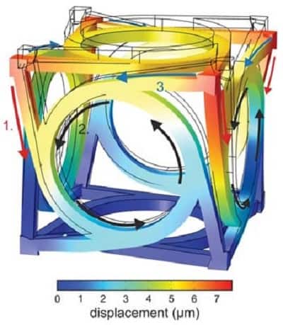
A computer-generated image showing the calculated deformation of a unit cell under compression.

A rationally designed metamaterial that twists in response to a linear force has been engineered by researchers at Karlsruhe Institute of Technology, Germany, and Université de Bourgogne Franche-Comté, France. The material consists of a repeating array of sub-millimetre units, each of which has a chiral structure.

In ordinary continuum mechanics, twisting motions never result from simple linear forces. Instead, squeezing an elastic object always causes it to expand at right angles to the direction of compression. By using precisely structured cells as the constituents, however, Tobias Frenzel and colleagues created a metamaterial crystal that can respond to pressure by deforming rotationally at a rate of more than 2° per % of shortening.

The researchers used numerical modelling to decide on a cubic form for the unit cells, and then demonstrated the configuration physically in a 3D-laser-printed polymer structure. When a given cell is compressed, rings in each face of the cube are made to rotate, pulling the corners of each cell around with them.

Frenzel and his team found that using a greater number of smaller cells – while keeping the overall size of the sample constant – caused the stiffness of the structure to increase and the twisting effect to diminish. This contrasts with the behaviour expected in classical continuum mechanics, in which rotational strain would be forbidden, and stiffness would be scale-independent.

The design of materials with custom elastic behaviour could allow engineers to create mechanical analogues of optical metamaterials. One potential application suggested by the researchers is the construction of passive or active structures to steer force fields or mechanical waves around obstacles.

The research is published in Science.

Smart bandage boosts healing

Researchers in the US have fabricated a flexible and wearable wound dressing that can help control the temporal and spatial release of different drugs. This could help fight the infection of damaged tissues and stimulate the healing process (Adv. Funct. Mater. doi: 10.1002/adfm.201702399).

The patch consists of a microcontroller and multiple fibres coated in a gel containing a drug agent. By sending a small electrical voltage, the microcontroller causes the fibres to heat up and release the agents. Each fibre can support a different cargo, which enables the delivery of multiple antibacterial drugs at the same time, while the microcontroller determines the dose and rate of distribution.

The study focusses on the engineering of the bandage and highlights the different steps undertaken in its manufacturing. Ali Tamayol and his colleagues at Brigham and Women’s Hospitalfirst ensured that the drug release rate and its temporal profile could be regulated via the microcontroller.

They realised that the more fibres were activated, the larger the administered dose. Higher temperatures, up to 45°C, caused a faster drug release and did not hinder the healing process.

Fighting infection and stimulating recovery

The researchers subsequently tested the patch in vitro by inserting the fibres into cell cultures containing bacteria. To showcase the patch’s potential to both fight infection and stimulate healing, they considered three settings: a control group where the fibres didn’t contain any drug; one where the patch contained an antibiotic; and a group with a patch that featured both the antibiotic and a vascular endothelial growth factor (VEGF).

The antibiotic eradicated the bacteria in the latter two cultures, while the VEGF helped capillaries to form. Conversely, the majority of the cells in the control culture were dead.

Three wounded mice where then given a patch loaded with VEGF while three others were administered dry patches. The results revealed that the animals treated with the manufactured bandage regrew three times as many blood-rich cells as the control ones.

An economic need for such bandages

Building from the group’s previous work, which showed that engineered textiles could monitor physiological informationsuch as glucose level and pH, this new platform has demonstrated the ability to release multiple drugs in a customized fashion. The new patch also has braiding and weaving that can be modified to fit different circumstances. It offers a personalized and multi-purpose alternative to a standard bandage.

This approach could be particularly useful to treat chronic wounds in which the healing process is impaired. For example, diabetic foot ulcers, a condition that can lead to amputation is a prime candidate. Foot ulcers represent an estimated expenditure of approximately $116 billion in direct medical costs and currently affect 25 million Americans – with the figure expected to double by 2050. This all-encompassing patch would hence help alleviate an economic burden. The bandage could be used to prevent infection of battlefield injuries and help trigger tissue recovery.

First light for SESAME synchrotron

The first monochromatic beam of X-rays has been produced at a major synchrotron in the Middle East. Yesterday, engineers at the Synchrotron-light for Experimental Science and Applications in the Middle East (SESAME), near Amman in Jordan, sent monochromatic light through to a beamline for the first time, marking the start of operations at the facility.

“After years of preparation, it’s great to see light on target,”
Messaoud Harfouche, SESAME

SESAME has eight members – Cyprus, Egypt, Iran, Israel, Jordan, Pakistan, the Palestinian Authority and Turkey. The facility is a third-generation synchrotron light source that will be used by scientists in the region for a range of experiments from condensed-matter physics to biology. The synchrotron features an 800 MeV pre-booster ring that sends a beam of electrons to a 133 m circumference main storage ring that in turn boosts their energies to 2.5 GeV – a feat that was achieved earlier this year.

Two beamlines

The initial research programme at SESAME will be carried out at two beamlines: the X-ray absorption fine structure/X-ray fluorescence (XAFS/XRF) beamline, which will operate in a energy range of 4.5–30 keV and the infrared spectromicroscopy (IR) beamline that will work between 1 meV and 3 eV. While XAFS/XRF is operating from the first day, IR is scheduled to start later this year. “After years of preparation, it’s great to see light on target,” says XAFS/XRF beamline scientist Messaoud Harfouche. “We have a fantastic experimental programme ahead of us, starting with an experiment to investigate heavy metals contaminating soils in the region.”

SESAME currently has a beam current of 80 mA in the main storage ring but over the coming months that will be gradually increased to its design value of 400 mA.

IceCube tests Standard Model with neutrinos from below

A study of Earth-transiting neutrinos detected by the IceCube Neutrino Observatory at the South Pole has determined the interaction cross-section for record-high neutrino energies. The measured value is 1.3 times that predicted by the Standard Model, but known sources of error in the analysis make the result consistent with theory.

Because neutrinos interact with matter only through gravity and the weak force, they are notoriously difficult to capture in laboratory-scale detectors. Experiments like the IceCube observatory can manage it only because of the huge target volumes that they encompass: at IceCube, more than 5000 optical sensors are spread throughout 86 vertical boreholes to monitor a cubic kilometre of ice. When neutrinos interact with nucleons within that volume, they produce muons travelling faster than the local speed of light. The faint flashes of Cherenkov radiation that result can be observed by multiple individual light sensors, revealing the direction from which the neutrino entered the experiment.

New energy range

Previous investigations of neutrino interaction have defined the particle’s behaviour up to only 370 GeV, which is the maximum accessible using accelerator-derived neutrino beams. Now, writing in Nature, the IceCube collaboration has reported the detection of naturally occurring, Earth-transiting neutrinos with energies between 6.3 and 980 TeV.

“Although only a first look, and not a precision measurement yet, for neutrinos, it is like commissioning an accelerator with more than 100 times higher beam energy,” explains Francis Halzen, principal investigator for the IceCube project. “That is a bigger step than from the Tevatron to the LHC.”

“That is a bigger step than from the Tevatron to the LHC,”
Francis Halzen, University of Wisconsin – Madison

Within the energy range investigated until now, the interaction cross-section increases linearly with particle energy. Even so, neutrinos at the top end are still exceedingly penetrating, and pass through the Earth with very little chance of being intercepted.

The significance of the higher range observed by IceCube lies in the fact that, above 10 TeV, the Standard Model predicts that the neutrino’s interaction cross-section starts to increase more slowly. Therefore, measurements in this region have the potential to pick holes in the theory through which new physics might be glimpsed.

Coming up from below

To test the theory, the researchers sifted through more than 100 million detection events and identified 10,784 muons that passed through the experiment at more than 90° from the zenith. Since these particles appeared to originate below the horizon, they represent a subset distinct from the vast majority that were created by cosmic ray collisions with the atmosphere. Muons in this subset can only have been produced by neutrinos that passed through the Earth before interacting with the detector.

The IceCube team then compared the measured flux of upward neutrinos with a reference set that arrived from shallower angles, having traversed less of the Earth’s bulk. Higher energy neutrinos, and those with angles of incidence farthest from horizontal, were attenuated to a greater degree. Using a model of the Earth’s density derived from seismic studies (and a well-constrained value for the planet’s overall mass), the researchers arrived at an energy–cross-section relationship that is consistent with the Standard Model’s predictions.

Early days

The results used in the reported analysis were gathered from observations made over just one year, in 2009 and 2010. “We have eight years’ worth of data by now and will come up with a much better measurement in the future,” says Halzen. “The number of events at high energy that are sensitive to absorption should accumulate linearly with time, so I anticipate a significant reduction of the error bars.”

These additional data will also be bolstered by observations from another site – the Cubic Kilometre Neutrino Telescope (KM3NeT), which is currently under construction. KM3NeT researcher Maarten de Jong, who was not involved in the recent work, said: “The report by the IceCube collaboration nicely shows what we can learn about these enigmatic particles by making use of cosmic particle accelerators. The KM3NeT detector will be located in the deep waters of the Mediterranean Sea instead of in ice. Once completed, it will significantly augment the sample of neutrinos recorded by IceCube, yielding a better resolution and a complementary field of view. With KM3NeT, we may be able to find out where these cosmic particle accelerators are and how they work.”

4D electron microscopy reveals nano eutectic reactions

Researchers at Caltech are the first to image the distinct stages of a eutectic transformation, where a liquid transforms into two solid phases upon cooling. They achieved this by using 4D electron microscopy, which has high resolution in both space and time dimensions, at the nanometre and nanosecond level. The ability to probe and control eutectic reactions at the nanoscale can enable profound industrial advances, such as designing nanostructural alloys and solders.

Atoms move at ultrahigh speeds in eutectic reactions, which can take as little as 100 nanoseconds to complete. Hence 4D electron microscopy with high resolution in both 3D space and time is needed to capture the eutectic reactions in nanostructures as they progress.

The Caltech scientists initiated the eutectic reaction with a laser heating pulse. They then followed this with photo-generated single electron pulses, which can capture the state of the nanostructure at certain times after the initiation. The single electron pulse images can reveal when the solid phases nucleate, grow and solidify during the eutectic transformation with nanosecond accuracy.

Au-Ga-As system

Lead author Bin Chen and his team chose to study GaAs nanowires each capped with a gold nanoparticle. There have been many studies on eutectic reactions involving two elements, but not for three or more elements. Hence investigating the Au-Ga-As system can provide insights for more complex material systems.

With repeated laser excitations, the GaAs nanowires shrunk in length while the gold nanoparticle caps grew larger. The researchers can distinguish between the two eutectic solids formed – AuGa and AuGa2 – using diffraction patterns in normal transmission electron microscopy mode. They could directly control the eutectic transformation process and the resulting nanostructure by varying the intensity, polarization and number of laser heating pulses.

Set up of 4D electron microscope experiment

To achieve the above results, the researchers had to set up the experiment in a clever way. They designed the growth of the GaAs nanowires such that the nanowires stand with their long axis perpendicular to the substrate. In the transmission electron microscope, the substrate was rotated until it was parallel to the electron beam. By growing the nanowires several microns apart, they were spaced out enough to make any overlap in their profiles unlikely, but close enough to each other for the researchers to study several nanowires at once in statistical investigations. In addition, the non-destructive sample preparation of the as-grown nanowires allows for both qualitative and quantitative studies of the anisotropic nanowire properties in their native environment, free from environmental disturbances.

Having captured eutectic reactions successfully, 4D microscopy has far-reaching applications in other complicated material systems, in fields from photochemistry to biology. More directly related to the results of this study, the laser heating pulses can be used to accurately control the bonding and welding in micro- and nanodevices.

The research is detailed in Proceedings of the National Academy of Sciences 10.1073/pnas.1708761114.

Shining light on colorectal cancer

A team of researchers from City, University of London, working with the National Bowel Research Centre, has developed a sensor that will aid during and after bowel resection surgeries for treating colorectal cancer. The team, led by Zaibaa Patel, is working on a photoplethysmography (PPG) sensor that measures physiological variables through the interaction of light with the tissue, to monitor the intestine’s wellbeing, during and after surgery. Currently, the gold standard for assessment of bowel surgery is visual inspection, which is not objective (EMBC doi: 10.1109/EMBC.2017.8037204).

The research group aims to provide a proof-of-concept for a device that will provide a continuous stream of measurements during and after bowel resection. They hope to implement this technology to help monitor the health of the large intestine, as the current method of supervision is subjective and can only be done before closing the patient. Further supervision is then limited to observing clinical signs and symptoms.

Designing the proof-of-concept

The researchers faced various challenges in designing a probe, and focused on minimizing the PPG technology to make a device suitable for use during surgery and comfortable after. The technology had to be consistent with standard functionalities of PPG sensors and safe to use inside the body. The team miniaturized the probe to have the LEDs and sensor fully contained in an area of 5.2 x 10.4 mm. To put this in perspective, a standard PPG sensor for the finger spans an area of twice that size.

To prove that the device was safe to use in soft tissue, the researchers designed an experiment to simulate the conditions in a patient intestine using a pig’s intestine. They then monitored closely the temperature inside and outside the intestine wall while the probe was in operation. The temperature of the tissue remained within a safe 37 °C.

Finally, Patel and collaborators tested the device’s performance in twelve test subjects, by placing the sensor inside the mouth and comparing the recorded signal to a signal acquired from the finger, as is traditionally done in PPG measurements. They obtained satisfactory readings that showed correlation between the signals from both locations.

Bringing the technology into the operating room

According to Cancer Research UK, colorectal cancer is the second most common cause of cancer deaths in the UK, where the only curative treatment is removing the tumour-bearing area. This involves surgeons cutting away the section of the bowel that is affected, and reattaching the two ends. A huge risk during (and after) the operation is that the bowel will not reattach properly, and the bowel content will leak into the abdomen. This is a difficult condition to diagnose within a short enough time-frame to save the patient. This latest research, therefore, aims to provide the tools to assess the patient’s bowel in real time.

Copyright © 2026 by IOP Publishing Ltd and individual contributors