Novel technology in healthcare based on fundamental physics saves millions of lives every year but while these machines have revolutionized medicine, the next generation must meet even greater challenges. Hannah Coleman and Matt Brookes are hoping that the University of Nottingham’s new quantum-enabled MEG scanner will herald a new dawn in the study of human brain function
In most types of medical imaging, the aim is to gain information on the internal structure of the body, looking for growths, tumours or other abnormalities. In doing so, medical practitioners gain critical information that can be used in treatment.
However, for many illnesses we must move beyond simple structure and learn about the way in which an organ functions. This is particularly important for assessing disorders of the brain – such as epilepsy, dementia or mental health problems – where a structural image, no matter how detailed, often looks “normal”, even in patients with profound difficulties. In such instances it is the function of the brain that is perturbed. To gain insight, we must therefore develop technologies that image what the brain is doing, which means developing ways to assess the electrical activity of its 90 billion or so neurons.
For many illnesses we must move beyond simple structure and learn about the way in which an organ functions
One method of imaging brain function is magneto-encephalography (MEG) – a relatively new technology in which we measure the magnetic fields generated by current flowing through neuronal assemblies. Mathematical modelling of these fields generates 3D images showing moment-to-moment fluctuations in neural current (figure 1a). In this way, MEG is a safe and non-invasive way of imaging brain networks as they form and dissolve to support cognition. By letting us probe brain function, MEG is used extensively in research to investigate and understand both the healthy brain, and its perturbation by disease (figure 1b).

Despite excellent promise, the current generation of MEG scanners are severely limited, preventing their widespread adoption. The fields generated by the brain are low amplitude (~100 fT – about a billionth of the Earth’s field) meaning high-sensitivity detectors are required. For many years the only viable option was the superconducting quantum interference device (SQUID) – a cryogenic sensor that relies on quantum tunnelling through an insulating gap between two superconductors (the Josephson effect). The tunnelling current is a function of magnetic flux through the SQUID.
Despite excellent promise, the current generation of MEG scanners are severely limited, preventing their widespread adoption
However, to maintain their superconductivity, SQUIDs must be cooled to –269 °C, which limits the design and deployment of MEG scanners. First, because they operate at such low temperatures, a thermally insulating gap must be maintained between the sensor and the patient’s head to prevent injury. Because magnetic field decays with distance squared, this gap limits sensitivity to the brain’s magnetic field. Second, cryogenics mean that sensors must be fixed in position above the head inside a cryogenic dewar, which means that if a patient moves their head relative to the scanner, the quality of the data goes down drastically. Just a 5 mm shift can render data useless, and many people cannot tolerate this environment. The fixed nature of the sensors also results in a one-size-fits-all helmet. This is a significant barrier to scanning young children and babies, since the helmet is much too large. Finally, the complex combination of SQUID sensors, control electronics and cryogenics makes MEG expensive (typically a price tag of more than £2m plus high running costs).
A quantum revolution
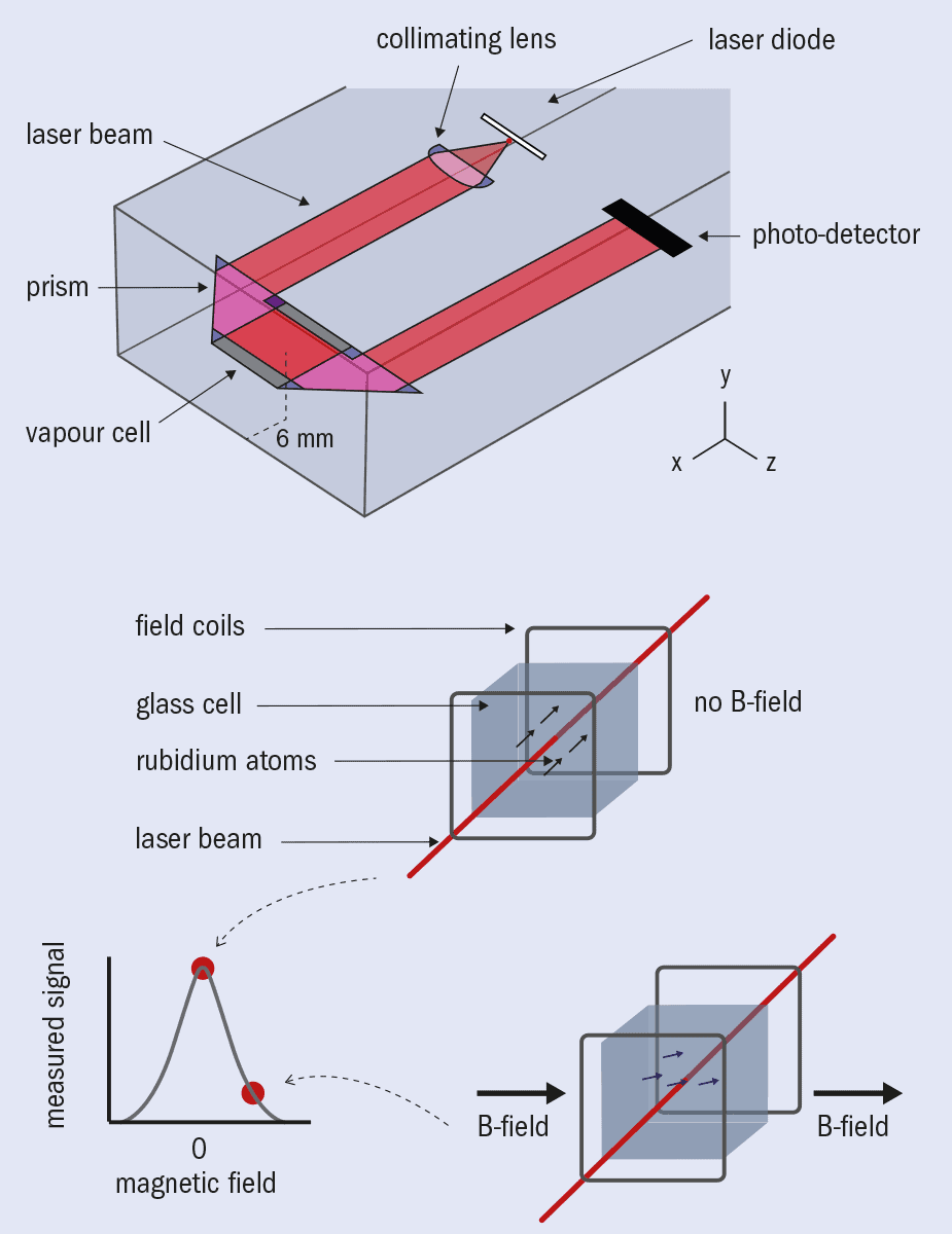
In recent years our research team has tackled MEG’s limitations by exploiting newly developed quantum technology. (This is a collaborative effort by the University of Nottingham’s Sir Peter Mansfield Imaging Centre and the Wellcome Centre for Human Neuroimaging at University College London, funded by the UK National Quantum Technologies programme and Wellcome.) In particular, the development of optically pumped magnetometers (OPMs) has been key. These are quantum enabled magnetic field sensors that offer similar sensitivity to SQUIDs but without the need for cryogenics (figure 2).
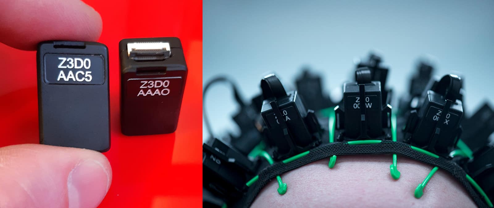
Recent commercialization by the US company QuSpin has made OPMs robust, easy to use and readily available, while miniaturization has made the most recent generation small and lightweight (similar to a Lego brick in both size and weight). Based on this new design, our team has integrated OPMs into a working prototype MEG device. Because they are so small and don’t need cryogenics, OPMs can be mounted directly on the surface of a human head, increasing sensitivity by removing the thermally insulating gap and getting the sensor closer to the brain.
Based on this new design, our team has integrated optically pumped magnetometers into a working prototype MEG device
This also allows the sensor array to move with the head, making the MEG measurement resilient to subject motion. Similarly, flexibility of OPM placement means an array can adapt to any head size, enabling babies and children as well as adults to be scanned with the same system. The lack of complex cryogenics also means that OPM-based MEG systems are ostensibly cheaper to produce and run. This technology therefore allows MEG to evolve, making systems more practical, more powerful, significantly cheaper and consequently much more suitable for clinical use.
However, significant barriers had to be overcome before OPMs could be used in practice, and one of the biggest was controlling background magnetic fields. The field from the brain is much smaller than the time-varying fields that exist all around us, generated by laboratory equipment, computers, passing cars and even our bodies. Such sources create a cacophony of magnetic interference that makes measuring magnetic fields from the brain akin to hearing a pin drop at a rock concert.
To complicate matters, there is also the Earth’s magnetic field. It has no impact on SQUIDs because they are sensitive only to time-varying fields – and of course the Earth’s field does not move. But with OPMs we want patients to move freely during a scan, and this means allowing magnetometers to move relative to the Earth’s field. The moving OPMs will therefore measure magnetic field changes that have nothing to do with the brain, because they are rotating in a uniform field (or translating in a field gradient). In some circumstances, this can even prevent OPMs from operating.
For these reasons, both the time-varying environmental magnetic fields and the Earth’s static magnetic field must be removed in the vicinity of the OPM measurement. We achieved this high-fidelity control through a combination of active and passive magnetic screening.
Passive screening involves placing the MEG system in a magnetically shielded room with walls made from multiple layers of high-permeability metal. However, even the most advanced passive shield cannot sufficiently suppress the remnant Earth’s field so that patient movement can be allowed, and for this reason active field compensation systems have been introduced.

These consist of intricate electromagnetic coil systems connected to a separate magnetometer array measuring the field close to the patient’s head. A feedback loop then uses these field measurements to generate an inverse field by controlling the currents in the coil array. In combination, these cancel the residual magnetic field almost entirely in the region of the patient’s head (figure 3). Working with industrial partner Magnetic Shields, we have built a unique hybrid active and passive shield in which the Earth’s field surrounding the patient is reduced from 50 μT to ~200 pT, a shielding factor of ~250,000. This technology generates a region of space around 0.5 m3 in which a patient can move freely during a scan.
This technology generates a region of space around 0.5 m3 in which a patient can move freely during a scan
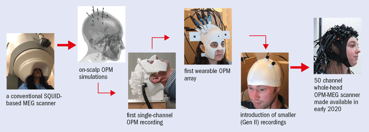
To make the OPM-MEG system a reality we also had to solve a number of other problems. The exact arrangement of sensors on the head was optimized by a combination of analytical analysis and computer simulation. This allows an OPM array to efficiently sample the brain’s magnetic field while also minimizing “cross talk” – an effect where the measurement of field at one sensor is disrupted by the presence of other sensors. Meanwhile, physically holding an array of OPM sensors on the head was achieved by advanced 3D printing. Electronic control and data acquisition systems were also developed to enable synchronized measurements from up to 50 OPMs, while separate control systems were needed to integrate OPM outputs with coil systems and the control of patient stimuli. Finally, mathematical modelling packages were redeveloped to allow imaging of current density in the brain based on the OPM scalp-level field measurements. These developments have led to the world’s first wearable OPM-MEG system, giving complete coverage of the whole brain (figure 4).
Wearable imaging
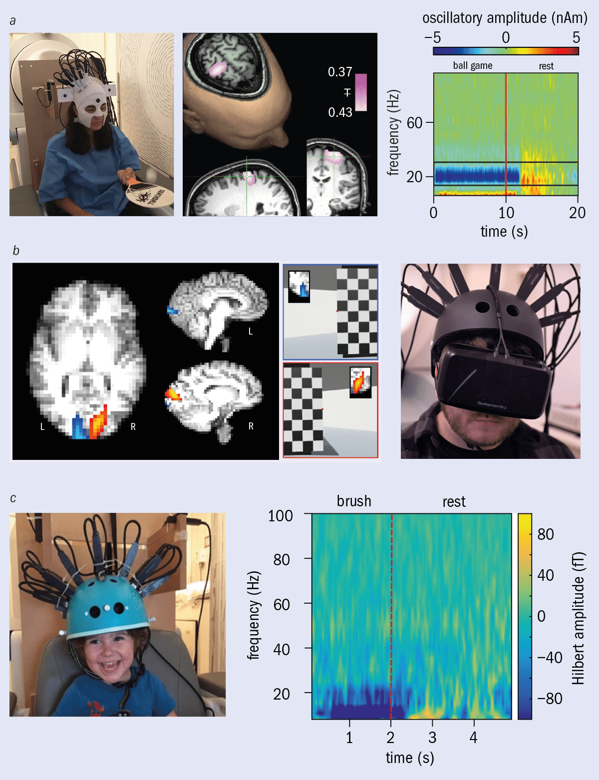
This unique combination of quantum technology and electromagnetic theory makes it possible to conduct previously unimaginable neuroimaging studies where subjects can move freely and interact naturally with the world. Even at this early stage, neuroscientific demonstrations have ranged from measuring brain activity as someone plays ping-pong, to making MEG recordings while a subject explores a virtual world (figure 5).
Laboratories around the world are trying to gain access to this new technology
With ongoing rapid exploitation, these examples are just the beginning; laboratories around the world are trying to gain access to this new technology, and a recently established spin-out company – Cerca Magnetics – is now bringing an integrated OPM-MEG system to the research market. Much of the ongoing technical development is now focused on imaging children’s brains, opening up the possibility of new studies on neurodevelopment. For example, imagine being able to see how a child’s brain function changes before and after they can walk, or talk. This opens a myriad of opportunities, prompting neuroscientists to conceive experiments in a completely new way.
Neuroscience aside, perhaps the ultimate marker of success for OPM-MEG will rest in its clinical applications. Conventional MEG can be used to diagnose epilepsy by identifying specific types of “spike and wave” activity. It is also already used when “resective” surgery (removing the region of the brain causing seizures) is required to treat epilepsy that cannot be controlled by drugs – a MEG scan locates the affected area, significantly increasing the chances of a successful surgery.
Understanding and managing human brain health is one of the major scientific challenges of the 21st century
MEG can also be used to map the “eloquent cortex” (areas of the brain that function normally) around the epileptic focus. This provides neurosurgeons with valuable information – for example, mapping the location in the brain of motor function allows a surgeon to avoid those regions, and so prevent paralyzing the patient. Now, however, the flexibility of OPM-MEG offers the chance to enable young children who suffer with epilepsy to also benefit as it avoids some of the limitations of conventional imaging techniques such as EEG and MRI, which can struggle to localize where the problem is. Motion robustness also ostensibly enables us to record brain activity during a seizure. For epilepsy patients, OPM-MEG is therefore extremely promising.
Do quantum effects play a role in consciousness?

But it could be applied to other disorders too. For example, this same practicality provides a better means of assessing individuals with Parkinson’s disease who struggle to remain still enough for conventional systems. Moreover, one in four people suffer from a mental health condition at some point in their lives and nascent recordings show that OPM-MEG can measure the connections in the brain that are thought to break down in severe mental health disorders. Meanwhile, characterization of “cortical slowing” (an effect where neural oscillations shift in frequency) in the elderly may generate new and early markers of the onset of dementia. These are just some of the brain disorders that may benefit from this new device.
Ultimately, understanding and managing human brain health is one of the major scientific challenges of the 21st century, and the steps needed to meet that challenge are far from clear. However, from X-rays to MRI, ultrasound to nuclear medicine, physics has always been able to deliver technology that changes lives. As we look to the future, perhaps this early success story of the UK’s quantum technology programme will become a cornerstone of the next generation of healthcare technology.