Nuclear medicine modalities such as positron emission tomography (PET) and single-photon emission computed tomography (SPECT) play a vital role in many areas of healthcare, including cancer diagnostics and cardiac imaging, among others. Alongside, innovative research projects aim to continually improve these molecular imaging techniques, by minimizing the amount of radioactive tracer needed, reducing the required imaging time or enhancing the image quality. At the recent Annual Meeting of the Society of Nuclear Medicine and Molecular Imaging (SNMMI), researchers presented the latest advances in PET and SPECT instrumentation.
CT-free PET reduces radiation dose
Total-body PET scanners with a long axial field-of-view can enable extremely low-dose PET scans. But the CT scan performed alongside to obtain attenuation maps can deliver a substantial radiation dose, negating these low-dose benefits. At the SNMMI Annual Meeting, Mohammadreza Teimoorisichani from Siemens Medical Imaging presented a fully quantitative PET imaging technique that does not require an accompanying CT scan and dramatically reduces the amount of radiation delivered to the patient. The approach could prove of particular benefit to paediatric patients and those requiring multiple scans.
“Most modern PET scanners use lutetium-based scintillators to detect gamma photons” explains Teimoorisichani in a press statement. “The lutetium in the scintillator contains a small amount – less than 3% – of the radioisotope 176Lu, which emits background radiation during the scan. In our study, we used this background radiation as a transmission source to simultaneously reconstruct attenuation maps and quantitative PET images without the use of CT.”
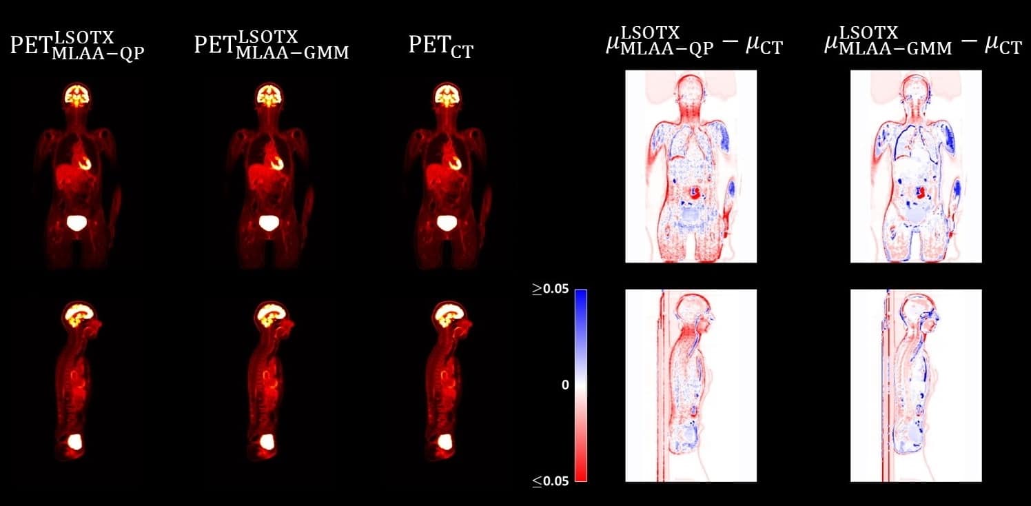
The researchers evaluated their proposed reconstruction technique using data from a clinical FDG-PET scan acquired with a Siemens Biograph Vision Quadra PET/CT scanner. The patient was injected with roughly 170 MBq of 18F-FDG and scanned 55 min post-injection for a duration of 10 min. Using the 202 and 307 keV gamma photons from 176Lu to reconstruct attenuation maps, they generated PET images using various CT-free reconstruction algorithms.
Comparing the results with standard PET/CT images showed that the largest quantification errors in the attenuation maps appeared around the patient boundary. Of the various organs examined, the brain had the largest quantitative error (activity underestimation of 15–21%). However, the CT-free reconstructed PET images showed average organ quantitative errors of 4.8% and 10% for two reconstruction techniques examined.
As well as reducing patient dose, the proposed method also eliminates potential attenuation map misregistration that can arise due to patient motion between the CT and PET scans. The approach could also provide a reliable technique for attenuation correction in hybrid PET/MR scanners.
“This study is an important step toward practical CT-less quantitative PET imaging,” notes Teimoorisichani. “In addition to reducing patient radiation exposure, a true low-dose quantitative PET scan can have a great impact on research studies that aim to better understand human physiology at the molecular level and on research involving the development of radiopharmaceuticals. The algorithm is currently being evaluated on a large number of patients to discover its full potential.”
Self-collimating SPECT offers rapid cardiac imaging
A team from Tsinghua University in Beijing has designed a cardiac SPECT system that performs scans 10 to 100 times faster than current SPECT devices. The new system employs active detectors in a multi-layer architecture that carry out the dual functionality of detection and collimation. This “self-collimation” concept improves upon conventional SPECT approaches to deliver dramatically shortened scan time, better image quality, increased patient throughput and reduced radiation exposure to patients.

“SPECT is an important non-invasive imaging tool for the diagnosis and risk stratification of patients with coronary heart disease,” says Debin Zhang in a press statement. “However, conventional SPECT suffers from long scan time and poor image quality as a result of relying on a mechanical collimator. The new SPECT system is capable of performing fast-framed dynamic scans with high quality.”
The self-collimating cardiac SPECT consists of three identical trapezoidal detector units, joined to form a half-hexagon that encloses a spherical field-of-view. Each detector unit comprises an inner tungsten plate containing many apertures, followed by four stacked detector layers, three containing scintillators sparsely arranged in a chessboard pattern and the outer one containing closely packed scintillators. These scintillators perform the dual functions of photon detection and collimation.

The researchers compared three aperture patterns in the metal plate (which also provides part of the collimation) and found that a random distribution of 140 apertures provided better signal-to-noise performance than 48 or 140 apertures in a grid pattern. Using this random configuration, the cardiac SPECT had an average sensitivity of 0.68 in the field-of-view.
In scans of phantoms, the system could separate 4 mm rods in a hot-rod phantom, and was able to identify a defect in a cardiac phantom in as little as 2 s.
The team concludes that the proposed detector design has potential to expand clinical applications of dynamic cardiac SPECT, by eliminating the impact of patient’s respiratory motion, increasing patient throughput, enabling ultra-low-dose imaging and precisely quantifying myocardial blood flow and coronary flow reserve.


