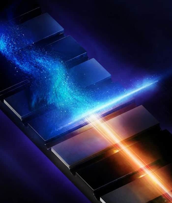
Hard X-ray light pulses lasting mere attoseconds have reached a record high intensity, concentrating 118.8 GW of power onto a focal spot measuring just 180 x 330 nm (2 x 10²⁰ W/cm2). This achievement, which comes from a team working at the European X-ray Free Electron Laser (XFEL) in Hamburg, Germany, clears the way for ultrafast structural imaging in materials as well as detailed studies of how electrons behave in atoms.
In the semi-classical Bohr model of a hydrogen atom, the electron takes roughly 150 attoseconds (150 x 10-18 s) to orbit the nucleus. One way of measuring this ultrafast motion is to send an attosecond light pulse through a sample. The electrons in the sample interact with this pulse and distort it. By monitoring this distortion, scientists can make 3D maps and even movies of electron motion.
Attosecond pulses are usually produced via a technique called high-harmonic generation (HHG) that was developed in the late 1980s. However, while this technique is good at producing attosecond pulses in the extreme ultraviolet and soft X-ray regions of the electromagnetic spectrum, study co-leader Jiawei Yan explains that extending this approach to shorter, hard X-ray, wavelengths has not been easy.
A few years ago, Yan and colleagues at XFEL and the nearby DESY facility developed a way of generating hard X-ray attosecond pulses at a scale of terawatts. The XFEL is a linear electron accelerator that generates coherent light via a process called self-amplified spontaneous emission, and the team was able to exploit this process to produce light at energies ranging from hundreds to many thousands of electron volts (eV).
Generating high energies at the source is only the first step, however. “For real-world experiments, what really matters is the intensity we can deliver to the sample,” Yan says. “We undertook this new study to see whether we could tightly focus these pulses to increase their intensity.”
Increasing pulse energy
The researchers generated their pulses using a scheme called self-chirping that begins by compressing a bunch of electrons into a short spike of current. As this bunch travels through the accelerator, its own electromagnetic fields imprint an energy variation along it. A magnetic arc then uses this variation to compress the bunch even further. “This ultrashort, high-current part of the bunch then emitted intense hard X-ray attosecond pulses in the accelerator’s undulator, with energies of up to a few hundred microjoules,” Yan explains. He points out that this value is more than 10 times higher than the energies of hard X-ray attosecond pulses produced by other methods.
The next step was to deliver these pulses to the XFEL’s Materials Imaging and Dynamics (MID) instrument. There, the researchers focused the pulses using a total-reflection optical system known as an Advanced Kirkpatrick–Baez (AKB) mirror that was developed by Yan’s Japan-based colleagues Jumpei Yamada and Ichiro Inoue, who are at the University of Osaka and the University of Tokyo, respectively.
New attosecond X-ray spectroscopy technique ‘freezes’ atomic nuclei in place

As well as revealing the behaviour of electrons, Yan suggests that the ultra-intense hard X-ray pulses the team created could be used to image the structure of materials on ultrafast time scales. “The pulses could allow us to collect diffraction signals before substantial electronic damage develops, improving structural measurements of samples that are sensitive to radiation, for example,” he explains.
Reporting their work in Optica, the researchers say they are now busy developing stabilized attosecond pulses and exploring ways to generate multiple pulses with controlled timing. “I would also like to see more experiments using these pulses to study nonlinear X-ray interactions and ultrafast electronic dynamics,” Yan tells Physics World. “We now have a source that can reach an extreme intensity regime, and I think the next important step is to find out what new physics we can do with it.”