structure on a nanoparticle surface for drug delivery. Courtesy ACS Nano")
Nanoparticles are promising vehicles for drug delivery in biomedical research. Their biggest limitation is that the immune system recognizes them as foreign and quickly removes them from the bloodstream. In a paper recently reported in ACS Nano, a team led by Hao Cheng at Drexel University in the United States explored solutions to this problem by creating a new type of polyethylene glycol (PEG) coating for nanoparticles. While previous coatings have focused on shielding nanoparticles from macrophages, this coating has the benefit of making the nanoparticles largely invisible to liver endothelial cells, which filter foreign objects out of the bloodstream. The team hopes that these new coatings can be used to improve drug delivery in clinical applications.
PEG stealth
Avoiding detection by the immune system is a key issue for drug delivery. Immune recognition is accomplished by proteins in the bloodstream that flag foreign objects (such as nanoparticles) by sticking to their surfaces. Cells of the immune system such as macrophages (immune cells which scavenge the body for foreign objects) can then engulf or take up the particles, preventing them from reaching their destination. One of the most popular methods of creating a “stealth particle” is by coating their surface with a neutral polymer such as polyethylene glycol (PEG).
Scientists have found that the shape of the PEG polymer affects how it prevents protein binding. For example, a high-density PEG layer – one that is closely packed together – prevents protein binding by crowding the surface and taking up potential protein binding sites. In contrast, low-density PEG, or PEG that has more space in between the bound polymer chains, hinders protein binding via its flexibility; each polymer chain has more freedom to rapidly wiggle around, making it difficult for proteins to squeeze past them to the nanoparticle surface.
Liver dodger
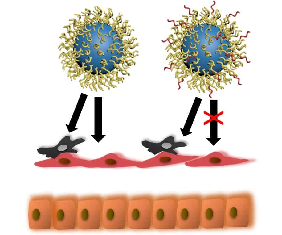
Cheng’s team has found a way to maximize PEG shielding of nanoparticles by combining the benefits of both high and low-density PEG. They were able to create a PEG coating with a dual layer: a short inner PEG layer that is highly dense and a longer outer layer with more flexibility.
Nanoparticles covered in the team’s specially designed PEG coatings could circulate in the bloodstream for roughly three times longer than previous nanoparticle designs. The reason for the increased circulation time was unexpected: the PEG coating prevented uptake by liver endothelial cells, but not macrophages. Macrophages in the liver still consumed the particles but because the liver endothelial cells ignored them, the particles circulation time was increased overall. Previously, the importance of preventing liver cell uptake was not well understood.
Because part of liver function is metabolizing drugs and filtering the blood, it is a primary route for nanoparticle removal. Making nanoparticles that are invisible to the liver is a big step towards producing a universal drug delivery vehicle.
Physical versus chemical paradigms play off in nanomedicine

The Cheng Lab is excited to see how this knowledge about PEG coatings on nanoparticles can be applied to benefit drug delivery applications, especially for cancer therapy. Future work will explore creating coatings that can block uptake by both liver cells and macrophages to produce ultra-long circulating nanoparticles.