Cherenkov imaging during radiotherapy enables real-time visualization and mapping of the radiation beams as they deliver dose to a patient’s body, providing a way to evaluate the accuracy of treatment delivery in real time. It is also being extensively tested in research laboratories worldwide as a tool to quantify the actual radiation doses delivered to patients, in a way that is unaffected by skin colour.
The optical imaging technique offers the benefits of high spatial resolution, high sensitivity and fast imaging speed compared with conventional methods for measuring radiation dose. But there are still challenges to overcome before all of its capabilities can be adopted for clinical use.
Cherenkov radiation is produced when charged particles travel at a speed greater than the phase velocity of light in tissue. The signal intensity is proportional to the delivered radiation dose and, in an ideal scenario, accurately indicates the dose delivered during radiotherapy treatment.
In reality, however, tissue attenuation reduces the intensity of the emitted Cherenkov radiation, and alters the linear relationship between deposited dose and observed Cherenkov emission. Because of this, the Cherenkov signal from human tissue is not yet accurately interpretable as fully proportional to the dose.
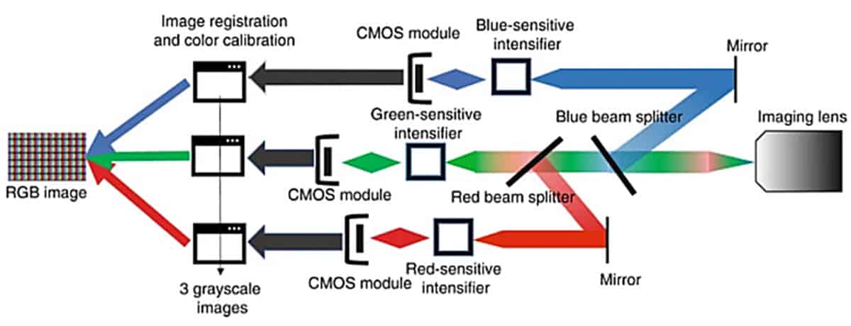
Researchers at Dartmouth College and the University of Wisconsin-Madison are working to make Cherenkov imaging a reliable indicator of radiation dose. In a recent study reported in the Journal of Biomedical Optics, they used a custom, time-gated three-channel intensified camera to image the red, green and blue wavelengths of Cherenkov emission from various tissue phantoms. They hypothesize that the intensity of Cherenkov emission changes with variations in biological absorption features – such as blood concentration within tissue and melanin concentration in human skin with different levels of pigmentation.

“Tissue absorption and scattering may cause a large variation among patients in detected Cherenkov emissions,” explains principal investigator Brian Pogue, of the University of Wisconsin-Madison School of Medicine and Public Health and Dartmouth’s Thayer School of Engineering. “We know that variation in skin colour can alter the signal level by up to 90%, and changes in blood or scattering content can cause up to 20% signal variation.”
“We conducted our study to better understand how tissue optical properties affect the emission colours of Cherenkov light, and to begin to identify ways to use the spectrum of the light for calibration or correction of tissue attenuation effects,” he explains.
For the study, Pogue and colleagues prepared tissue and blood phantoms with varying levels of melanin and blood volume. They created synthetic 0.1 mm-thick epidermal layers containing seven different concentrations of synthetic melanin that match those in human skin, and then placed these layers on top of thick bulk tissue phantoms. The researchers also tested seven blood phantoms with blood concentrations ranging from that of fatty tissue up to highly vascularized muscle tissue.
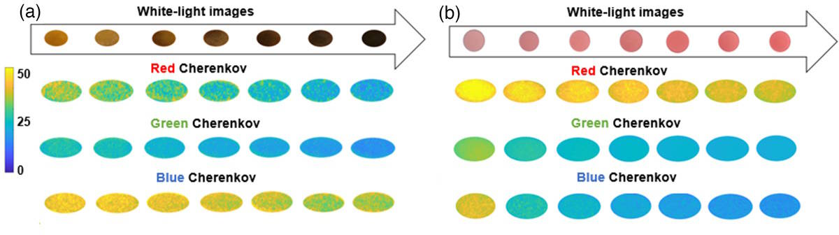
The researchers irradiated the phantoms with a 3 Gy dose using 6 MV photon and 6 MeV electron beams and acquired images for each colour channel. The acquisitions were time-gated to the linac, to capture Cherenkov emission only during the microsecond radiation pulses without background ambient light. They note that for both beams, there was no observable Cherenkov emission for melanin above 0.0076 mg/ml (a medium high-level).
The team reports that Cherenkov emission from the phantoms decreased as melanin concentration increased. Extremely high melanin levels caused a significant reduction in Cherenkov emission, making it challenging to perform imaging in individuals with the darkest skin tones.
Colour also made a difference when imaging blood phantoms, with greater attenuation as blood concentration increased. The red channel attenuated to a lesser extent than the blue and green channels, due to the absorption of blue and green colours by oxyhaemoglobin in the blood. “These findings suggest that imaging in the red and near-infrared wavelengths will be better,” comments Pogue. “In addition, having characterized the amount of attenuation in each colour band will facilitate calibration for skin colour.”
“Our findings support the idea that colour or spectral imaging of Cherenkov might provide an experimental methodology for separation of biological attenuation of the intensity from the physical generation of Cherenkov with dose deposition. The goal would ideally be to use the Cherenkov intensity as an indicator of the dose delivered in the tissue, independent of the blood volume within it or skin colour, using colour correction,” the researchers write.
Cherenkov imaging for visualizing radiotherapy: one year of clinical use

The team has started a clinical trial with collaborators at Moffitt Cancer Center, to image patients with a larger range of skin colour variation, and hopes to expand the trial to UWHealth in Madison “This will allow us to test this type of imaging in patients who better represent the normal range of cancer patient populations,” Pogue tells Physics World. “We really want to better understand how the images look, and if we can rely upon Cherenkov imaging to show us the pattern of radiation delivery to all patients, independent of their skin colour.”
“So far, the data look encouraging,” he adds. “Because less light is emitted as the melanin content of the skin gets higher, we are also using colour imaging to correct for this. We are hopeful that we can make the system largely independent of the skin colour. We believe that spectroscopic interpretation may help to better relate Cherenkov emission to ionizing radiation dose delivered during radiotherapy.”


