Early detection of metastasis, in which cancerous cells spread through the body, could turn the tide on cancer and enable clinicians to provide suitable therapies. Importantly, the introduction of artificial intelligence (AI) and enhanced image analysis has helped improve diagnostic accuracy. To assist pathologists in identifying metastatic lymph nodes (MLNs), researchers at Xidian University and Changhai Hospital in China have developed a computational approach to predicting the clinical outcomes of patients with gastric cancer.
Stomach cancer, also known as gastric cancer, occurs when cells in the inner lining of the stomach begin to grow abnormally. If left untreated for several years, these abnormal cells may develop into a tumour. Traditionally, experienced pathologists examine excised lymph nodes for the presence of gastric cancer metastases and evaluate the tissue morphology with the aid of an optical microscope. While this is an acceptable standard of practice, the process can be tedious and may lead to human errors.
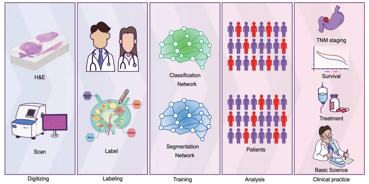
The multidisciplinary group, led by Guanzhen Yu and Xiyang Liu, developed a deep-learning framework for identifying and analysing micrometastases (with a diameter of less than 2 mm) in lymph nodes. The framework was designed to uncover the tumour-area-to-MLN-area ratio (T/MLN) from whole slide images. The team tested the approach on two independent datasets of gastric cancer patients.
Diagnostic accuracy with AI assistance
The researchers point out that while pathologists possess better specificity in the detection of tumour tissues, AI offers scalability of performance due to its sensitivity and speed. Combining the two could provide the most clinically meaningful outcome.
The researchers first digitized MLN pathology samples and annotated them to create a training dataset. They then subjected this dataset to a deep-learning algorithm for classification and segmentation. These steps resulted in a precise calculation of the proportions of tumour components and lymph nodes in the samples.
Clinical pathologists reviewed the deep-learning results and reported a 94.5% consistency in lymph node detection between the AI diagnosis and the original diagnosis. Importantly, the researchers demonstrated that, with AI assistance, a pathologist required an average of 2–6 min to diagnose a patient’s lymph node. Without AI assistance, the diagnosis required 3–15 minutes.
This potential to enhance performance with AI-assisted analysis will improve patient prognosis and shorten the time required for making therapeutic decisions, say the researchers, who report their findings in Nature Communications.
Gastric cancer prediction
One challenge when predicting cancer prognosis is the insufficient information acquired during diagnostic evaluation. However, the deep-learning architecture developed in this study was able to efficiently identify MLNs, thereby reducing the rate of missed diagnosis by human pathologists. The researchers note that cancer patient’s outcomes were correlated with the area of metastatic tumour in the MLN. They could therefore use the AI algorithm to calculate the precise number of tumour cells within the MLN and deploy this as a prognostic marker for gastric cancer.

The researchers point out that the prediction results in this study are representative of a gastric cancer cohort from an individual nation. However, they propose that the AI should be tested in a large-scale clinical trial across several countries. They believe that this would validate the algorithm and enable clinicians to improve treatment outcomes.


