Researchers within Elekta’s MR-Linac Consortium are opening up new frontiers in cancer treatment through the collaborative development of biological image-guided adaptive radiotherapy (BIGART)

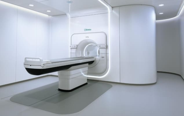
The Elekta Unity MR-Linac is among a new generation of MR-guided radiotherapy (MR/RT) systems transforming patient care and treatment outcomes in the radiation oncology clinic. Think online image guidance and adaptive radiotherapy tailored to the unique requirements of each patient – adjusting radiation delivery “on the fly” to address the daily variation in the tumour and surrounding healthy tissue while allowing adaptation of the plan for tumours that respond rapidly to treatment (as well as those that prove unresponsive to standard doses of radiation).
If that’s the back-story, it’s already evident that MR/RT is poised to drive ongoing innovation and transformation along the continuum that is treatment planning, delivery and management. Most notably, while enabling the clinician to visualize a tumour target, and its adjacent anatomy, with exceptional soft-tissue contrast both prior to and during treatment, MR/RT systems also have the capacity to acquire functional and quantitative images. It’s this capability, in turn, that points the way to the long-anticipated end-game: the fusion of biological targeting and adaptive radiotherapy – otherwise known as biological image-guided adaptive radiotherapy (BIGART).
Think big, think BIGART
So what might BIGART look like in terms of a next-generation radiation oncology workflow? Put simply, with the help of frequent anatomical and functional imaging, it is hoped that MR/RT systems will be able to monitor changes in the volume, shape and biological characteristics of the tumour so that the treatment plan can be updated regularly in line with the observed treatment response. “A new era in cancer treatment is coming into view,” according to Uulke van der Heide, group leader at the Netherlands Cancer Institute (NKI) and professor of imaging technology in radiation oncology at the Leiden University Medical Center. “In that sense,” he adds, “MR/RT technology is a game-changer, giving us the platform we need to undertake clinical studies of biological targeting for adaptive radiotherapy.”
Van der Heide, for his part, is at the clinical sharp-end of the BIGART development effort. Within the Elekta MR-Linac Consortium, for example, he heads up a working group on quantitative imaging biomarkers (QIBs), a range of metrics spanning tumour morphology, biology and function that could one day inform routine assessment of treatment response during radiotherapy. Currently, the QIB working group comprises more than 15 cancer treatment centres across Europe, the US, Canada and Asia – all of them Unity clinics and all of them aligned with the broader Consortium remit to drive improved patient outcomes through the application of MR-Linac technology.

In terms of specifics, the QIB collaboration is active in developing clinical trial strategies, quality assurance programmes and data acquisition/analysis methods to fast-track BIGART research on the Unity MR-Linac platform. “The starting point, of course, is to identify QIBs that show changes early during treatment and in turn are predictive of treatment outcome,” explains van der Heide. “The first pilot studies are encouraging and show that repeated QIB measurements are feasible using a range of quantitative MRI [qMRI] techniques during patient treatment on the Unity system.”
The opportunity, it seems, lies in the diversity of qMRI options available to clinicians – and, by extension, the matrix of radiobiological insights that could over time support online adaptation of radiotherapy treatment planning. Diffusion-weighted imaging (DWI) is the most studied qMRI technique in this regard, yielding data on the cellular density of tumour tissues (with reductions in density linked to the breakdown of cell membranes and necrosis during radiotherapy). Another qMRI modality showing early promise is intravoxel incoherent motion (IVIM), which has the potential to track changes in tissue perfusion and vascular permeability in the tumour microenvironment.
Taken together, there are already multiple – and proliferating – lines of qMRI enquiry with significant clinical potential. “The combination of qMRI techniques reflects the richness and complexity of tumour tissues,” says van der Heide, “and is definitely something we will be pursuing in a clinical setting. One qMRI modality is not going to cut it for biological targeting – a multimodal strategy will be key.”
A collective endeavour
Over the longer term, however, several conditions need to be met if researchers are to maximize the clinical benefits of qMRI. For starters, it’s important to know how to relate measurements from the MR-Linac to measurements taken on diagnostic MR scanners outside the MR/RT domain – knowledge that will ultimately enable clinical studies to include measurements from before treatment when considering the patient treatment response.
To achieve this goal, the QIB working group has adjusted the qMRI measurement protocols on the MR-Linac to consider the detailed differences in MR hardware implementation. “We have done a set of multicentre studies – using digital and physical phantoms – to demonstrate accuracy and reproducibility of the MR-Linac qMRI measurements,” notes van der Heide. “The accuracy is similar to the previously reported literature for diagnostic scanners.”
Equally important is ensuring a high level of confidence that measurements performed on one MR-Linac can be related to any other MR-Linac. As such, the underlying hardware needs to be consistent across different systems, while different research teams must also use standardized qMRI measurement protocols. The QIB working group is currently developing these protocols for its network of cancer treatment centres.
Another focus of the working group is to understand, by assessing the repeatability of qMRI measurements using test-retest studies, which changes in qMRI values can be linked to the effects of radiotherapy. Finally, van der Heide and his colleagues are in the process of establishing QA best practice for the QIB programme. “The MR-Linac Consortium is growing and we expect more cancer centres to get involved in these studies,” adds van der Heide. “It’s therefore essential to have a managed and standardized QA programme to support the qMRI development effort across all participating centres.”
In the same way, the QIB working group clearly benefits from the fact that all members are Elekta Unity users. “The common MR-Linac platform will take away a lot of the multicentre variability during clinical trials,” van der Heide concludes. “Over time, though, we hope to join up our QIB development efforts with those of the ViewRay MRIdian community.”
The future’s bright, it seems, for qMRI and BIGART. Clinical trials are on the MR-Linac Consortium’s development roadmap in the next two to three years, with the first step likely to involve an investigation of daily changes in qMRI values in different tumour sites across multiple oncology clinics.
- For further information, readers can register to access the on-demand Physics World webinar Quantitative MRI for Biological Image-Guided Adaptive Radiotherapy. See also the Frontiers in Oncology mini review Quantitative Magnetic Resonance Imaging for Biological Image-Guided Adaptive Radiation Therapy


