
Artificial intelligence (AI) may soon have a central role to play in the global battle against COVID-19. A European campaign is underway to develop a deep learning-based model for the automated detection of abnormalities on chest CT and for quantifying lung involvement.
“In these unprecedented circumstances, we must find ways of helping doctors in their fight against the virus,” noted Erik Ranschaert, president of the European Society of Medical Imaging Informatics (EuSoMII), who is leading the initiative with fellow radiologist Laurens Topff, from the Netherlands Cancer Institute (NKI) in Amsterdam. “The value of AI also comes into play, by reducing the burden on clinicians. While a manual read of a CT scan can take up to 15 minutes, AI can finish reading the image in 10 seconds.”
Around 30 partners have already expressed their willingness to share data and to support the plan to train the algorithm. These include academic and non-academic hospitals located in the most affected areas of Italy and Spain, and also in Germany, Belgium, the Netherlands and the UK.

Each hospital will transfer the data directly and securely to the servers of Quibim. Based in Valencia, Spain, it specializes in machine learning and image processing technologies for medical images, and it will provide a research platform for the development and deployment of the deep-learning model. For data preparation, annotation and algorithm training, the Robovision AI (RVAI) software will be used. The data will only be used for research purposes.
Automated image analysis with AI techniques can optimize the role of CT in the assessment of COVID-19 by supporting clinical decision-making, improving workflow efficiency, and allowing accurate and fast diagnosis of infection in a large number of patients, Ranschaert explained.
“We believe that it’s possible to train an accurate AI algorithm with the wealth of data already available since the outbreak of the virus in Europe, with the main aim of helping doctors to make this diagnosis in time,” he added.
A team from the NKI will assess and statistically analyse the performance of the deep-learning model, which will be made freely available as a research solution to participating hospitals.
Growing use of CT
The clinical presentation of patients with COVID-19 ranges from asymptomatic to severe pulmonary infection. The most specific method and reference standard to diagnose infection is the reverse transcription polymerase chain reaction (RT-PCR) test, but due to the varying levels of sensitivity of viral testing, shortage of viral testing kits and longer turnaround times to provide results, lung CT scans are attracting attention, Ranschaert said.
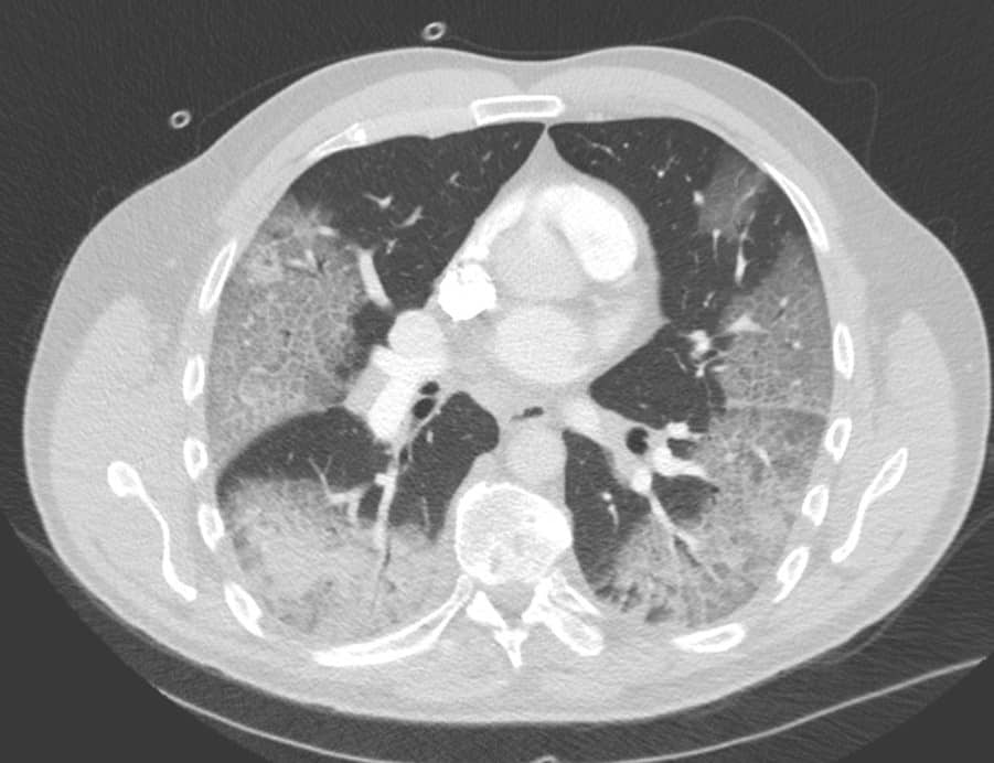
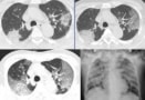
“COVID-19 causes a wide variety of findings on these scans, most typically ground-glass type of densities located on the outside of both lungs (see white areas in figure),” he continued. “The accuracy of chest CT to diagnose COVID-19 has been reported as high and can predate a positive classic serological RT-PCR test. Therefore, in endemic areas where the healthcare system is under pressure, hospitals with a high volume of admissions are using CT for rapid triage of patients.”
There is a role for chest CT to assess COVID-19 infection in patients with severe and worsening respiratory disorders. Based on the images, doctors can evaluate how severely the lungs are affected and how the patient’s disease is evolving, which is helpful in making treatment decisions, according to Ranschaert.
Also, pulmonary abnormalities caused by COVID-19 can be found by chance in exams carried out for other reasons – for example, abdominal CT scans for bowel problems – in patients without respiratory complaints.
“Patients are coming in with different types of complaints and therefore sometimes get other examinations. Some radiologists are even proposing to do a standard CT chest with every CT abdomen,” he noted. “If a CT abdomen needs to be done because of nonspecific complaints, just scan higher to assess more lung tissue, leading to more nonspecific COVID-19 patients being detected.”
In areas of widespread coronavirus outbreak, many hospitals are installing special scanning units to enable efficient screening of the steadily growing number of victims. If CT is used for screening, there will be so many studies that radiologists will be overwhelmed and AI will be urgently needed.
CT traces disease progression in novel coronavirus
“The scans run full-time and the number of doctors to assess all these scans is sometimes insufficient, partly due to the fact that doctors are also more often exposed to infected patients and therefore themselves become victims of the virus,” Ranschaert pointed out, adding that 300 Chinese doctors had to be flown into Italy to cope with the growing number of patients.
The danger of cross-infection via the CT scanner is an important consideration.
“Some hospitals are very prudent and do a complete disinfection of the CT scan suite, taking about an hour,” he said. “Others only clean the contact surfaces of the scanner, while also protecting the radiographers of course with an adapted outfit – this on advice of their microbiologists. These hospitals usually have a dedicated scanning suite for COVID-19 scans. Some even have a mobile unit outside the hospital to provide these scans.”
A LinkedIn post last week was viewed more than 100,000 times with over 2000 likes. The website for the project is active now.
- This article was originally published on AuntMinnieEurope.com ©2020 by AuntMinnieEurope.com. Any copying, republication or redistribution of AuntMinnieEurope.com content is expressly prohibited without the prior written consent of AuntMinnieEurope.com.


