
For the first time, researchers have measured the mechanical stresses and strains brought about by foetal movements during the second half of gestation, using computational modelling based on MRI scans acquired in utero. This exciting work, performed in collaboration between Imperial College London, Kings College, UCL and Great Ormond Street Hospital, quantified foetal kick and muscle forces to show that stress and strain increased consistently until term (J. R. Soc. Interface doi: 10.1098/rsif.2017.0593). This is important, since changes in or lack of foetal movement late in pregnancy can be indicative of poor outcomes. This research fills in a gap of knowledge regarding the link between skeletal biomechanics and malformations.
Foetal movements such as kicking are thought to be important for healthy musculoskeletal development, with the stresses and strains (deformations) of the bones providing the relevant stimulation. The absence of movement, or little movement in the later stages of pregnancy has been linked to thin bones, and spinal and joint abnormalities, such as developmental dysplasia of the hip (DDH), for example. While previous studies have recognised the importance of foetal movement, tracking these changes and quantifying them within the uterus has never been performed before.
Imaging and modelling
Cine-MRI is typically used in cardiac motion studies to visualize the biomechanics and movement of fluid in the heart. Fast, dynamic movement can be measured by acquiring lots of images very quickly across time (such as a heart beating or, in this case, a foetus kicking). For this study, researchers chose four subsets of five MR scans at 20, 25, 30 and 35 weeks of gestation to track the movements of individual foetal joints.
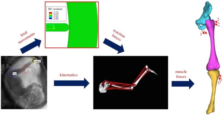
The researchers calculated foetal kick force by simulating a mechanical uterus environment and then inputting the observed kick displacements from the cine-MRI data. Next, they combined these predicted reaction forces with the cine-MRI tracking in order to predict forces for 19 muscles. Finally, they used post-mortem MRI scans (from a separate cohort) to create a stress/strain model. The forces calculated previously were used to quantify the amount of stress and strain in foetal lower limbs.
Pumped up kicks
The researchers found that uterine wall displacement was unchanged between 20–30 weeks, but dropped at 35 weeks. However, this did not result in a reduced stress or strain, possibly because later in gestation higher muscle forces are expected (due to the larger foetus and cramped position when kicking). Foetal kick force steadily increased, showing a similar drop at 35 weeks.
Maximum stress was observed in the femur shaft and tibia, and at joint surfaces. Since the stresses occurred at the interface between mineralized and unmineralized regions, the researchers suggested that mechanical stress is linked to new bone tissue formation (ossification). Maximum levels of strain occurred near the joints, indicating a role for joint shaping (joint morphogenesis). Both stress and strain increased from 20 to 35 weeks in all regions.
Stresses in the foetal skeleton increased steadily throughout gestation, and hence the authors suggested that movement restrictions, even at the later stages of pregnancy, may have an impact on normal musculoskeletal development. This may result in different biomechanical stimulation, and possibly deleterious implications for severely pre-term babies.
This work provides a unique glimpse into the development of a foetus’ skeleton, combining imaging and modelling to provide grounded estimates of the mechanical forces produced in utero. Insights from this work will further research on foetal biomechanical development and skeletal malformations.
- A wonderful GIF of foetal movement in the uterus can be seen on the Royal Society website.


