Better brain imaging or even drug delivery could be possible thanks to researchers at the Polytechnic University of Valencia in Spain. They have developed a technique for 3D printing holographic lenses that focus ultrasonic sound waves in the brain.
Penetrating the skull
Ultrasound, an acoustic wave with frequencies greater than those audible to humans, is widely used in diagnostics and for imaging soft tissue, such as a developing fetus. Furthermore, it can be used as a non-invasive therapeutic technique, which operates by focusing high intensity waves to ablate fibroids or destroy cancerous tissue. In the case of the brain, however, this method is challenging because the skull blocks and distorts the ultrasound, preventing it from focusing on brain tissue.
Previously scientists have tried using phased arrays to control the incoming ultrasound to correct for aberrations on penetrating the skull but these have limited numbers of pixels and can be quite costly. Led by Noé Jiménez the Polytechnic University of Valencia researchers have now 3D printed a lens that can generate complex patterns to help refocus the beam upon penetrating the skull, allowing it to effectively target brain regions such as the hippocampus, and image more clearly.
Making waves
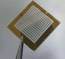
The lenses comprise of a block of plastic with varying voxel sizes. Each voxel diffracts the ultrasonic wave in a slightly different way. The interference of these waves creates a hologram as they focus onto a 3D target volume within the brain.
Testing the invention
Jimènez and colleagues used a four-step process to test out their approach. Firstly, they extracted the geometry and acoustic properties of the human skull using open source X-ray images from computerized axial tomography (CAT) scans. They then also examined soft tissue information from the brain itself, using data from magnetic resonance imaging (MRI). This allowed them to produce a computer model of the patient’s skull and brain.

Following this step, the team devised a method to bend ultrasonic waves inside the skull. They used three different types of holographic focusing, with increasing complexity: one that focuses the waves to a point, another that results in a curved wave path and a third that guides the waves to flood the whole of the right hippocampus in the brain.
Next, the scientists modelled the sound waves required to create an ultrasonic hologram within the brain. By back propagating virtual acoustic fields in the brain to a point outside the skull they could calculate the phase and amplitude of the source waves needed and design a lens to produce them.
Finally, using the previously acquired CAT scan and MRI data, they manufactured a 3D printed skull phantom (a realistic brain replica), which they used to test the holographic lens. The data they obtained from the skull phantom showed good agreement with theory and simulations.
Impact
Despite the good fit between the experimental data and theory and simulations, differences did arise due to the disparity in density between the printed material and bone. However, the researchers show by using full-wave simulations, that the same ultrasonic focusing would still be possible in a realistic situation.
Short ultrasound pulses improve drug delivery to the brain

This new concept will hopefully lead to low cost therapy, imaging of the central nervous system or even ultrasound-triggered neuromodulation. Perhaps, most importantly, it could also have implications for new drug delivery techniques. For example, it has the potential to open the blood-brain barrier, which typically blocks therapeutic drugs in the treatment of Alzheimer’s disease.
Full details of the research are documented in Phys. Rev Applied.