
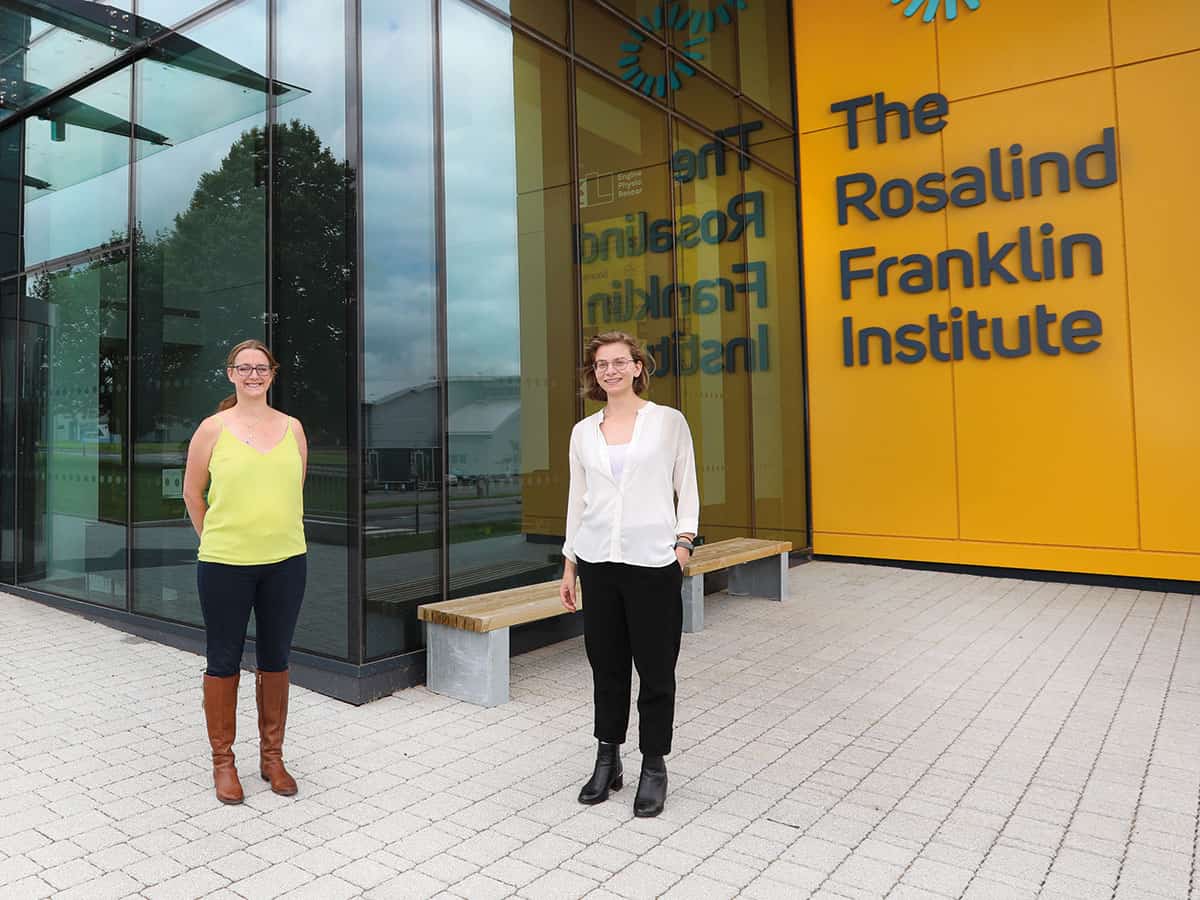
From improving the sensitivity of ion sources to boosting image resolution, Felicia Green and Anna Simmonds unveil the ambitious biological mass spectrometry programme at the Rosalind Franklin Institute to image molecular interactions in tissues
As scientists, one of the most frustrating things we can be told is “nice idea, but the technology to achieve it doesn’t exist yet”. Academic researchers rarely have enough time and resources to push the development of such technology forward, so these ideas invariably get shelved.
One such nice idea would be to use mass spectrometry to “see” metabolism – the chemical processes that underpin life itself – as they take place, to help our understanding of diseases. As you might imagine, the technology to do this doesn’t yet exist. But for once, a team of physicists, to which we belong, has been given both the time and resources to try and change that.
Mass spectrometry – where a sample is ionized to separate its components and measure them, based on the combined masses of its atoms – is one of the most sensitive and versatile techniques for studying the nature and interactions of molecules in a biological sample. The use of this century-old technique for biological applications has grown exponentially over the last 20 years, thanks to the discovery of “soft” ionization techniques, which allow researchers to analyse much bigger molecules than was ever possible before. Combined with the use of today’s improved computational power, mass spectrometry is now able to study everything from antibodies to viruses. Indeed, it allows researchers to identify, characterize and quantify proteins and other molecules present in biological samples.
Our team, based at the Rosalind Franklin Institute in the UK, is primarily interested in two areas of mass spectrometry: mass spectrometry imaging and structural mass spectrometry. Mass spectrometry imaging allows images of biological tissue samples to be produced by collecting mass spectra in a spatially resolved manner. Structural mass spectrometry methods make the technique increasingly useful for studying the structure of biological molecules, not just their identity. However, to study everything that is happening in a biological tissue sample at the molecular level at one time requires a degree of complexity that isn’t currently possible.
What we need is the ability to measure things at a systems level, to see all the molecules in one place, even in something highly heterogenous, like a tumour sample. The goal is to be able to measure and identify all the metabolic substances, the lipids and many of the proteins, in their original spatial location. This would create a complete picture of metabolism in situ, alongside the key proteins that govern that metabolism at a cellular level. If specific molecular interactions can be related to a disease state, it could make a fundamental impact on our understanding of diseases and how they might be treated.
Setting the challenge
Our team, led by analytical chemists Josephine Bunch and Zoltan Takats, has set itself quite a challenge: to develop new mass spectrometry instrumentation that can make molecular maps of biological tissues at unprecedented sensitivity, chemical depth and spatial resolution.
To do this, we are tackling the three Achilles heels of mass spectrometry for biological imaging: size of molecules, speed and molecular identification. The goal is to be able to study both large and small molecules using the same instrument, while also making measurements faster and increasing their sensitivity. And not just that: the machine will also need to have subcellular spatial resolution and swiftly identify the structures of all species detected.
We are tackling the three Achilles heels of mass spectrometry for biological imaging: size of molecules, speed and molecular identification
As a first step, the various groups involved in the biological mass spectrometry project have, with industry partners, been developing instruments that use new technologies – such as a new type of ion source – and combining existing technologies for the first time. The goal set by the Franklin for technology development is to improve by a factor of 10 on what’s currently possible – but in reality, we’re looking at much more than that just for these initial individual instruments. If we are finally able to combine them all into one mass spectrometer, it’s almost impossible to calculate quite how extraordinary that could be. Even if, ultimately, creating that one mass spectrometer with all these capabilities just isn’t possible, those individual improvements we will have made in sensitivity, image resolution, ion mobility resolution, mass accuracy, mass resolution and speed will still have wide-ranging benefits.
Shape and structure
One of the planned instruments is a hybrid that incorporates two mass analysers, a time-of-flight (TOF) analyser and a Fourier-transform ion cyclotron resonance (FT-ICR) analyser. The team working on this brings together scientists from our institution, along with those from the National Physical Laboratory and Imperial College London in the UK, as well as the global analytical instrumentation company Bruker. This hybrid instrument is being developed in parts, with the team currently spread across three locations and two countries. Ensuring that each piece works well independently is important, but we also have to make sure that all the pieces will fit together when we start to combine them, so we’re in frequent communication with each other to share our progress and ideas.
The high throughput of the TOF analyser in the hybrid instrument allows the rapid spectral acquisition necessary for high spatial-resolution MSI experiments, while the FT-ICR allows unparalleled mass resolution and access to many ion-manipulation techniques that will make it possible to probe the structure of biological molecules, not just their identity. The instrument will also include a tandem trapped-ion mobility spectrometry (TIMS) device, which can in addition analyse the structure of biomolecules as well as separate out the components of biological samples based on their shape (Analyst 143 2249). We are currently working on modelling multiple possible geometries of the device, to explore how ions travel through it, as well as studying how the device would be able to direct ions into either one of the mass analysers.

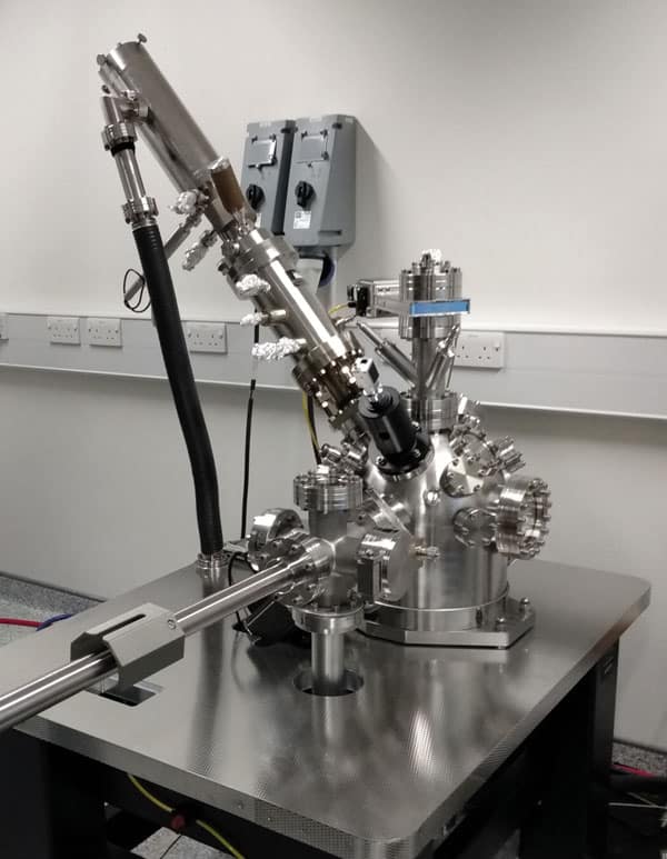
To generate ions from biological samples, we are also designing a multimodal imaging source that will be coupled to the hybrid mass spectrometer. This source will include multiple ionization techniques to provide choice and flexibility over a number of aspects, including the pixel size of the resultant mass spectrometry image; which classes of biomolecules are ionized; and how much preparation needs to be performed on the sample prior to its analysis. We still need to make significant improvements to obtain the required sensitivity, while at the same time producing data that can be interpreted. With this in mind, we’re planning to include post-ionization techniques to increase the number of molecules detected from each sampling location, as well as to improve the range of biomolecules detected from tissues.
A prototype of the multimodal ion source has been developed and installed on a Bruker timsTOF fleX mass spectrometer. The current iteration of this ion source includes atmospheric pressure “matrix-assisted laser desorption/ionization” (MALDI) – an ionization technique that uses a laser-absorbing matrix to create ions from molecules, with minimal fragmentation – in both transmission and reflection modes. The set-up has successfully demonstrated an enhancement of detected ion intensity, by plasma post-ionization (Analytica Chimica Acta 1051 110). This set-up has the ability to complement the molecular information collected by commercially available MALDI systems, by allowing a number of different chemical components to be analysed.
We currently use in-house software to control the source, which allows real-time processing of data to create a live mass spectrometry image, as the data are being acquired. It also provides feedback control of experimental parameters in an on-the-fly manner. As the software is highly modular, it will allow us to easily incorporate new additions, as the instrument evolves. Work is also under way on a new atmospheric-pressure interface that will improve the transmission of ions from the multimodal ion source into the mass spectrometer. Traditional interfaces are based on simple capillaries, which tend to have significant transmission losses when transporting ions and charged clusters into vacuum. By exploring novel inlet designs, we aim to create a new inlet that is customisable to each mode of the source, thereby significantly increasing our transfer yields, and improving the overall sensitivity of the set-up.
Protein problems
Analysing proteins, in particular, using mass spectrometry imaging is a key challenge for scientists, as proteins are often large and challenging to extract from biological samples. “Bottom-up” techniques aim to study proteins by digesting them (almost always enzymatically) into smaller fragments, which can then be “reconstructed” into the original protein. Unfortunately, these techniques often use liquid-phase reactions (such as “liquid chromatography” – a technique used to separate and analyse a mix of proteins) that are incompatible with mass spectrometry imaging, and require significant sample preparation and time.
With this in mind, we have developed an “atmospheric pressure glow discharge device” that can digest proteins and other large molecules in situ. A glow discharge is a plasma that forms when an electric current flows through a gas; and such plasmas have long been used as ionization sources in mass spectrometry. By developing this device, we aim to produce unique, consistent and spatially resolved markers from proteins, which could be subsequently analysed by mass spectrometry imaging. This would represent a dramatic reduction in sample preparation and, crucially, would retain the valuable spatial information in native samples. Another mass spectrometer we have planned will exploit new developments in the use of water cluster beams for molecular desorption, which can enhance sensitivity 100-fold while reducing and controlling the fragmentation during surface sampling. It will help us to retain sensitivity at very low pixel sizes and ensure full coverage across the types of biological molecules detected.
Snapshot in time
Yet another instrument we’re developing as part of the theme will use mass spectrometry in “microscope mode” and is based on secondary-ion mass spectrometry (SIMS), in which the specimen’s surface is sputtered using a focused primary ion beam, and the analysis is done by collecting ejected secondary ions.
Our “stigmatic” (microscope) SIMS instrument allows for rapid molecular mapping of biological tissues at unprecedented speed, as it decouples acquisition time from spatial resolution. Typically, mass spectrometry imaging works by scanning across a surface, and taking a mass spectrum at each spot, to build up the pixels of the image. In this case, however, the whole surface is imaged simultaneously using state-of-the-art cameras that operate as an array of position- and time-sensitive detectors, recording a mass spectrum for each pixel in the camera image (Rapid Commun. Mass Spectrom. 27 2745).
This instrument design is a joint endeavour between the Franklin, chemists at the University of Oxford and staff from the ion-beam technology company Ionoptika. The ion source was built, tested and installed at Oxford by Ionoptika. Typically, SIMS is used for microprobe analysis, which uses a highly focused (~500 nm) primary ion beam, so the next stage required us to produce a uniform defocused beam (~2 mm) for microscope mode. This involved detailed modelling and simulation of the primary ion dynamics, and experimental measurement to ensure beam size and uniformity at the sample, before we could consider secondary ions and imaging. The Oxford team’s expertise in adapting complementary metal oxide semiconductor (CMOS) sensors into pixelated time-sensitive cameras (Phys. Chem. Chem. Phys. 16 383) will allow us to develop spatially sensitive detection systems that record the arrival position and time of each secondary ion with nanosecond timing resolution. By combining new ion beam technology (J. Am. Soc. Mass Spectrom. 31 1903) and fast detectors, our team aims to improve both mass and spatial resolutions while maintaining rapid imaging. This would mean a mass spectrometry image of a standard tissue biopsy would take seconds rather than hours or days, paving the way for routine analysis.
The next stage of development will take place at the Franklin’s new hub building, which opened on the Harwell Campus near Oxford this year. The Franklin’s mission is to develop new technologies that can have a major impact on the life sciences, by pushing the boundaries of physical sciences. The building has been designed to house the new technologies being developed under each of the institute’s key research areas – which include artificial intelligence and structural biology, among others – as well as providing space for the teams to work together on further innovations. Bit by bit, we’ll be moving our novel instruments, developed at different sites around the UK, into their new home. Then we’ll need to see if we can combine them all together, turning that “nice idea” into reality.


