
PET imaging with radiotracers to accurately identify molecules in cancer cells that prevent the immune system from attacking a disease, called immuno-PET, could become an effective tool in assessing inflammatory bowel disease (IBD), according to a preclinical study published in the Journal of Nuclear Medicine.
Australian researchers have shown that immuno-PET has potential in detecting IBD in mice. If successful in human trials, immuno-PET could provide the dual theranostic objective of diagnosing inflammatory diseases while also helping clinicians follow up precision treatment (J. Nucl. Med. 10.2967/jnumed.118.219287).
“Our results provide further evidence of the clinical utility of immuno-PET for IBD diagnosis and monitoring,” wrote lead author Nicole Dmochowska, a doctoral student, and colleagues from the South Australian Health and Medical Research Institute in Adelaide. “Development of these techniques would be of particular benefit to patients who are difficult to scope, including the paediatric population and those that have inflamed or fibrotic regions of the small intestine.”

The diagnosis and monitoring of IBD, a disease category that includes Crohn’s disease and ulcerative colitis, are critical for many patients. A lack of definitive indications makes it difficult to know when symptoms will reappear, and patients with a long history of IBD run the risk of developing colon cancer.
Currently, IBD assessment is dependent upon endoscopy and colonoscopy, but neither procedure can reach all areas of the small intestine, nor can these procedures provide real-time information on the efficacy of treatment and drugs.
“Therefore, new technologies are required for imaging IBD that are less invasive than endoscopy and can provide information on specific pathologic processes in real-time,” the authors wrote.
What is known is that IBD inflammation activates the body’s natural immune system, which contains immune cells that have the surface receptor CD11b. Those cells secrete IL-1β to generate immune responses.
“However, a direct comparison of antibodies targeted to innate immune mediators and cells has not been done,” Dmochowska and colleagues added. “We aimed to compare immuno-PET of antibodies to IL-1β and CD11b against standard F-18 FDG and MRI approaches to detect colonic inflammation.”
For this study, the researchers looked at two sets of mice: those with ulcerative colitis and a group of age- and weight-matched healthy mice. The team watched the first group daily for signs of acute colitis. They then compared the ability of immuno-PET with zirconium-89 (Zr-89) conjugated antibodies against IL-1β, CD11b, and F-18 FDG, along with MRI, to detect inflammation in colitic mice.
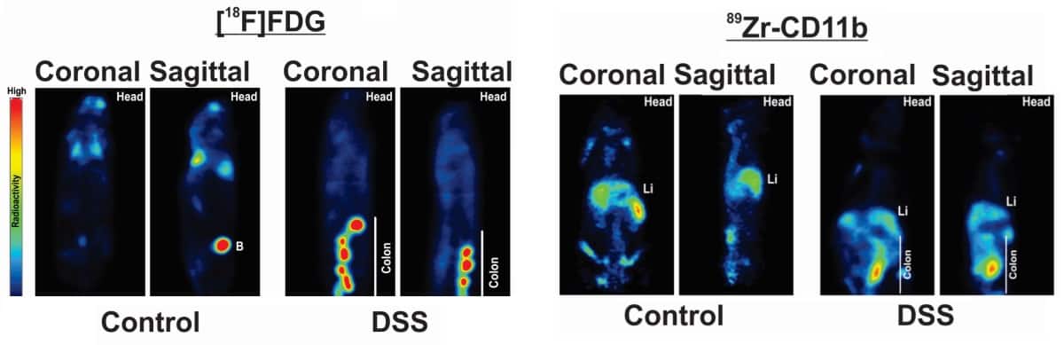
Immuno-PET images showed that in colitic mice, distal colonic uptake of Zr-89 IL-1β increased by approximately threefold; uptake of Zr-89 CD11b was five times greater, and uptake of F-18 FDG rose by around 3.5-fold. MRI achieved an approximately twofold increase in the T2 signal intensity ratio in colitic mice.
In addition, an ex vivo PET analysis showed the uptake of Zr-89 IL-1β and Zr-89 CD11b increased throughout the entire gastrointestinal tract of the colitic mice, compared with the control mice. Zr-89 IL-1β also was distributed mainly in the gastrointestinal tract, while Zr-89 CD11b spread to more tissue types. Zr-89 IL-1β also correlated with colitis severity, while Zr-89 CD11b showed no such association.
“Our comparison demonstrates the strong potential that immuno-PET of innate immune mediators has for diagnosing and monitoring IBD,” the researchers concluded.
- This article was originally published on AuntMinnie.com. ©2019 by AuntMinnie.com. Any copying, republication or redistribution of AuntMinnie.com content is expressly prohibited without the prior written consent of AuntMinnie.com.


