Spectral-domain optical coherence tomography (OCT) is the gold standard for diagnosing retinal diseases such as macular degeneration and diabetic retinopathy, as well as glaucoma. OCT is particularly useful as a screening tool for early detection of disease. But widespread screening is hindered by the large size and expense of OCT systems, which can cost more than $100,000.
Biomedical engineers at Duke University have now developed a portable OCT scanner that’s 15 times lighter than current commercial systems and is expected to be sold for less than $15,000. The team hopes that the new scanner, which delivers comparable image quality to current systems, could increase access to retinal imaging and help bring this vision-saving technology to underserved regions throughout the world (Trans. Vis. Sci. Tech. 10.1167/tvst.8.3.61).
“Once you have lost vision, it’s very difficult to get it back, so the key to preventing blindness is early detection,” says lead author Adam Wax. “Our goal is to make OCT drastically less expensive so more clinics can afford the devices, especially in global health settings.”
System redesign
OCT is an interferometric technique that works by sending near-infrared light into tissue and detecting the backscattered signal. A spectrometer compares the phase of this returning light with that of a reference beam that has travelled the same distance but not interacted with tissue, and uses the time delay information to create a depth profile of the tissue structure.
To build a smaller, less expensive OCT device, Wax and his former graduate student Sanghoon Kim redesigned the spectrometer. While traditional spectrometers direct light through a W-shaped series of lenses, mirrors and diffraction slits, in Wax’s spectrometer, the light travels on a circular path within a housing made mostly from 3D-printed plastic. This means that any mechanical shifts arising from temperature changes occur symmetrically, balancing out to keep the optical elements aligned. The device also uses a large detector at the end of the light path to minimize misalignments.
The handheld scanner, which contains the light source, reference arm and sample arm, weighs less than 500 g. The system body, which houses the custom-designed spectrometer and an integrated mini-PC, weighs 1.8 kg. The team also incorporated a touchscreen into the system body to enable data acquisition control and display of the retinal images.
“Right now, OCT devices sit in their own room and require a PhD scientist to tweak them to get everything working just right,” says Wax. “Ours can just sit on a shelf in the office and be taken down, used and put back without problems. We’ve scanned people in a Starbucks with it.”
Clinical comparisons
In the first clinical trial of the new OCT scanner, retina surgeon J Niklas Ulrich from the University of North Carolina tested it’s performance against a commercial Heidelberg Spectralis OCT system. Ulrich imaged 60 eyes in healthy volunteers and 60 eyes with known retinal disease. Although the low-cost scanner is designed as a handheld device, for this comparison, it was mounted on a chin rest.
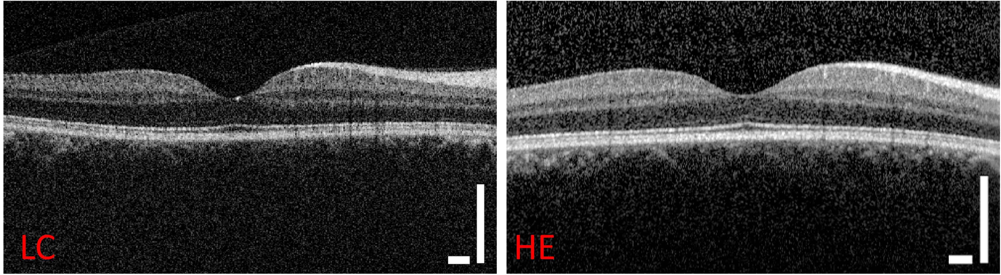
The low-cost OCT demonstrated an axial resolution of 8.0 µm, a lateral resolution of 19.6 µm and an imaging depth of 2.7 mm for a 6.6 mm field-of-view. The images could clearly resolve relevant layers of the retina, comparable to those from the Heidelberg Spectralis system. The mean contrast-to-noise ratio (CNR) of images from the portable scanner was only 5.6% lower than that of the commercial machine, which is good enough for clinical diagnostics.
“I have been very impressed by the quality of images from the low-cost device, it is absolutely comparable to our standard commercial machines,” says Ulrich. “It allows for accurate diagnosis of structural retinal disease as well as monitoring of treatment success. The setup is quick and easy with a small footprint, allowing the device to perform well in smaller satellite offices.”
The researchers also imaged five patients using the scanner in handheld mode. They were able to obtain high-quality retinal images with an operator holding the scanner without a chin rest. There was no significant difference in mean CNR between images acquired in handheld mode and those acquired using a chin rest.
Wax is commercializing the device through the start-up company Lumedica, which is already producing and selling first-generation instruments for research applications. “There’s a lot of interest from people who want to take OCT to new parts of the globe as well as to underserved populations right here in the US,” he explains.
“With the growing number of cases of diabetic retinopathy in places like the United States, India and China, we hope we can save a lot of people’s sight by drastically increasing access to this technology,” says Wax.


