Diagnosing skin cancer is a tricky task – even highly skilled dermatologists rely on magnifying dermatoscopes to examine suspicious blemishes and excise tissue for analysis. Now, researchers at Stevens Institute of Technology have developed a millimetre-wave imaging technique that can detect skin lesions and determine whether they are cancerous or benign. In future, they plan to incorporate the technology into a handheld device that will rapidly diagnose skin cancers without the need for biopsy (IEEE Trans. Med. Imag. 10.1109/TMI.2019.2902600).
The non-invasive diagnostic method, developed by Negar Tavassolian, director of the Stevens Bio-Electromagnetics Laboratory, and postdoctoral fellow Amir Mirbeik-Sabzevari, offers the potential to halve the number of unnecessary biopsies. “This could be transformative,” says Mirbeik-Sabzevari. “No other technology has these capabilities.”
The imaging system uses millimetre waves (30–300 GHz), which penetrate up to 1.3 mm into tissue, making them highly effective for sensing pathological changes in different skin layers. To improve the resolution of the acquired images, the team developed an approach called “synthetic ultrawideband millimetre-wave imaging” to create an imaging system with a synthetic bandwidth of 98 GHz.
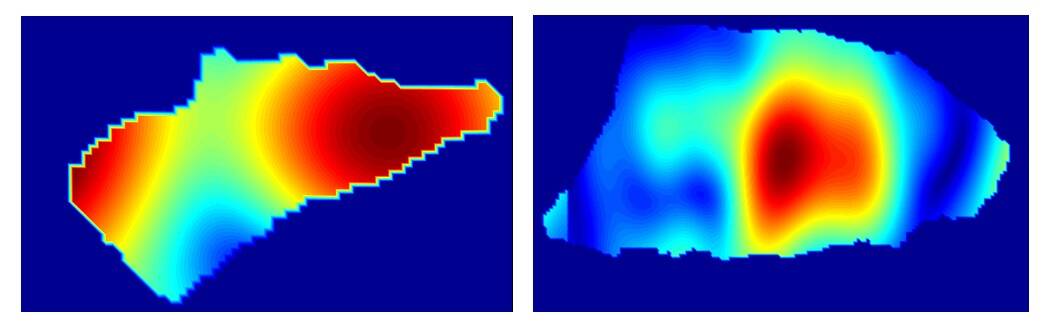
The researchers used their system to examine 21 skin cancer samples: 13 basal cell carcinoma (BCC) and eight squamous cell carcinoma (SCC) specimens. They found that millimetre-wave imaging could produce high-contrast, high-resolution 3D images of the skin, which identified the locations of the tumours as accurately as histological imaging.
Both types of cancerous cells showed higher reflectivity than normal skin tissue, enabling identification of diseased tissue by looking for reflectivity hotspots. Over all 21 samples, the average percentage reflectivity was 74% and 30%, for tumour and normal regions, respectively, indicating that millimetre-wave reflectivity is a reliable marker for cancerous tissue.
“We’ve shown proof-of-concept that this technology can be used for rapidly detecting skin cancer,” says Tavassolian. “That’s a major step forward toward our ultimate goal of developing a handheld device, which would be safe to use directly on the skin for an almost instant diagnostic reading of specific kinds of skin cancer – including lethal melanomas – based on their individual reflectivity signatures.”
Since millimetre-wave imaging does not require tissue processing or staining, it can be performed promptly, enabling diagnosis of tumours at an early stage. A handheld scanner could also be used to generate real-time 3D images of tumours to guide surgeons, eliminating the need for multiple biopsies to fully remove cancerous tissue.
The devices could also be configured to interpret images automatically and deliver basic diagnostic information without needing a trained operator. “We could place these devices in pharmacies, so people can get checked out and go to a doctor for a follow-up if necessary,” says Tavassolian. “People won’t need to wait weeks to get results, and that will save lives.”
Crucially, the underlying technology is inexpensive. “It should be possible to keep manufacturing costs below $1000, even at low production volumes,” says Tavassolian. “That’s about the same as the magnifying tools already used by dermatologists, and an order of magnitude cheaper than laser-based imaging tools, which also tend to be slower, bulkier and less accurate than millimetre-wave scanners.”
Mirbeik-Sabzevari, who has been working on the technology for five years, is confident that this invention will prove a hit and plans to launch a start-up to commercialize the scanners.


